Reading...
![]()
Play button
![]()
Play button
![]()
Use LEFT and RIGHT arrow keys to navigate between flashcards;
Use UP and DOWN arrow keys to flip the card;
H to show hint;
A reads text to speech;
66 Cards in this Set
- Front
- Back
|
When does mesoderm develop?
|
third week
|
|
|
When does primitive streak appear?
|
third week
|
|
|
What is the name given for the third layer?
|
mesoderm
|
|
|
What layer gives rise to embryonic connective tissue of the facial region and to the cartilages of the branchial arches?
|
Ectomesenchyme
|
|
|
What is mesenchyme?
|
At the end of the third week the paraxial mesoderm forms two solid rods of tissue one on each side of the central notochord.
These rods undergo segmentaion and form somites. During 4th week the somites begin to differentiate further forming dermatome, myocoels, myotome and sclerotome. Sclerotome differntiates into connective tissues called mesenchyme. |
|
|
When does mesoderm develop?
|
third week
|
|
|
When does primitive streak appear?
|
third week
|
|
|
What is the name given for the third embryological layer?
|
mesoderm
|
|
|
What layer gives rise to embryonic connective tissue of the facial region and to the cartilages of the branchial arches?
|
Ectomesenchyme
|
|
|
What is mesenchyme?
|
At the end of the third week the paraxial mesoderm forms two solid rods of tissue one on each side of the central notochord.
These rods undergo segmentaion and form somites. During 4th week the somites begin to differentiate further forming dermatome, myocoels, myotome and sclerotome. Sclerotome differntiates into connective tissues called mesenchyme. |
|
|
What structures do the neural crest cells become into?
|
components of peripheral nervous system including sensory ganglia, sympathetic neurons, Schwaan cells
meninges, and pigment cells |
|
|
How is ectomesenchyme differ to mesenchyme?
|
mesenchyme is derived from mesoderm but ectomesenchyme is derived from neuro crest cells.
|
|
|
What is Treacher Collin's Syndrome?
|
interference in the migration of neural crest cells in the facial area
|
|
|
Where is the origin of facial and neck muscles?
|
Somites/somitomeres which migrate into the branchial arches or the frontonasal process
|
|
|
What is the name for the developing oral cavity?
|
Stomatodeum
|
|
|
When does Stomatodeum appear?
|
in the 4th week
|
|
|
At first what is stomatodeum lined with?
|
oral ectoderm
|
|
|
What becomes of oral ectoderm later?
|
give rise to the teeth and oral epithelium
|
|
|
Which feature represents the wall that separates the gut from the mouth?
|
buccopharyngeal membrane
|
|
|
When does the neck start to develop?
|
Fourth week of development
|
|
|
What is the origin of the neck?
|
brachial arches and the primitive pharynx
|
|
|
Describe the origins of oesophagus, larynx and trachea.
|
Caudal part of primitive pharynx forms oesophagus
Ventral outgrowth leads to larynx and trachea |
|
|
How many brachial arches are there originally?
|
6
|
|
|
How many brachial arches survive?
|
5
|
|
|
What are the components of a brachial arch?
|
primitive striated muscle tissue
some nervous tissue some vascular tissue bar of cartilage in its mesodermal core |
|
|
What lines the branchial arches?
|
ectoderm outside
endoderm inside |
|
|
Which arches are related to the formation of the face and which arches are for the neck?
|
first two arches for the face
and the rest for the neck |
|
|
What is the name given for the 1st branchial arch?
|
mandibular arch
|
|
|
What is the name for the cartilage found in first pharyngeal arch?
|
Meckel's cartilage
|
|
|
What does 1st branchial arch become?
|
bone: incus and malleus bones, maxilla, mandible
ligament: anterior ligament of malleus and the sphenomandibular ligament muscles: muscles of mastication, tensor tympani, tensor veli palatini, mylohyoid and anterior belly of the digastric nervous tissue: mandibular division of trigeminal nerve vascular tissue: degenerates |
|
|
What is the name given to 2nd branchial arch?
|
Hyoid arch
|
|
|
Name the cartilage of the 2nd branchial arch.
|
Reichert's cartilage
|
|
|
What does 2nd branchial arch become?
|
bone: Stapes, styloid process of the temporal bone, and some of the hyoid bone including the upper part of its body and the lesser cornu
ligament: stylohyoid ligament muscles: Muscles of facial expression, stapedius, stylohyoid and the posterior belly of digastric nervous: Facial nerve(VII) vascular: degenerates |
|
|
What happens to the cartilage of third arch?
|
contributes to the hyoid bone
|
|
|
What happens to the muscles of third arch?
|
stylopharyngeus muscle
|
|
|
What happens to the Nerves of third arch?
|
glossopharyngeal nerve(IX)
|
|
|
What happens to the artery of third arch?
|
becomes common carotid artery and part of internal carotid artery
|
|
|
Which two arches fuse together?
|
4th and 6th
|
|
|
What happens to the cartilages of 4th and 6th arch?
|
contribute most of the laryngeal cartilages
|
|
|
What are Laryngeal cartilages?
|
thyroid
cricoid arytenoid corniculate cuneiform |
|
|
What happens to the nervous tissues of 4th and 6th arch?
|
4th: superior laryngeal branch of the vagus nerve (X)
6th: recurrent laryngeal branch of vagus (X) |
|
|
What happens to the arteries of 4th and 6th arch?
|
4th: arch of the aorta on the left side and contributes to the right subclavian and brachiocephalic arteris
6th: contributes to the pulmonary arteries |
|
|
What happens to the 5th arch?
|
it degenerates
|
|
|
Differentiate between pharyngeal cleft and pouch.
|
Pouch is the fold seen in the inside aspect and cleft is the fold seen from outside.
|
|
|
What becomes of the first pharyngeal pouch?
|
auditory tube and middle ear
|
|
|
What becomes of the first pharyngeal cleft?
|
external auditory meatus, and the tympanic membrane
|
|
|
What becomes of the second pharyngeal pouch?
|
tonsillar fossa between the palatoglossal and palatopharyngeal folds
which eventually becomes palatine tonsil |
|
|
What are the processes that are responsible for the facial development?
|
frontonasal process
maxillary process mandibular process |
|
|
The processes (frontonasal, maxillary, mandibular) of facial development originate from which pharyngeal arch?
|
the first pharyngeal arch
|
|
|
Describe the fusion of the arches to form the face.
|
Frontonasal process forms nasal pits bilaterally. Both maxillary swellings move towards the center fuse with the frontonasal process superior to stomatodeum to form the upper lip. Mandibular swelling becomes the lower lip and the mandible.
|
|
|
When is the secondary palate formed?
|
week 7 or 8
|
|
|
The secondary palate arise from which process?
|
maxillary process
|
|
|
What is the nerve of the primary palate?
|
incisive branch of the long nasopalatine branch of the maxillary division of trigeminal nerve (V)
|
|
|
What is the nerve of the secondary palate?
|
Greater palatine branch of the maxillary division of V
|
|
|
What is the name of the swelling in the midline of the mandibular process of the first brachial arch? What does it become?
|
tuberculum impar
becomes the anterior 2/3 of the tongue |
|
|
What are the swellings are involved in the formation of the tongue?
|
tuberculum impar
Lateral lingual swellings |
|
|
What do the swellings develop into?
|
They merge together to become mucous membrane of 2/3 of the tongue.
|
|
|
What nerve innervates the anterior 2/3 of the tongue?
|
lingual nerve of mandibular division of V (sensory)
chorda tympani VII (taste) |
|
|
The nerve of the second arch contributes taste fibres to the anterior two-thirds of the tongue, via the ____ ____.
|
chorda tympani
|
|
|
The posterior part of the tongue arises from the ____ ____, a large midline swelling from the ____ pharyngeal arch, the nerve of which becomes the ____
|
hypobranchial eminence
third glossopharyngeal (IX) |
|
|
What nerves innervate the posterior part of the tongue?
|
Glossopharyngeal (IX)
|
|
|
How is the mandible formed?
|
Two mandibular processes fuse in the midline to form a mandibular arch. The bone begins to form in the mesenchymal tissue lateral to the cartilage. The cartilage begins to disappear.
|
|
|
How is Meckel's cartilage involved in the formation of the mandible?
|
very little. It may contribute to positioning of the mental foramen.
|
|
|
What are the five environmental factors that can affect the embryo development?
|
infectious agent
ionising radiation drugs hormones nutritional deficiencies |
|
|
The most common birth defect in the oral cavity is called...
|
orofacial clefts
|
|
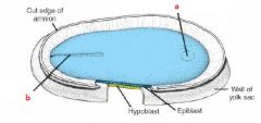
Label
|
a. buccopharyngeal membrane
b. primitive streak |