Reading...
![]()
Play button
![]()
Play button
![]()
Use LEFT and RIGHT arrow keys to navigate between flashcards;
Use UP and DOWN arrow keys to flip the card;
H to show hint;
A reads text to speech;
8 Cards in this Set
- Front
- Back
|
EM of a CNS tumor showed intracellular lumens lined by microvilli with specialized junctions between cells and
glial features. |
ependymoma.
|
|
|
EM of a CNS tumor shows prominent interdigitating processes without a basal lamina. There are well formed
desmosome junctions and prominent cytoplasmic inclusions. |
meningioma
|
|
|
EM of a tumor shows rhomboid crystalline inclusions. Cytogenetics also showed a t(X;17)
|
Rhomboid crystalline inclusions are characteristic of alveolar soft part sarcoma.
|
|
|
This EM depicts an adenocarcinoma with short mircovilli protruding from the cell surface. Mesothelioma
would have longer microvilli. |
|

|
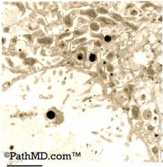
These EM photos illustrate the “granule in granule” appearance, which characterizes a granular cell
tumor. These are also refered to as autophagasomes. Melanoma is a thought, but the dense bodies are not characteristic of a melanosome. Granular cell tumors are of nerve sheath orgin (myoblastomas) and express S-100. They are notorious for overlying psuedoepitheliomatous hyperplasia. ( |
|
|
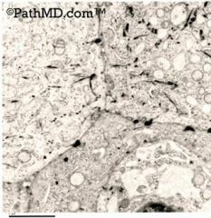
Squamous cell carcinoma
These EM photographs show tonofilaments (the black irregular fibril-like densities). Some of these can be seen arising from desmosomes, which anchor cells to cells. |
|
|
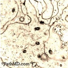
These images are best associated with a meningioma. The “interdigitating” cell membrane combined with
the desmosomes are the most characteristic finding. Lymphoma would not have so much cytoplasm or desmosomes. Adenocarcinoma usually will have evidence of microvilli, and squamous cell carcinoma will have tonofilaments. |
|
|
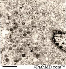
The features in these EM photos best illustrate an oncocytoma with numberous mitochondria in the
cytoplasm. A chrmophobe RCC will have 150-750nm vesicles in the cytoplasm. |