Reading...
![]()
Play button
![]()
Play button
![]()
Use LEFT and RIGHT arrow keys to navigate between flashcards;
Use UP and DOWN arrow keys to flip the card;
H to show hint;
A reads text to speech;
40 Cards in this Set
- Front
- Back
|
Principles of functional tissue engineering (2)
|
(1) understand structure function relationships
(2) the mechanical properties of the Native tissues must be established for subfailure and failure conditions |
|
|
Epithelial Tissue function
|
Skin epidermis, thyroid follicles. Lines, covers, protects, absorbs, and secretes
|
|
|
Connective tissue
|
Skin dermis, tendon, bone marrow, cartilage, bone, blood cells. Scaffolds for cells, nerves, blood vessels, energy storage, defence mechanism, oxygen and nutrient transport, mechanical support
|
|
|
Muscle tissue
|
contraction. regulates blood flow, regulates flow in the GI tract, motor functions
|
|
|
ECM
|
represents a complex alloy of variable members of diverse protein families defining structural integrity and various physiological functions. The primary function of ECM is to endow tissues with their specific mechanical and biochemical properties and also effects cell functions.
|
|
|
What does the ECM do to cell function? (4)
|
(1) Promotes cell attachment and migration (2) regulate or promote cell differentiation and gene expression levels (3) establishment of tissue microenvironment (4) sequestering, storage, and presentation of soluble regulatory molecules
|
|
|
What is the ECM made of?
|
Fibrous proteins (collagen, elastin, fibronectin), large aggregating sugars (proteoglycans- cement material), water
|
|
|
What does collagen do?
|
forms the fibrillar and microfibrillar networks of ECM basement membranes. Type I is most abundant- in skin, tendon, bone, ligaments. Type II in cartilage.
|
|
|
Collagen structure
|
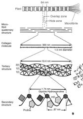
3 strands of procollagen form a helix made which is stabilized by hydrogen bonds. Hydroxylation and Glycosylation occurs to the procollagen. Telopeptides on the ends are cleaved and tropocollagen is formed. Multiple tropocollagen mols form collagen fibrils and are crosslinked by lysyl oxidase.
|
|
|
Tendon structure
|
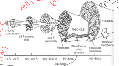
The crimping pattern structure helps with shock absorption, Allows some facicles to move without moving others. endotendon binds facicles together and allows some sliding.
|
|
|
Ligaments vs. Tendons
|
Ligaments: Connects bones together, augment joint stability, constrain joint motion. Tendons: Attach muscle to bone. Transmit muscle force to bone. Absorb and release energy- important for protecting from injury
|
|
|
Tendon & ligament biochemistry
|
-70% water, dry mass is mostly collagen with some elastin and proteoclygan. The collagen is mostly made of type I collagen. The proteoglycans include decorin, which helps the assembly of collagen into larger fibers, and aggrecan, which mediates the viscoelastic response.
|
|
|
Tendon vs. ligament biomechanics
|
Tendons contain parallel collagen fibrils which provide high unidirectional strength. Parallel alignment of collagen fibers is less in ligaments which enables them to withstand loads in different directions.
|
|
|
Tendons can behave like...
|
Springs
|
|
|
Stress, strain, and hooke's law
|
stress = sigma = F/A;
strain = epsilon = delta L/L0; Hooke's law for elastic materials = sigma = E * epsilon where E is the elastic modulus |
|
|
Stress strain curve of Tendons and Ligaments
|
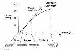
area under the curve is proportional to toughness. Stiffness is the slope of the force/deformation curve.
|
|
|
Doubling the area leads to...
Doubling the length leads to... |
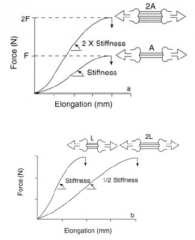
...2x the stiffness, aka greater slope.
...1/2 the stiffness, smaller slope. |
|
|
Mechanical traits of tendons (4)
|
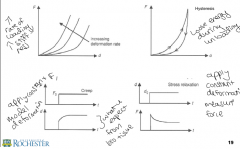
(1) mechanics are dependent on the strain rate. Faster loading leads to stiffer tendons with higher capacity for energy storage.
(2) A small amount of energy is lost when they are strained, so different stress-strain curves for loading and unloading (hysteresis) (3) The fluid within them reduces oscillations and shock by absorbing kinetic energy or dissipating it slowly (damping). (4) if a tendon is held in a stretched position, the energy stored within the tendon will decrease over time (creep). |
|
|
the mechanical spring model
|
a linear elastic solid; the stress is linearly proportional to the strain
|
|
|
Mechanical dashpot model
|
a linear viscous fluid element; the rate of deformation or flow is proportional to the force, where the proportionality constant is the viscosity. Also analogous to the shear stress in a viscous fluid where shear stress is linearly proportional to the rate of shear deformation and the constant is called the shear modulus (mu).
|
|
|
Maxwell model of a viscoelastic fluid
|
spring in series with a dashpot. This undergoes creep indefinitely- initially there is a lot of deformation but then there is none after a while.
|
|
|
Kelvin model of a viscoelastic solid
|
spring in series with a dashpot. The spring deformation is confined by the dashpot, there is 0 initial deformation until the limit of the spring is released.
|
|
|
Stiffness mismatch
|
Tendon has to overcome stiffness mismatch which is when two mechanically different tissues are joined, resulting in stress concentrations where injury is most likely to occur
|
|
|
Musculo-tendinous junction
|
the link between myofibers that generate the force in muscle and collagen fibers that transmit the forces to bone to effect locomotion. There isn't a clear cut change from muscle to tendon- increases SA toreduce stress
|
|
|
Osteo-tendinous junction
|
tendon inserts into bone across a fibrocartilaginous transition
|
|
|
Which has a higher elastic modulous, bone or tendon?
|
Bone (20 GPA). tendons or ligaments have about 200 MPa
|
|
|
Functions of cartilage (5)
|
Joint lubrication, low-friction surface, load distribution, joint congruity, shock absorption
|
|
|
What is cartilage made of?
|
water, Type II collagen, also Type 9 which is a crosslinker and type 11 which helps maintain spacing between type II collagens.
|
|
|
Structure of articular cartilage
|
Superficial zone- horizontal collagen fibers, high water content. Middle- mixed collagen fiber orientation, high proteoglycan content. Deep- closest to bone, vertical collagen fibers
|
|
|
Mechanics of the different zones in articular cartilage
|
Surface- high flow in & out, fluid pressure/tension, large compressive strains, tensile surface strains.
Transitional zone- low fluid flow, mainly fluid pressure, moderate compressive strains. Radial zone- little fluid flow, fluid pressure, small compressive strains. Noncompressible. |
|
|
Anisotropic
|
There are different properties of the material in different directions. For cartilage, collagen fibers resist tension in the same direction but break and perpendicular tensile forces.
|
|
|
Proteoglycans structure and functions
|
Made up of a hyaluronan backbone, glycosaminoglycans are attached wit hlink proteins. They are branch like structures which are negatively charged and can trap water. The water binds with electrostatic charges and it takes a lot of force to push the water out.
|
|
|
Properties of Articular cartilage (6)
|
(1) resists tensile (2) compressive (3) shear stresses, (4) Permeability of water (5) swelling, and (6) lubrication which can be tested with friction tests
|
|
|
Models of articular cartilage (4)
|
(1) AC is elastic- time dependent properties (2) single phase viscoelastic- flow independent and time-dependent (3) Biphasic- accounts for the interactions between the solid and interstitial fluid phases of the ECM, flow dependent and time dependent behaviors. (4) multiphasic- biphasic effects plus: electrical effects, chemical swelling, and transport of nutrients
|
|
|
3 biphasic testing configurations
|
(1) (Confined- determines bulk properties- piston pushes down on a cartilage sample which is enclosed and water can only escape in one direction- makes the problem into 1-D. (2) Unconfined- cartilage sample in between two plates, water can escape from sides and bulging can occur- not representative of tissue. (3) Indentation- allows to measure localized properties of cartilage- can create a topographical map of properties
|
|
|
Confined compression of cartilage
|
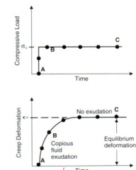
Can measure creep. Force is held constant over time, monitor deformation. This relates to the permeability of cartilage and compressibility of tissue. Normal AC has low permeability, diseased has high . During creep, the load at the surface is equal to the compressive stress + frictional drag due to fluid exudation which is releated to permeability as water leaves. at equilibrium, this is only due to compressive stress, which is related to aggregate compressive modulus (Ha) which is representative of the elasticity of the material.
|
|
|
Creep problem set up
|
k * Ha * u''(z) = u'(t). where k is the permeability, Ha is the aggregate modulus, and u (z,t) is the creep deformation. Boundary conditions: (z=0 at the top, z=h at the bottom) fluid pressure is 0, u(h,t) = 0 (no deformation at the bottom) and u(z,0)=0. As you increase the depth (towards the bottom) creep decreases. creep is mostly due to permeability
|
|
|
How do you calculate Ha from creep model?
|
set z=0, t=infinity. u(0, infinity) is equal to F0 *h/ (A* Ha)
|
|
|
As Ha increases...
|
the material is stiffer, and deformation decreases
|
|
|
As k (permeability) increases...
|
...the ability of water to leave increases and creep increases.
|