Reading...
![]()
Play button
![]()
Play button
![]()
Use LEFT and RIGHT arrow keys to navigate between flashcards;
Use UP and DOWN arrow keys to flip the card;
H to show hint;
A reads text to speech;
21 Cards in this Set
- Front
- Back
|
What are two common causes of intracerebral hematoma (ICH)?
|
Hypertension
Amyloid Angiopathy |
|
|
What's Amyloid Angiopathy?
|
It's a common cause of ICH. Amyloid protein deposits and accumulates on blood vessel walls, leading to their breakdown and thus ICH. You can see the hemorrhages anywhere in the brain.
|
|
|
What's characteristic of arteriovenous malformations (AVM)?
|
Nidus - tangle of small vessels that directly connect arteries with veins without a normal capillary bed.
This can lead to blood steal where the brain is less perfused, and also can increase risk of hemorrhage due to high pressure in the receiving veins. |
|
|
If after a surgery to fix a hemorrhage, the size of the hemorrhage does not decrease, what might this indicate?
|
POSSIBLE NEOPLASM! Enhancing MRi will show a thick rim of contrast to indicate a tumor.
|
|
|
What causes most subarachnoid hemorrhages?
|
Aneurysms, especially in basal cisterns.
traumatic brain injury |
|
|
What do patients with subarachnoid hemorrhages complain of?
|
"brick to the head" sudden terrible headaches
|
|
|
What are some complications of subarachnoid hemorrhages in basal cisterns?
|
Hydrocephalus - blood can clog the reuptake of CSF
Vasospasm - blood vessels can spasm down and get small if there's blood in the area, leading to secondary cerebral ischemia |
|
|
Where do most saccular aneurysms occur?
|
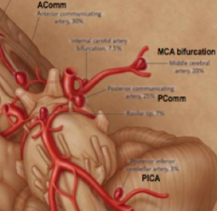
They tend to occur at arterial bifurcations in the circle of willis.
|
|
|
What are common causes of ischemic stroke?
|
Cardiogenic embolism
large-artery atherosclerosis (eg. internal carotid artery) Small-artery (<1mm) occlusion --> lacunar strokes |
|
|
What do we look at for ischemic stroke in imaging? Hint: 2 P's.
|
Parenchyma - presence and size of infarction
Pipes - vessels - occlusions in vessels |
|
|
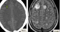
What signs show on the CT if there is infarcted matter?
|
Hypodensity - darker area. Can be due to cytotoxic edema
Note: if the hypodensity is large (more than 1/3 the area of MCA distribution), then poor outcome and more likely to hemorrhage after reperfusion. |
|
|
On a DWI, how does an acute infarct show? Brighter or darker?
|
Brighter - less diffusion.
Darker is a chronic ischemic stroke. |
|
|
How do you treat ischemic stroke?
|
IV-tPA if arrive in ER within 3 hours - NCCT is sufficient for eligibility of this treatment.
Intra-arterial (IA) thrombolysis - going in to the artery with a catheter and injecting tPA directly in there. - depends on "core" and "penumbra" mismatch. |
|
|
How does a cardioembolism show on imaging?
|
It generally shows as multiple, bilateral infarctions.
|
|
|
During the ictus of a seizure, does SPECT show increased or decreased blood flow?
What about inter-ictal or late post-ictal? |
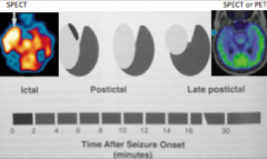
Increased blood flow during the ictal phase.
decreased blood flow in late or inter. |
|
|
During the inter-ictal or late post-ictal phases of a seizure, does PET show increased or decreased glucose metabolism?
|

Decreased glucose metabolism.
|
|
|
What are common anatomic lesions of epilepsy?
|
Mesial temporal sclerosis
Congenital disorders of cortex formation neoplasm less common: cortical gliosis, neurocysticercosis |
|
|
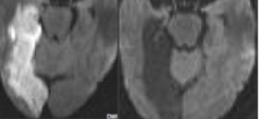
What brain area is involved in mesial temporal sclerosis?
|
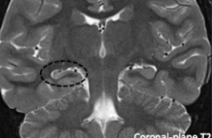
Temporal lobe, more specifically atrophy and gliosis in the HIPPOCAMPUS.
|
|

Is this scarring (gliosis) or edema? How can you tell?
|
This is gliosis! Mass effect shows PULLING toward lesion.
|
|
|
What can we use to demonstrate areas of eloquent cortex?
|
fMRI and diffusion tractography (preferential flow of water in white matter axons).
|
|
|
What is neurocysticercosis?
|
Infection caused by pork tapeworm. Leads to multiple cysts with larvae in the brain.
|