![]()
![]()
![]()
Use LEFT and RIGHT arrow keys to navigate between flashcards;
Use UP and DOWN arrow keys to flip the card;
H to show hint;
A reads text to speech;
49 Cards in this Set
- Front
- Back
- 3rd side (hint)
|
Doppler frequency / shift |
Increase in pitch on approach and decrease in pitch on departure. Frequency of sound changes when sound source & receiver move CLOSER together or FARTHER apart. |
|
|
|
Demodulation |
Process of extracting the low doppler frequency from transducers carrier frequency. |
|
|
|
Doppler shift created by ... |
Transmitted sound waves striking moving RBC. |
|
|
|
Positive Doppler shift |
Blood cells move toward the transducer. Reflected frequency higher than transmitted frequency. |
|
|
|
Negative Doppler shift |
Blood cells move away from transducer. reflected frequency lower than transmitted frequency. |
|
|
|
Speed |
Magnitude only. Indicates distance a RBC moves in 1 second. |
distance ÷ time (cm/s) EX/ 50 miles/hour |
|
|
Velocity |
Magnitude & direction. Think length of travel indicates magnitude, and angle indicates direction. |
EX/ 50 miles/hour WEST |
|
|
Doppler Equation |
2 x velocity of blood x transducer frequency x cos angle ÷ propagation speed |
|
|
|
Velocity of blood and Doppler shift |
Directly related. Faster velocity = greater doppler frequency. |
↓↓ or ↑↑ |
|
|
X-axis of Doppler system represents ... |
Time |
|
|
|
Y-axis of Doppler system represents ... |
Velocity |
|
|
|
Frequency of transmitted sound and Doppler shift ... |
Directly related. If transducer freq. doubled = Doppler shift doubled. |
↓↓ or ↑↑ |
|
|
Doppler shift measured in Hertz |
* directly related to velocity * directly related to transducer frequency |
↓↓ or ↑↑ |
|
|
Sound beam direction vs. flow direction |
Parallel for entire velocity to be measured. Directly toward or away transducer for 100% accuracy. |
|
|
|
Percentage of true velocity depends on ... |
Cosine of the angle between sound beam and direction of motion (flow). |
|
|
|
Angle (degree) * 0° * 60° * 90° |
Cosine * 1 * 0.5 * 0 |
|
|
|
Only portion of true velocity returned at angles other than ... |
0° and 180° |
|
|
|
Bidirectional Doppler |
distinguishes direction of flow toward or away from transducer. |
|
|
|
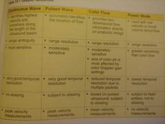
Continuous Wave Doppler |
2 crystals in transducer. One that constantly sends and one that constantly receives ultrasound energy. |
→ ← |
|
|
Advantage of CW Doppler |
Ability to accurately measure very high velocities. |
|
|
|
Primary disadvantage of CW Doppler |
Exact location of moving RBC cannot be determined. (Range ambiguity) |
|
|
|
Dedicated CW transducer |
No anatomic image can be produced (only sound) . Uses NO backing material which makes it high quality & highly sensitive |
EX/ Pedoff |
|
|
Pulsed Wave Doppler |
Only 1 crystal that alternates between sending & receiving ultrasound pulses. |
→ then ← |
|
|
Advantage of PW Doppler |
Ability to select exact location to measure velocities. (Range specificity) |
|
|
|
Primary disadvantage of PW Doppler |
Inaccurate measurement of high velocity signals. (aliasing) |
|
|
|
Aliasing |
(False Identity) the most common error associated with Doppler ultrasound. Only when PW is used. |
|
|
|
Aliasing spectral Doppler display |
RBC velocities reach top of spectral display & wrap to appear at the bottom.
|
|
|
|
Nyquist Frequency / limit |
the highest Doppler freq. or velocity that can be measured without appearance of aliasing. (shown at the top of spectral display) |
|
|
|
Nyquist formula |
Nyquist (Hz) = PRF (Hz) ÷ 2 |
|
|
|
2 ways to avoid aliasing |
* raise the nyquist limit * reduce Doppler shift (Adjust: scale/sample depth/transducer freq./baseline) |
|
|
|
Less aliasing ... |
* slower blood velocity * lower frequency transducer * shallow gate (high PRF) |
|
|
|
More aliasing ... |
* faster blood velocity * higher freq. transducer * deep gate (low PRF) |
|
|
|
Gray shades on Doppler spectrum are related to ... |
* amplitude on reflected signal * number of RBC creating reflection |
|
|
|
Color Doppler measures MEAN (average) velocity |
Spectral Doppler (PW/CW) measures PEAK velocity |
Color vs. Spectral |
|
|
Color Map (look-up table) |
Converts measured velocities into colors that appear on image. Displayed as verticle bar with black as center region. |
|
|
|
2 most common used color map |
* velocity mode * variance mode |
|
|
|
Velocity mode |
Info on direction & velocity. Above black = toward / positive Below black = away / negative Multicolor closer to black = slow Multicolor farther off black = fast |
Color changes always up or down, never side to side |
|
|
Variance mode |
Indicates laminar or turbulent flow by color location on map. Left side = laminar flow Right side = turbulent flow (above/below black same toward/away) |
|
|
|
Power Doppler (energy mode) |
Non-directional color doppler, only shows presence of a shift. |
|
|
|
3 advantages of power mode |
* increased sensitivity to low flow. Venous flow/small vessels. * unaffected by angles, except 90°. * no aliasing, since velocity is ignored. |
|
|
|
3 disadvantages power mode |
* no velocity or direction measurement. * lower frame rates, reduced temporal resolution. * affected by motion of patient, tissues, & transducer. |
|
|
|
2 Doppler artifacts |
Clutter = low frequency Doppler shift artifacts. Ghosting = with color Doppler. |
Scary mess |
|
|
Crosstalk |
Special form "mirror image" artifact, arises only from spectral Doppler. Identical above & below baseline. |
|
|
|
Spectral analysis |
Tool that breaks the complex signal into basic blocks & identifies each velocity that makes up reflected signal. |
|
|
|
2 methods of spectral analysis |
* fast Fourier transform (FFT) * autocorrelation |
Four fast autos |
|
|
FFT |
Digital technique used to process both PW & CW. |
|
|
|
2 advantages of FFT |
* exceedingly accurate * displays all individual velocities that make up reflected signal. |
|
|
|
Autocorrelation |
Digital technique used to analyze color flow Doppler. |
|
|
|
Helpful summary |
Doppler modes |
|