![]()
![]()
![]()
Use LEFT and RIGHT arrow keys to navigate between flashcards;
Use UP and DOWN arrow keys to flip the card;
H to show hint;
A reads text to speech;
19 Cards in this Set
- Front
- Back

|
Tonsilitis
1. Inflammatory Cells in the Epithelium 2. Lymphoid Hyperplasia
|
|

|
Tonsilitis
- arrow notes inflammatory cells (lymphocytes here) - note that the tonsil is a non encapsulated lymph organ |
|

|
Chronic Lymphadenitis
1. Sinuses 2. Germinal Centers and Histocytes
|
|
|
Caseating Granuloma: Classically in TB and fungal infections. Omaha Histoplasmosis is most likely cause. |
|

|
Caseating Granuloma 1. Lymphocytes 2. Macrophages and Epithelioid Cells 3. Caseous Material (amorphous and eosinophilic) |
|

|
Tuberculous Granuloma
- Note the Langhans Giants cell in the lower left - Langhans cells are large multinucleated cells formed by the fusion of macrophages and contain nuclei in a horseshoe shape.
|
|

|
Epithelioid Cells: Located around the center of a granuloma. Named bc they have lots of pink cytoplasm similar to squamous epithelial cells. Nuclei tend to be elongated |
|

|
Non-Necrotizing Granuloma: - Generally indicative of a non-infectious etiology (Chrohn's Disease, hypersensativity, drug reactions) - May occur with Necrotizing Granulomas (which do indicate an infectious agent)
|
|

|
Non-Necrotizing Granuloma: Note there is no casseation and the elongated nuclei within the histocytes. |
|

|
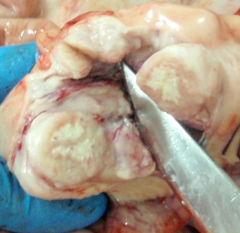
Metastatic Carcinoma in Subcapsular Sinus |
|

|
Metastatic Carcinoma in Subcapsular Sinus |
|

|
Thymoma: note classic jigsaw pattern |
|

|
Thymoma: |
|

|
Thymoma: proliferation of the epithelial cells (these are NOT lymphoid tumors) |
|

|
Thymoma: proliferation of the epithelial cells (these are NOT lymphoid tumors) |
|

|
Splenic Infarcts with coagulation necrosis. Note the triangle shape of the infarcts |
|

|
Splenomegaly: Normal Spleen is 150g while this spleen was 1500g. Again note the triangle shaped infarcts. |
|

|
Perisplenitis: Here we have a "sugar icing" of thickened fibrous tissue on the capsule. This is not very clinically significant. |
|

|
Lacerated Spleen: Trauma is a common cause for splenectomy. Spleen is much more susceptible if the it is already enlarged. |