![]()
![]()
![]()
Use LEFT and RIGHT arrow keys to navigate between flashcards;
Use UP and DOWN arrow keys to flip the card;
H to show hint;
A reads text to speech;
108 Cards in this Set
- Front
- Back
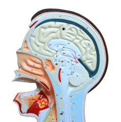
|
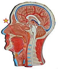
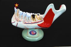
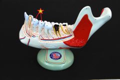
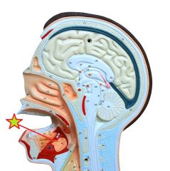
Hard Palate |
|
|
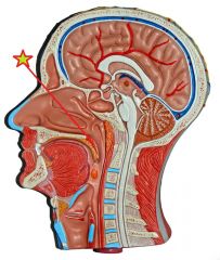
Soft Palate |
|
|
Uvula |
|
|
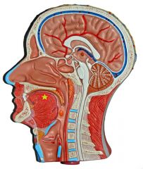
Tongue |
|
|
Incisors |
|
|
Incisors |
|

|
Canines (Cuspids) |
|
|
Canines (Cuspids) |
|
|
Premolars (Bicuspids) |
|

|
Premolars (Bicuspids) |
|
|
Molars |
|
|
Molars |
|
|
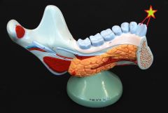
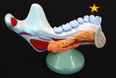
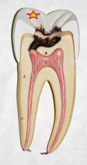
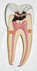
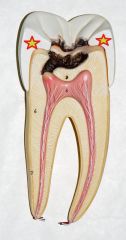
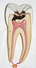
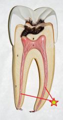
Apical Canal |
|
|
Cementum |
|
|
Crown |
|
|
Dentin |
|
|
Enamel |
|
|
Nerves and Vessels |
|
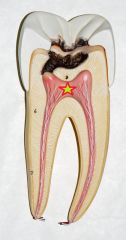
|
Pulp |
|
|
Root Canals |
|
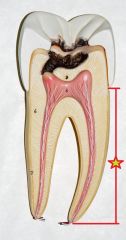
|
Root |
|
|
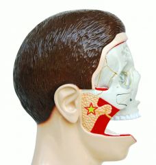
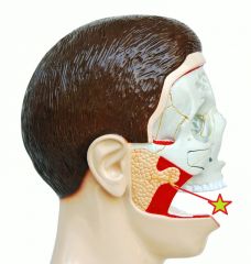
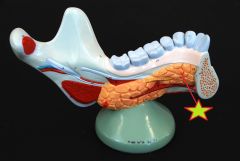
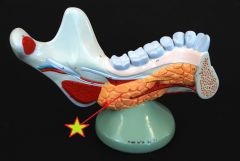
Parotid Gland |
|
|
Parotid Duct |
|
|
Sublingual Gland |
|

|
Sublingual Gland |
|
|
Submandibular Gland |
|
|
Submandibular Gland |
|

|
Submandibular Duct |
|
|
Submandibular Duct |
|
|
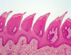
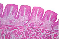
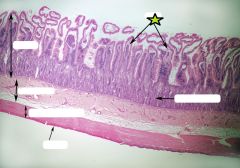
Filiform Papillae |
|
|
Fungiform Papillae |
|
|
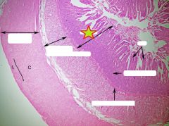
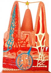
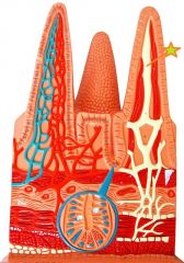
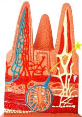
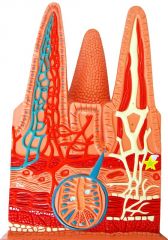
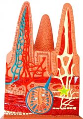
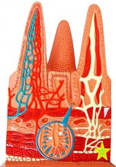
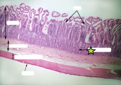
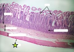
Mucosa |
|
|
Muscularis Mucosa |
|
|
Submucosa |
|
|
Muscularis Externa |
|
|
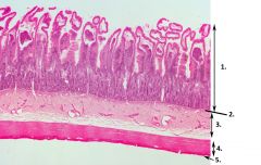
1. Mucosa 2. Muscularis Mucosae 3. Submucosa 4. Muscularis Externa 5. Serosa |
|
|
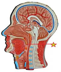
Esophagus |
|

|
Cardiac Sphincter |
|

|
Cardiac Sphincter |
|

|
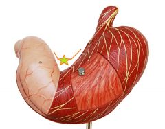
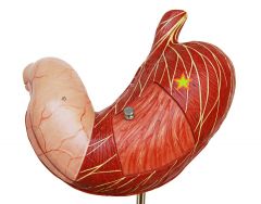
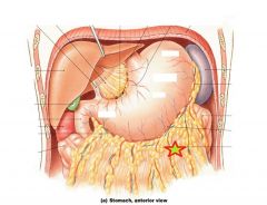
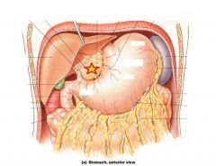
Cardia |
|

|
Fundus |
|

|
Body |
|
|
Pylorus |
|

|
Pyloric Sphincter |
|
|
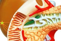
Rugae |
|
|
Greater Curvature |
|
|
Lesser Curvature |
|

|
Longitudinal Muscle |
|
|
Circular Muscle |
|

|
Oblique Muscle |
|
|
Greater Omentum |
|
|
Lesser Omentum |
|
|
Duodenum - Small Intestine - connected to pyloric sphincter - mixing bowl, receives chyme from stomach, digestive secretions from pancreas and liver |
|

|
Jejunum - Small Intestine - abrupt bend at duodenojejunal flexure - bulk of chemical digestion and nutrient absorption |
|
|
Ileum - Small Intestine - 3rd and longest segment - ends at ileocecal valve |
|
|
Duodenal Papilla - Small Intestine - pancreatic and bile duct entrances in duodenum |
|
|
Duodenal Papilla - Small Intestine - pancreatic and bile duct entrances in duodenum |
|
|
Plicae Circulares - Small Intestine - transverse folds that increase the surface area for absorption |
|
|
Plicae Circulares - Small Intestines - transverse folds that increase the surface area for absorption |
|
|
Villus - Small Intestine - fingerlike projections of mucosa that increase surface area |
|
|
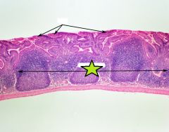
Lacteal - Small Intestine - mucosa layer - transport materials that can't enter capillaries |
|
|
Intestinal Crypts - Small Intestine - mucosa layer - pockets extending deep into lamina propria - location of stem cell division |
|

|
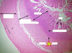
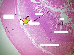
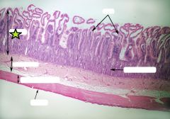
Mucosa - Small Intestine |
|
|
Muscularis Mucosa - Small Intestine |
|
|
Submucosa - Small Intestine |
|
|
Muscularis Externa - Small Intestine |
|

|
Serosa - Small Intestine |
|
|
Ileocecal Valve - Large Intestine - regulates passage from ileum to cecum |
|

|
Ileocecal Valve - Large Intestine - regulates passage from ileum to cecum |
|
|
Cecum - Large Intestine - expanded pouch at beginning of large intestines - collects and stores arriving material before compaction |
|
|
Cecum - Large Intestine - expanded pouch at beginning of large intestines - collects and stores arriving material before compaction |
|
|
Ascending Colon - Large Intestine - right lateral abdominal wall - cecum, ascends to hepatic flexure - haustra w/ taeniae coli |
|

|
Transverse Colon - Large Intestine - crosses abdomen right to left - hepatic flexure to splenic flexure - haustra w/ taeniae coli |
|
|
Descending Colon - Large Intestine - descends along left side of abdomen - from splenic flexure to sigmoid flexure - haustra w/ taeniae coli |
|

|
Sigmoid Colon - Large Intestine - s-shaped segment - sigmoid flexure to rectum |
|

|
Rectum - Large Intestine - waste deposited from sigmoid colon - temporary storage of fecal matter - movement triggers urge to defecate - exits to anal canal |
|

|
Hepatic Flexure - Large Intestine - ascending colon to transverse colon |
|

|
Splenic Flexure - Large Intestine - transverse colon to descending colon |
|

|
Haustra - Large Intestine - series of pouches w/in the large intestine - allows for distension and elongation |
|
|
Taenia Coli - Large Intestine - longitudinal ribbons of smooth muscle |
|
|
Appendix - attached posteromedial surface of cecum |
|
|
Spleen |
|
|
Spleen |
|
|
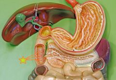
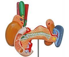
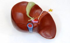
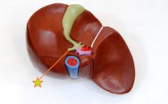
Cystic Duct - Gall Bladder - exits gall bladder, joins common hepatic duct to form common bile duct |
|
|
Gall Bladder - hollow, pear shaped muscular organ - stores and concentrates bile before excretion |
|
|
Gall Bladder - hollow, pear shaped muscular organ - stores and concentrates bile before excretion |
|

|
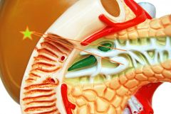
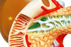
Head - Pancreas |
|

|
Body - Pancreas |
|
|
Tail - Pancreas |
|
|
Pancreatic Duct - delivers digestive enzymes and buffers to duodenal ampulla |
|
|
Accessory Pancreatic Duct - may exist - empties in lesser duodenal ampulla |
|

|
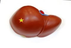
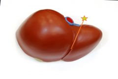
Left Lobe - Liver - falciform ligament marks division between left and right lobes |
|
|
Right Lobe - Liver - falciform ligament marks division between left and right lobes |
|

|
Caudate Lobe - Liver - between left lobe and inferior vena cava |
|

|
Quadrate Lobe - Liver - between left lobe and gall bladder |
|
|
Falciform Ligament - Liver - divides left and right lobes - |
|

|
Round Ligament - Liver - fibrous band marks path of degenerated fetal umbilical vein |
|
|
Left Hepatic Duct - Liver - collect bile from all bile ducts of the liver lobes |
|

|
Right Hepatic Duct - Liver - collect bile from all bile ducts of the liver lobes |
|

|
Common Hepatic Duct - Liver - union of left and right hepatic ducts - leaves liver and flows into common bile duct to duodenum or cystic duct to gall bladder |
|
|
Common Bile Duct - Liver - leads to duodenal ampulla |
|
|
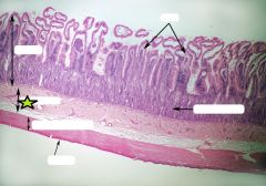
Villi - Small Intestine |
|
|
Intestinal Crypts - Small Intestine |
|
|
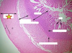
Mucosa - Small Intestine |
|
|
Submucosa - Small Intestine |
|
|
Muscularis Externa - Small Intestine |
|
|
Serosa - Small Intestine |
|
|
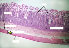
Peyer's Patch - Ileum |