![]()
![]()
![]()
Use LEFT and RIGHT arrow keys to navigate between flashcards;
Use UP and DOWN arrow keys to flip the card;
H to show hint;
A reads text to speech;
31 Cards in this Set
- Front
- Back
|
Regions |
1. Epithalamus: Pineal gland, Habernacular nucleus 2. Dorsal thalamus (Thalamus) 3. Subthalamus: nuclei involved in BN circuits (motor circuits) 4. Hypothalamus: endocrine regulation, homeostasis, motivation |
|
|
Thalamic nulcei: main goupings |
Thalamic nuclei are divided into three main groups by the internal medullary lamina: 1. Anterior group 2. Lateral group 3. Medial group In addition there are the internal medullary lamina nuclei and reticular nuclei Metathalamic nuclei (MGN, LGN) NB the reticular thalamus is not continuous with the reticular formation and functionally differs from the rest of the thalamus |
|
|
Anterior subdivision nuclei |
Anterior nucleus |
|
|
Lateral subdivision nuclei |
Dorsal tier: contains, from caudal to rostral, the pulvinar, the lateral posterior (LP) and the lateral dorsal (LD) nuclei. Ventral tier: ventral lateral (VL), ventral anterior (VA), ventral posterior nuclei. The ventral posterior is further divided into the ventral posteromedial (VPM) and ventral posterolateral nuclei (VPL). |
|
|
Medial subdivision nuclei |
Medial dorsal nucleus (MD; also called the dorsal medial nucleus - DM) as well as smaller midline nuclei (located right beneath the wall of the third ventricle). |
|
|
Thalamic reticular nucleus |
Thin shell of neurons covering the entire lateral aspect of the thalamus. This is separated from the thalamus by the external medullary lamina. No projections to cortex receives inputs from cortex/thalamic projection neurons and sends inhibitory (GABA) projections back to thalamus All thalamocortical/corticothalamic fibers must pass through this structure, and thus send off collaterals to it The output of each part of the reticular nucleus is to the thalamic region from which it received inputs Thus functions as important source of regulatory input to thalamus |
|
|
Metathalamus |
Includes nuclei that protrude from the posterior aspect of the pulvinar of the thalamus. These include the medial geniculate body (an auditory relay nucleus) and the lateral geniculate body (the principal visual relay). |
|
|
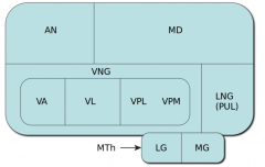
Thalamic nuclei basic structural arrangement (line diagram) |
|
|
|
Thalamic region anatomy |

|
|
|
Relay nuclei |
Receive very well defined inputs and project this signal to functionally distinct areas of the cerebral cortex. These include the nuclei that relay primary sensations (VPL, VPM, LGN, MGN), nuclei involved in limbic relays (Anterior nucleus/LD nucleus) and also the nuclei involved in feedback of cerebellar signals (VL) and in feedback of basal gangliar output (part of the VL and the ventral anterior nucleus - VA), |
|
|
Association nuclei |
Receive most of their input from the cerebral cortex and project back to the cerebral cortex in the association areas where they appear to regulate activity. There are two great areas of association in human brains; prefrontal cortex, parietal-occipital-temporal association cortex -DM nucleus; interconnected with the PFC and is involved in PF functions (affect, foresight) -Pulvinar-LP complex: interconected with POT |
|
|
Non-specific nuclei |
Project quite broadly through the cerebral cortex, may be involved in general functions such as alerting. Intralaminar and midline thalamic nuclei |
|
|
Intralaminar nuclei |
Two largest are: -centromedian (CM) -parafascicular (PF) |
|
|
Anterior nucleis I/O |
I: mammillothalamic tract, hippocampus O: cingulate gyrus Limbic relay nucleus; part of Papez circuit |
|
|
LD nucleis I/O |
I: hippocampus O: cingulate gyrus Limbic relay nucleus |
|
|
VA/VL nuclei I/O |
I: basal nuclei, cerebellum O: motor areas BN outputs go mostly to VA and cerebellar outputs mostly to VL but generally they are considered together as a combined motor relay MOTOR relay nuclei |
|
|
VPL nucleis I/O |
I: medial lemniscus (body), spinothalamic tract (body) O: somatosensory cortex |
|
|
VPM nucleus I/O |
Medial lemniscus (face), spinothalamic tract (face), central tegmental tract (taste) O: somatosensory cortex, insula |
|
|
MGN I/O |
I: brachium of the inferior colliculus O: auditory cortex (superior temporal gyrus->Heschl's gyrus) |
|
|
LGN I/O |
I: optic tract O: visual cortex |
|
|
DM nucleus I/O |
I: PF cortex, olfactory and limbic structures (eg amygdala) O: PF cortex Involved in PF functions-affect, foresight Is PFC association complex |
|
|
Lateral posterior (LP) I/O |
I: parietal lobe O: parietal lobe Part of Pulvinar-LP complex (nuclear complex for POT association cortex) |
|
|
Pulvinar I/O |
I: PTO lobes O: PTO lobes Part of Pulvinar-LP complex (nuclear complex for POT association cortex) |
|
|
Intralaminar nuclei inputs and projections |
DO NOT have diffuse and non specific projections as long supposed Inputs: diverse sources including multiple cortical areas, basal nuclei, cerebellum, brainstem reticular formation, spinothalamic/spinoreticulothalamic fibers Collectively the nuclei have widespread projections to multiple cortical areas, BN, limbic structures BUT each individual nucleus has specific projections -CM: putamen/motor cortex PF: caudate/PFC Midline/intralaminar nuclei probably affect the multiple parallel circuits in BN/Limbic structures. As these structures collectively affect most cortical functions collective changes in activity of the midline/intralaminar nuclei would be expected to have widespread effects on cortical activity |
|
|
Blood supply to Thalamus |
Mostly from small perforating branches of the posterior cerebral artery Blood supply is consistent enough that thalamic strokes often affect predictable sets of nuclei |
|
|
Thalamic lesion |
Most often as a result of vascular accidents Thalamic pain: poorly understood. Lesions causing it are almost exclusively involving VPL/VPM Extsnive damage to posterior thalamus also causes total/near total loss of somatic sensations in contralateral head/body Thalamic syndrome: combination of thalamic pain, hemianaesthesia, sensory ataxia (all contralateral) |
|
|
Anatomical extensions of thalamus |
Anterior to the IV foramen Supriorly to the transverse cerebral fissure/floor of lateral ventricle Inferiorly to hypothalami sulcus Posteriorly it overlaps the midbrain |
|
|
External medullary lamina |
Lateral surface of thalamus Second curved sheet of myelinated fibers Intervenes between the thalamus and thalamic reticuclar nucleus |
|
|
Reglulatory inputs to Thalamus |
Broadly similar for all nulei Most come from the cerebral cortex (mainly cortical area to which nucleus projects), some come from the thalamic reticular nculeus and the remainder come from diffuse cholingeric, noradrenergic, serotonergic and dopaminergic endings from the brainstem RF Regulatory inputs outnumber specific inputs (eg LGN fewer than 10% of synapses on projection neurons are from optic tract) |
|
|
Specific inputs to Thalamus |
Those conveying the information that a given thalamic nucleus may pass on accurately to the cerebral cortex eg medial lemniscus is the specific input to VPL, optic tract is specific input to LGN Nuclei are defined functionally by their specific inputs and ouputs: -Relay -Association -Intrlaminar/midline nuclei |
|
|
Two physiological states of Thalamic projection neurons |
1. Tonic mode: neurons slightly DP and behave like typical neurons; can faithfully transmit infromation using trains of AP whose frequency match the frequency of inputs 2. Burst mode: neurons HP beyond the tonic range, characterized by availability of special voltage-gated Ca channels. DP causes transient activation of these channels (followed by their inactivation) leading to a burst of Na based AP. Duration of Ca channel opening is short (100ms) so burst of AP can only occur a few times a second. Neurons in this mode are very sensitive, but are unable to transit info about specific inputs Focusing attention, whether on a stimulus. a task, or a thought. presumably involves placing thalamic Projection neurons in tonic mode Burst mode may have more than one function-most neurons are in burst mode during sleep, many are in burst mode during wakefulness as well |