Reading...
![]()
Play button
![]()
Play button
![]()
Use LEFT and RIGHT arrow keys to navigate between flashcards;
Use UP and DOWN arrow keys to flip the card;
H to show hint;
A reads text to speech;
36 Cards in this Set
- Front
- Back

How does the nephrogenic cord arise?
|
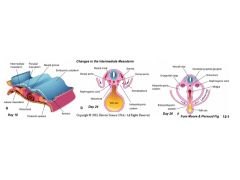
Intermediate mesoderm separates from the middle mesoderm layer and ends up on the dorsal aspect of the coelom.
|
|
|
What derivates (organs) arise from the nephrogenic cord (or UG ridge)?
|
Kidneys, ureters, gonads, genital ducts, and adrenal cortex
|
|
|
Not all components of UG comes from the intermediate mesoderm. What tissue do the other components arise from and what are these components>
|
These come from the Endoderm:
Urinary Bladder Urethra Lower half of vagina vestibule |
|
|
The walls of bladder, vagina, etc come not from intermediate mesoderm or endoderm - they derive from:
|
Splanchnic mesoderm
|
|
|
What is the cloaca and what is its main function at about 4 weeks?
|
It is a dialated part of the primitive hindgut that functions as a transient common outlet for the UG and GI systems. [Sewer]
|
|
|
What are the mesonephric tubules8?
|
Immature nephrons
|
|
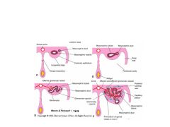
Describe the four steps required to form Mesonephric Tubules
|

1. Mesonephric duct sends signals to mesenchyme - induces vesicle formation
2. Vesicle grows, it eventually units with duct 3. Vesicle continues to elongate and finds a tuft of vessels 4. Vesicle eventually forms a small cup around the vessel BAM - you've got an immature nephron |
|
|
In the male, components of the mesonephric kidney become:
In the female? |
Male = reproductive system. Female, not much except uterine mesentery.
|
|
|
Mesonephric tubules take the embryo from ? wks to about ? wks.
|
4th to 8 or 10th week.
|
|
|
What is the early definitive kidney called? Where does it form on the nephrogenic cord?
|
METAnephric kidney. It forms at the most caudal end of the nephrogenic cord.
|
|
|
The metanephric kidney has two precursors. What are they?
|
Metanephric diverticulum (ureteric bud) and the metanephrogenic mesenchym (blastema)
|
|

Name tissue types
|

1. Metanephric mesenchyme
2. Metanephric diverticulum |
|
|
About how many generations of metanephric diverticulum branches are there? Which branch generations form the calyces, pelvis and ureters?
|
There are about 15 generations. Expansion of the 3rd to 6th generation form the calyces, etc.
|
|
|
Describe hypoplastic kidneys.
|
May be small & normal kidneys that may be small bc of abnormal development
|
|
|
Describe dysplastic kidneys
|
Often contain cysts or "primitive" tissues. Most common cystic anomaly of the kidneys. Fatal if bilateral.
|
|
|
Describe duplications re kidney anomalies.
|
Duplications of ureter or kidney may be partial or complete.
|
|
|
How might an extra artery form on a kidney?
|
As ascends to its definitive location, it breaks and forms new arteries. Sometimes ones that are supposed to break, don't.
|
|
|
If the two kidneys form too close to each other, they might form a horseshoe kidney. In this case, where does it get caught as it ascends?
|
Under the IMA.
|
|
|
What is the difference between Autosomal Recessive PKD and Autosomal Dominant PKD?
|
Recessive forms fine cycts and age at onset varies. Dominant forms large cysts and onset is usually bn 30's and 50's
|
|

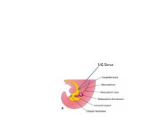
Recall that the cranial or vesicle part of the Primary UG sinus enlarges. What does this become? What is it lined with and what is its wall made of? (see back for image)
|
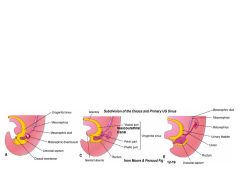
It becomes the bladder and part of the urethra. It is lined with endoderm and the wall's smooth muscle & CT is derived from the surrounding splanchnic mesoderm.
|
|
|
Remodeling of the posterior wall of the bladder results in the formation of (see back for diagram):
|
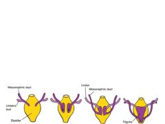
Trigone Region
|
|
|
The trigone region is associated with:
|
Bladder Ureter entrance and Urethra exit
|
|
|
In males, differential growth of the mesonephric duct results in the formation of the:
|
Urethra
|
|
|
What is the extent of the urethra formed from the vesicle part of the UG sinus in Males and Females?
|
Males: Vesicle part forms proximal portion of prostatic urethra. In Females, it forms most of the urethra.
|
|
|
The trigone region is associated with:
|
Bladder Ureter entrance and Urethra exit
|
|
|
In males, differential growth of the mesonephric duct results in the formation of the:
|
Urethra
|
|
|
What is the extent of the urethra formed from the vesicle part of the UG sinus in Males and Females?
|
Males: Vesicle part forms proximal portion of prostatic urethra. In Females, it forms most of the urethra.
|
|

How does the UG sinus form?
|
The anorectal canal subdivides the cloaca into the UG sinus.
|
|
|
What are the three subdivisions of the UG sinus?
|
Vesicle, Pelvic, and Phallic
|
|
|
The UG sinus has three subdivisions - describe which part elongates, narrows, and expands.
|
The vesicle part elongates, the pelvic part narrows, and the phalic part expands.
|
|
|
In the male, describe each of the fates of the UG sinus subdivisions.
|
Vesicle part becomes the bladder. Pelvic portion becomes distal prostatic and membranous urethra. Phallic portion becomes penile urethra.
|
|
|
In the female, describe each of the fates of the UG sinus subdivisions.
|
Vesicle part becomes the bladder. Pelvic portion becomes lower half of the vagina. Phallic portion become the vestibule.
|
|
|
The bladder, as it forms, is connected to:
|
The Allantois (makes sense - this helps embryo handle waste from almost day one)
|
|
|
As the bladder develops, it is connected to the allantois to help it manage wasted. As the allantois disappears, it can cause various problems. What are the three he described in class?
|
1. Cyst formation
2. A sinus forms at the umbilicus that tracks down to where allantois was 3. A fistula can form between umbilicus and bladder and urine can flow out. |
|
|
As the allantois disappears, it ultimately becomes the:
|
Median umbilical ligament
|
|
|
What is exstrophy of the bladder?
|
The bladder wall forms on the outside of the body and what you see is the inside of the bladder.
|