Reading...
![]()
Play button
![]()
Play button
![]()
Use LEFT and RIGHT arrow keys to navigate between flashcards;
Use UP and DOWN arrow keys to flip the card;
H to show hint;
A reads text to speech;
16 Cards in this Set
- Front
- Back
|
Macule
|
Small, flat discoloration, not palpable
|
|
|
Patch
|
Large, flat discoloration (>2cm)
Example: Bruise |
|
|
Papule
|
Small Bump, palpable lesion (<1cm)
Example: Small Wart |
|
|
Nodule
|
Large Bump, palpable (> 1 cm)
Example: Nevus |
|
|
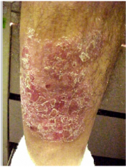
Plaque
|
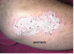
Large raised flat topped lesion, usually rough/scaly, greater than 2 cm
Lesion is greater in diameter than it is high; may be a bunch of nodules or papules Example: Psoriasis |
|
|
Vesicle
|
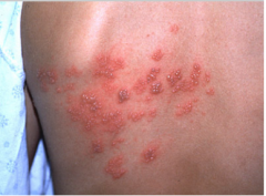
Small Blister, elevated epidermis containing clear fluid (< .5 cm in diameter)
Example: Herpes |
|
|
Bulla
|
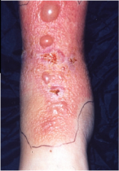
Large Blister, elated epidermis containing clear fluid (> .5 cm)
Example: Bullous Pemphigoid |
|
|
Pustule
|
Pus filled bump, papular or nodular
Example: Acne |
|
|
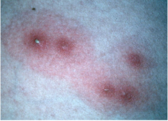
Wheal
|
Raised smooth topped lesion; sometimes called "welt," often with erthematous borders and place centers
Example: Urticaria |
|
|
Scale
|
Roughness at surface; represents hyperkeratosis
Example: Psoriasis |
|
|
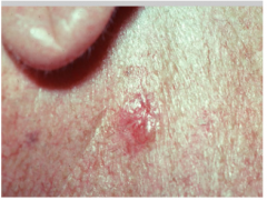
Crust
|
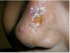
Dried plasma at surface; represents epithelial disruption
Example: Eczema |
|
|
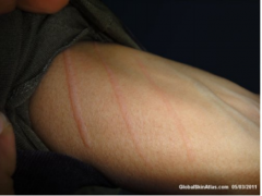
Excoriation
|
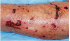
Scratched area; often linear, scratched, picking, burns
Example: Eczema |
|
|
Lichenification
|
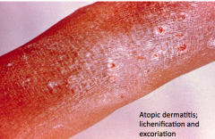
Leathery thickening with accentuated skin markings, often caused by chronically rubbed or scratched skin
Example: Chronically rubbed skin of eczema |
|
|
Telagiectasia
|
Dilated Capillaries visible through the skin surface
Example: Sun-damaged face |
|
|
Onycholysis
|
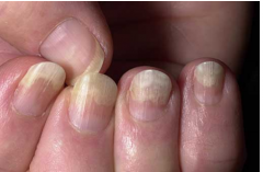
Separation of nail from bed
Example: Onchomysosis |
|
|
Atrophy
|
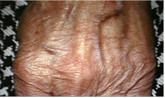
Sunken thinned area of skin, microscopically shows loss of rete ridges in epidermis, occurs with age
Example: Corticosteroid induced. |