Reading...
![]()
Play button
![]()
Play button
![]()
Use LEFT and RIGHT arrow keys to navigate between flashcards;
Use UP and DOWN arrow keys to flip the card;
H to show hint;
A reads text to speech;
62 Cards in this Set
- Front
- Back
- 3rd side (hint)
|
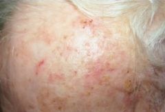
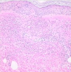
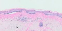
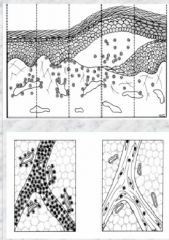
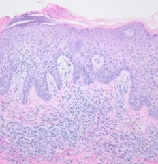
actinic keratosis
rough, scaling, reddish, ill-defined patch or plaque |
UV radiation induced, can spontaneously regress or progress to SCC
may form horn-like excrescences (need to biopsy). inflammatory infiltrate and solar elastosis (sun damage) common full thickness dysplasia = SCC in situ |
|
|
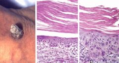
actinic keratosis
variable atypia (less than full thickness), solar elastosis, chronic dermal inflammation |
UV radiation induced, can spontaneously regress or progress to SCC
may form horn-like excrescences (need to biopsy). inflammatory infiltrate and solar elastosis (sun damage) common full thickness dysplasia = SCC in situ |
|
|
angiocentric
|
associated w/ urticaria and necrotizing vasculitis
|
|
|
angiocentric
|
associated w/ urticaria and necrotizing vasculitis
|
|
|
atrophy
dermal/subcutaneous |
|
|
|
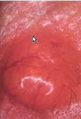
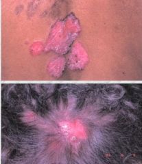
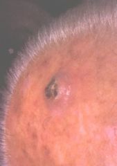
BCC
erythematous patch w/ central erosion/ulceration and rolled pearly border w/ telangiectasia or pearly papule or nodule w/ telangiectasias |
sun exposed skin, most common skin cancer (most common human malignancy)
rarely metastasizes, locally aggressive |
|
|
BCC
enlarged basaloid keratinocytes mitosis, individual cell necrosis, peripheral palisading, separation artifact |
sun exposed skin, most common skin cancer (most common human malignancy)
rarely metastasizes, locally aggressive |
|
|
BCC
superficial type |
sun exposed skin, most common skin cancer (most common human malignancy)
rarely metastasizes, locally aggressive |
|
|
dermatitis herpetiformis (celiac dz)
microabcesses and neutrophils |
IgA-TeG (antiendomysial Ab) deposition from gluten sensitivity causes blistering and herpetiform
Tx: Dapsone (skin symptoms), gluten free diet (systemic symptoms) |
|
|
compound melanocytic nevus
melanocytes in dermis and epidermis |
|
|
|
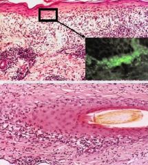
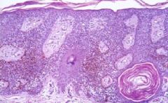
discoid lupus
localized to skin (5-10% have SLE), pigment change, telangiectasia, alopecia, follicular plugging |
autoimune interface dermatitis
may be entirely localized to skin chronic atrophy of skin, pigment change due to damage to basal layer |
|
|
discoid lupus
epidermal atrophy, hyperkeratosis, lymphocytes attacking DEJ, follicular destruction, dilated vessels, positive "lupus band" |
autoimune interface dermatitis
may be entirely localized to skin chronic atrophy of skin, pigment change due to damage to basal layer |
|
|
dysplastic nevus
irregular nests, bridging, shoulder, cytologic atypia, lamellar fibroplasia |
melanoma precursor
|
|
|
dysplastic nevus
irregular nests, bridging, shoulder, cytologic atypia, lamellar fibroplasia |
melanoma precursor
|
|
|
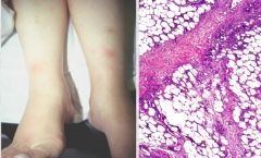
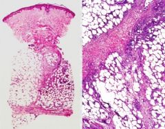
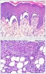
Erythema Nodosum
panniculitis chronic inflammation (lymphocytes, histiocytes, eos, granulomas) septal firborsis |
often provoked by infection, drug ingestion, or associated systemic inflammatory disorder (most common cause is idiopathic)
tender subc nodules |
|
|
Erythema Nodosum
panniculitis chronic inflammation (lymphocytes, histiocytes, eos, granulomas) septal firborsis |
often provoked by infection, drug ingestion, or associated systemic inflammatory disorder (most common cause is idiopathic)
tender subc nodules |
|
|
inflammation
|
angiocentric/spongiotic/interface
|
|
|
inflammation
|
vesiculobullous/panniculitic
|
|
|
interface infiltrated
Discoid Lupus Erythematosis |
|
|
|
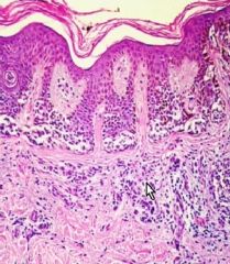
Lichen Planus
pink purple pruritic polygonal papules or plaques often on wrists/ancles |
interface dermatoses
most cases idiopathic, koebner phenom, lymphocytes attacking keratinocytes |
|
|
Lichen Planus
Interface dermatoses w/ lymphocytes obscuring keratinocytes |
interface dermatoses
most cases idiopathic, koebner phenom, lymphocytes attacking keratinocytes |
|
|
melanocytes
|
|
|
|
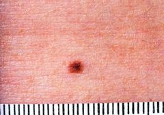
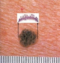
melanocytic nevus
|
most acquired btwn 6 and 35
variable but uniform appearance brown to flesh colored, <5mm diameter |
|
|
melanocytic nevus
|
most acquired btwn 6 and 35
variable but uniform appearance brown to flesh colored, <5mm diameter |
|
|
melanocytic nevus
|
most acquired btwn 6 and 35
variable but uniform appearance brown to flesh colored, <5mm diameter |
|
|
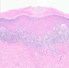
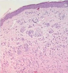
melanoma in situ
|
melanoma cells confined to the epidermal layer
|
|
|
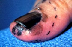
acral melanoma
|
most common type of melanoma in dark skinned pts
nail beds and soles of feet |
|
|
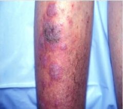
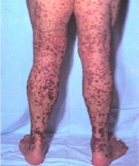
necrotizing vasculitis
palpable purpura |
can be limited to skin or systemic, non blanching (extravasated RBCs)
Drugs, infection, connective tissue Dz, underlying malignancy may cause Typically IgG Henoch-Schonlein Purpura an IgA variant more common in peds requiring serial UAs |
|
|
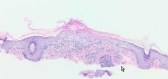
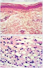
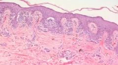
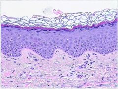
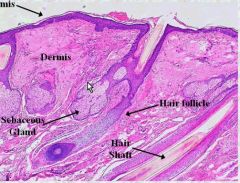
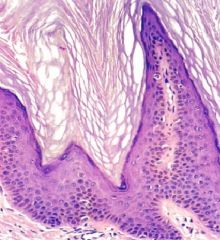
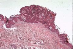
normal skin
|
|
|
|
normal skin
|
|
|
|
normal skin
|
|
|
|
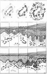
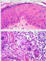
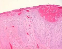
Hyperplasia (Acanthosis)/Dysplasia
|
|
|
|
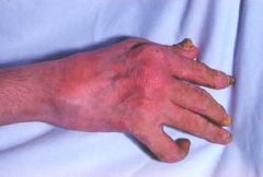
Psoriatic Arthritis
|
gross
|
|
|
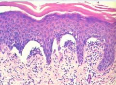
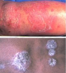
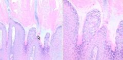
psoriasis
erythematous plaques w/ silvery scales |
Koebner Phenom, Auspitz sign
HLA types (genetic factor) neutrophils arthritic lesions may present autoreactive T Cells? |
|

|
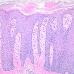
psoriasis
"regular" acanthosis parakeratosis neutrophils loss of granular layer suprapapillary dermal plate prominent dilated capillaries |
Koebner Phenom, Auspitz sign
HLA types (genetic factor) neutrophils arthritic lesions may present autoreactive T Cells? |
|
|
psoriasis
"regular" acanthosis parakeratosis neutrophils loss of granular layer suprapapillary dermal plate prominent dilated capillaries |
Koebner Phenom, Auspitz sign
HLA types (genetic factor) neutrophils arthritic lesions may present autoreactive T Cells? |
|
|
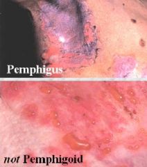
Pemphigus Vulgaris
flacid easily ruptured blisters Nikolsky sign |
genetic predisposition, 40s-50s
autoimmune attack of IgG against desmoglein 3 uniformly fatal before steroid therapy available |
|
|
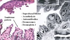
Pemphigus Vulgaris
tombstone pattern, basal layer still attached |
genetic predisposition, 40s-50s
autoimmune attack of IgG against desmoglein 3 uniformly fatal before steroid therapy available |
|
|
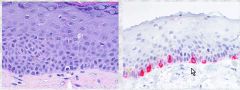
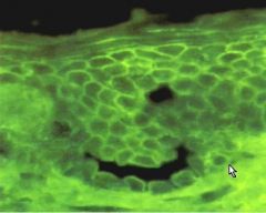
Pemphigus Vulgaris
immunofluorescence for IgG against Desmoglein 3 Lace of fish scale pattern differentiates from lupus or dermatitis herpetiformis |
genetic predisposition, 40s-50s
autoimmune attack of IgG against desmoglein 3 uniformly fatal before steroid therapy available |
|
|
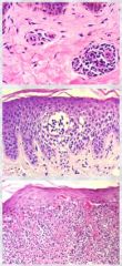
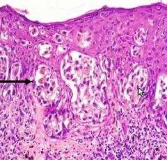
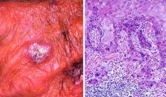
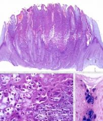
Squamous Cell Carcinoma
scaling erythematous plaque or nodule |
occurs often on sun-exposed skin or mucous membrane or sites of chronic injury
locally invasive small potential to metastasize |
|
|
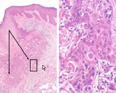
Squamous Cell Carcinoma
atypical keratinocytes, growth into underlying dermis, keratin pearls, hyperkeratosis, parakeratosis |
occurs often on sun-exposed skin or mucous membrane or sites of chronic injury
locally invasive small potential to metastasize |
|
|
Squamous Cell Carcinoma
atypical keratinocytes, growth into underlying dermis, keratin pearls, hyperkeratosis, parakeratosis |
occurs often on sun-exposed skin or mucous membrane or sites of chronic injury
locally invasive small potential to metastasize |
|
|
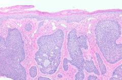
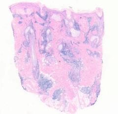
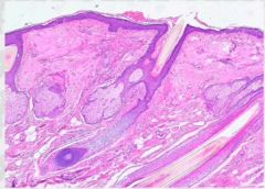
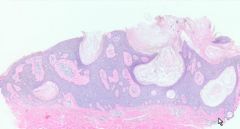
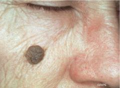
seborrheic keratosis
"stuck on" plaque or papule rough surface |
middle age and older
Sign of Leser-Trelat = many SKs in short time a result of internal malignancy possible environmental cause |
|
|
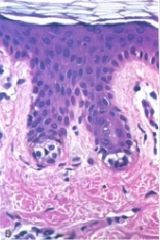
seborrheic keratosis
no melanocytic incr horn cytsts of onion like laminated whorls |
middle age and older
Sign of Leser-Trelat = many SKs in short time a result of internal malignancy possible environmental cause |
|
|
seborrheic keratosis
no melanocytic incr horn cytsts of onion like laminated whorls |
middle age and older
Sign of Leser-Trelat = many SKs in short time a result of internal malignancy possible environmental cause |
|
|
seborrheic keratosis
no melanocytic incr horn cytsts of onion like laminated whorls |
seborrheic keratosis
no melanocytic incr horn cytsts of onion like laminated whorls |
|
|
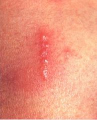
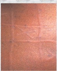
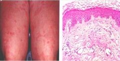
Allergic contact dermatitis
pruritic well demarcated erythematous edematous papules and plaques with vesicle formation often linear |
blister in 1-2 days
scaly w/ chronicity type IV hypersensitivity reaction (cell-mediated, delayed) (Langerhans present to naive T cells --> itchy) initial type I reaction may also be present initially |
|
|
Allergic contact dermatitis
spongiotic reaction intercellular edema, vesicles may form btwn keratinocytes perivascular infiltrate of lymphocytes, histiocytes, and eos |
blister in 1-2 days
scaly w/ chronicity type IV hypersensitivity reaction (cell-mediated, delayed) (Langerhans present to naive T cells --> itchy) initial type I reaction may also be present initially |
|
|
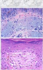
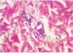
Storage deposition
solar elastosis/amyloid |
|
|
|
Allergic contact dermatitis
spongiotic reaction intercellular edema, vesicles may form btwn keratinocytes perivascular infiltrate of lymphocytes, histiocytes, and eos |
blister in 1-2 days
scaly w/ chronicity type IV hypersensitivity reaction (cell-mediated, delayed) (Langerhans present to naive T cells --> itchy) initial type I reaction may also be present initially |
|
|
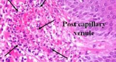
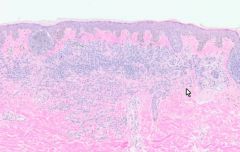
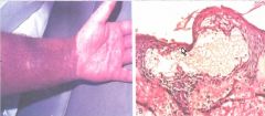
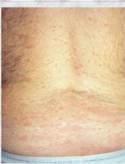
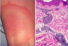
urticaria
angiocentric inflammation dermatographism, tempurature sensitive vascular dilation, dermal edema |
often in young adults
<24 hrs, intensly pruritic, angioedema chronic forms: urticarial vasculitis, underlying dz IgE/mast cell/PG reaction angioedema in airways a immediate life threat |
|
|
urticaria
angiocentric inflammation dermatographism, tempurature sensitive vascular dilation, dermal edema |
often in young adults
<24 hrs, intensly pruritic, angioedema chronic forms: urticarial vasculitis, underlying dz IgE/mast cell/PG reaction angioedema in airways a immediate life threat |
|
|
urticaria
angiocentric inflammation dermatographism, tempurature sensitive vascular dilation, dermal edema |
often in young adults
<24 hrs, intensly pruritic, angioedema chronic forms: urticarial vasculitis, underlying dz IgE/mast cell/PG reaction angioedema in airways a immediate life threat |
|
|
urticaria
angiocentric inflammation dermatographism, tempurature sensitive vascular dilation, dermal edema |
often in young adults
<24 hrs, intensly pruritic, angioedema chronic forms: urticarial vasculitis, underlying dz IgE/mast cell/PG reaction angioedema in airways a immediate life threat |
|
|
urticaria
angiocentric inflammation dermatographism, tempurature sensitive vascular dilation, dermal edema |
often in young adults
<24 hrs, intensly pruritic, angioedema chronic forms: urticarial vasculitis, underlying dz IgE/mast cell/PG reaction angioedema in airways a immediate life threat |
|
|
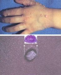
Verruca Vulgaris
|
HPV
benign squamoid proliferation, koilocytosis |
|
|
verruca vulgaris
|
HPV
benign squamoid proliferation, koilocytosis |
|
|
verruca vulgaris
|
HPV
benign squamoid proliferation, koilocytosis |
|
|
seborrheic keratosis
"stuck on" plaque or papule rough surface |
middle age and older
Sign of Leser-Trelat = many SKs in short time a result of internal malignancy possible environmental cause |
|
|
seborrheic keratosis
"stuck on" plaque or papule rough surface |
middle age and older
Sign of Leser-Trelat = many SKs in short time a result of internal malignancy possible environmental cause |