![]()
![]()
![]()
Use LEFT and RIGHT arrow keys to navigate between flashcards;
Use UP and DOWN arrow keys to flip the card;
H to show hint;
A reads text to speech;
56 Cards in this Set
- Front
- Back
|
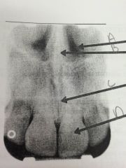
A: nasal cavity
B: nasal septum C: median palatal suture D: outline of lip |
|
|
hole in bone located in the midline of anterior portion of the hard palate |
incisive foramen |
|
|
small ovoid radiolucency between the roots of maxillary central incisors |
incisive foramen |
|
|
an immovable joint between the two palatine processes of the maxilla |
median palatal suture |
|
|
thin radiolucent line between the maxillary central incisors |
median palatal suture |
|
|
pear shaped compartment of bone located superior to the maxilla |
nasal cavity |
|
|
large, radiolucent line between the maxillary central incisors |
nasal cavity |
|
|
vertical bony wall or partition that divides the nasal cavity into the right and left nasal fossae |
nasal spetum |
|
|
vertical, radiopaque partition that divides the nasal cavity |
nasal septum |
|
|
sharp projection of the maxilla located the anterior and inferior portion of the nasal portion of the nasal cavity |
anterior nasal spine |
|
|
v-shaped radiopaque area located at the intersection of the floor of the nasal cavity and the nasal spetum |
anterior nasal spine |
|
|
paired compartments of bone located within the maxilla located above the maxillary premolar and molar teeth |
maxillary sinus |
|
|
radiolucent area superior to the apices of the maxillary posterior teeth |
maxillary sinus |
|
|
borders that outline and or divide the maxilary sinus |
floor of the maxillary sinus |
|
|
thin radiopaque lines that surround/divide the sinus |
floor of the maxillary sinus |
|
|
the intersection of the maxillary sinus and the nasal cavity |
inverted Y |
|
|
rounded prominence of bone that extend posterior to the third molar region |
maixllary tuberosity |
|
|
a small, hook like projection of bone located posterior to the maxillary tuberosity region |
hamulus |
|
|
bony projection of the maxilla that articulates with the zygoma |
zygomatic process of the maxilla |
|
|
J or U shaped radiopacity located superior to the maxillary first molar region |
zygomatic process of maxilla |
|
|
cheek bone; composed of dense cortical bone |
zygoma |
|
|
diffuse, raidopaque band extending posterior form the zygomatic process of the maxilla |
zygoma |
|
|
tiny bumps of bone that serve as muscle attachment sites |
genial tubercles |
|
|
ring shaped radiopacity below the apices of the mandibular incisors |
genial tubercles |
|
|
hole in the bone located on the internal surface of the mandible near the midline |
lingual foramen |
|
|
small, radiolucent dot surrounded by the genial tubercles |
lingual foramen |
|
|
tube like passagways that house nerves and blood vessels |
nutrient canals |
|
|
vertical radiolucent lines |
nutrient canals |
|
|
linear prominence of bone extending from the premolar region to the midline |
mental ridge |
|
|
thick radiopaque band that extends from the premolar to the incisor region |
mental ridge |
|
|
scooped out depressed area of bone located above mental ridge |
mental fossa |
|
|
hole in bone lacated on the external surface of the mandible in the region of the mandibular premolars |
mental foramen |
|
|
tube like passageway through bone that travels the lenght of the mandible |
madnibular canal |
|
|
linear prominence of bone located on the internal surface of the mandible that extends downward and forward from the ramus |
interal oblique ridge |
|
|
radiopaque band extending downward from the ramus; may continue on as the mylohyoid ridge |
internal oblique ridge |
|
|
linear prominence of bone located on the external surface of the body of the mandible |
external oblique ridge |
|
|
radipaque band extending downward from the ramus; typically ends in the third molar region |
external oblique ridge |
|
|
scooped out depressed area of bone located on the internal surface of the mandible |
submandibular fossa |
|
|
radiolucent area in the mandibular molar region below mylohyoid ridge |
submandibular fossa |
|
|
marked prominence of bone on the anterior ramus of the mandible |
coronoid process |
|
|
rectangular radiopacity superimposed over the maxilary tuberosity region ; only mandibular landmark to appear on maxillary films |
coronoid process |
|
|
completely radiopque |
amalgam gold |
|
|
radiopque but not as dense as amalgam |
stainless steel and chrome crowns |
|
|
do not appear to fit the tooth well "see through" areas |
stainless steel and chrome crowns |
|
|
radiolucent to radiopque |
composite restorations |
|
|
seen in edodontically treated teeth |
post and core |
|
|
resembles prepped protion of a tooth |
core |
|
|
extends into the pulp cnal |
post |
|
|
slightly radiopaque |
porcelain |
|
|
metal component is completely radiopaque the pocelain components slightly radiopaque |
porcelain fused to metal crown |
|
|
slightly radiopaque to radiolucent endodontic material |
gutta percha |
|
|
very radiopaque endodontic material |
silver points |
|
|
radiopaqe posts placed within bone to support restorations for missing teeth |
dental implants |
|
|
used to ensure strength and stability with a larger restoration; generally radiopaque |
retention pins |
|
|
fixed bridge in which the pontic is supported only on one side |
cantilevered bridge |
|
|
space between the palate and tongue; horizontal radiolucent band |
palatoglossal air space |