Reading...
![]()
Play button
![]()
Play button
![]()
Use LEFT and RIGHT arrow keys to navigate between flashcards;
Use UP and DOWN arrow keys to flip the card;
H to show hint;
A reads text to speech;
100 Cards in this Set
- Front
- Back
|
primate spacing
|
spacing between each anterior primary tooth
|
|
|
primate spaces are usuallly located
|
between the:
primary maxillary lateral incisor and canine primary mandibular canine and 1st molar |
|
|
comparing primary & permanent dention
|
-all primary anterior teeth resemble permanent but they are smaller
-1st molars have serveral cusps, the do not look like any permanent tooth -2nd molars resemble perm 1st molars -more pronounced cervical ridge -posterior roots more flared -cervix appears more constricted -crowns have less enamel -pulp horns extend more occlusally -pulp is closer to surface |
|
|
Maxillary incisors
|
labial crown is smooth with a straight incisal edge
no mamelons crown is wide cingulum marginal ridges on lingual side |
|
|
maxillary canine
|
broad cervical ridge (cervix appears constricted)
cusp tip pointed but short root is long & slender |
|
|
maxillary first molar
|
no definite anatomy
number of cusps varies from 2-4 no groove on buccal surface to divide cusps occlusal surface has central fossa & mesial triangular fossa connected by central groove 3 roots on all maxillary molars |
|
|
maxillary 2nd molar
|
same as permanent maxillary 1st molar
2 buccal cusps divided by buccal groove 2 lingual cusps with a cusp of carabelli on mesiolingual cusp 3 roots: 2 buccal, 1 lingual |
|
|
mandibular incisors
|
labial & lingual surfaces are smooth
slight cingulum & marginal ridges on lingual |
|
|
mandibular canines
|
buccal surface has a pronounced cervical ridge
lingual surface has a cingulum & lingual ridges |
|
|
mandibular 1st molar
|
no definite anatomy
usually 2 buccal cusps divided by a depression 2 lingual cusps 2 roots: long, slender & divergent occlusal surface has a central groove crossed by the buccal & lingual groove |
|
|
mandibular 2nd molar
|
identical to that of the permanent mandibular 1st molar
grooves divide 3 buccal cusps and 2 lingual cusps may be more supplemental grooves 2 long thin divergent roots |
|
|
eruption of primary dentition
|
begins around 6 months, completed between 2 1/2 and 3 years
|
|
|
the deciduous mandibular 2nd molar has the same anatomy as the permanent:
|
mandibular 1st molar
|
|
|
the first deciduous tooth erupts at age:
|
6 months
|
|
|
which of the following teeth are not part of the deciduous arch?
a. 4 incisors b. 2 canines c. 2 premolars d. four molars |
c. 2 premolars
|
|
|
which of the following characteristics of the deciduous teeth is different from the permanent teeth?
a. smaller b. more pronounced cervical ridge c. more divergent roots d. all of the above |
d. all of the above
|
|
|
the normal spacing between deciduos anterior teeth is referred to as:
|
primate spacing
|
|
|
human life begins with an embryo which has 3 layers, what are the layers?
|
ectoderm
mesoderm endoderm |
|
|
ectoderm
|
forms covering of the body and also the lining of the oral cavity
|
|
|
mesoderm
|
the skeletal & muscular systems as well as the cementum, dentin and pulp of the tooth
|
|
|
endoderm
|
lining of the internal organs
|
|
|
what happens during the 3rd week of fetal life?
|
the face begins to develop
at one end of the embryo there is an invagination of the ectoderm forming the stomodeum |
|
|
stomodeum
|
"primitive mouth"
forms at one end of the embryo forms from ectoderm later becomes the oral & nasal cavities |
|
|
primitive mouth is..
beneath it is... |
is lined with ectoderm
becomes oral epithelium beneath this is the mesenchyme developed from mesoderm which becomes connective tissue |
|
|
what happens between the 5th &6th week in utero?
|
the first sign of tooth development is evident
teeth are formed from the ectoderm and mesoderm |
|
|
formation of teeth
|
formation of teeth begins the 5th or 6th week in utero with the formation of primary mandibular anterior teeth followed by primary maxillary anterior teeth
and continues posteriorly |
|
|
odontogenesis
|
origin of tooth
|
|
|
initiation- bud stage
|
1st stage, odontogenesis
initiation takes place 10 buds= 10 teeth |
|
|
initiation
|
when the tooth begins formation from the dental lamina
|
|
|
dental lamina
|
a growth from the oral epithelium that gives rise to the tooth budss
|
|
|
molar development
|
the last 3 molars in each quadrant develop behind the primary dentition
6- year molar begins developing at birth 12-year molar begins developing when the baby is 6 months old 3rd molars (wisdom teeth) start when the child is about 5 years old |
|
|
proliferation- cap stage
|
-the bud of the tooth grows and changes shape
-primary embryonic ectoderm layer matures into the enamel of the developing tooth -process of proliferation, histodifferentiation, morphodifferentiation -during this process the primary embryonic mesoderm layer develops into connective tissue (mesenchyme tissue) -connective tissue forms a dental sac & further matures into dentin, cementum, and pulp of the tooth |
|
|
proliferation
|
cells multiply
|
|
|
histodifferentiation
|
cells develop into different tissues
|
|
|
morphodifferentiation
|
the cells begin to outline the future shape of the developing organ
|
|
|
morphodifferentiation/ histodifferentation- bell stage
|
further specialization of the cells (histodifferentation)
-inner epithelium of the enamel organ becomes ameloblasts (enamel forming cells) -peripheral cells become odontoblasts (cells that form dentin) -cementoblasts (cementum forming cells) form from the dental sac continues morphodifferentiation |
|
|
define:
1.amenoblasts 2.odontoblasts 3.cementoblasts |
1. enamel forming cells
2. dentin forming cells 3. cementum forming cells |
|
|
apposition- maturation state
|
-odontogenesis reaches completion
-tissues of enamel, dentin and cementum are formed in layers and fused together -process of depositing calcium salts and other minerals in the tooth takes place during apposition (calcification) -eruption -attrition |
|
|
name the stages of tooth development in order
|
1. initiation (bud stage)
2. proliferation (cap stage) 3. histodifferentiation (bell stage) 4. morphodifferentiation 5. apposition (maturation stage) 6. calcification 7. eruption 8. attrition |
|
|
eruption
|
the movement of the tooth from its position within the jaw to its position in the oral cavity
passive & active |
|
|
active eruption
|
the crown of the tooth 1st moves from within the jaw into the oral cavity
begins when the crown of the tooth is complete and a portion of the root has started to form |
|
|
how long does the entire process of permanent tooth development take from initiation to completion
|
10 years
1 1/2 - 3 years for completion of a deciduous root 3 years after eruption for the completion of a permanent root |
|
|
passive eruption
|
increase in the length of the clinical crown caused by gingival recession
wear from use or trauma causing attrition or breakdown on the periodontium |
|
|
anomaly
|
deviation in the development of the teeth
|
|
|
supernumerary tooth
|
extra tooth that has formed
|
|
|
which embryonic layer forms the teeth
|
mesoderm
|
|
|
the primitive mouth begins formation at approximately
|
3 weeks in utero
|
|
|
the enamel organ forms during
|
morphodifferentiation
|
|
|
the final stage of tooth formation is
|
apposition
|
|
|
the process whereby the tooth moves into the oral cavity until it meets its antagonist is:
|
active eruption
|
|
|
occlusion
|
occurs when the maxillary and mandibular teeth contact each other in any functional relationship
also includes areas such as growth and development of the entire masticatory system |
|
|
masticatory system
|
includes the teeth and surrounding structures, jaws, temporomandibular joint (TMJ), muscles,lip, tongue, and related nerves and blood vessels
|
|
|
what determines the final occlusal relationship of the permanent dentition
|
influence of hereditary and environmental factors such as trauma, oral habits or faulty dental treatment
|
|
|
ideal occlusion
|
complete harmonious relationship of the teeth, as well as other structures involved in the masticatory system
|
|
|
interdigitation
|
intercusp relationship
|
|
|
how are the teeth when their in ideal position
|
-max teeth facially overlap the mandibular teeth by 1/3
-each max tooth has a distal relationship to its mandibular counterpart by the distance of about 1/2 a tooth -lingual cusps of max post teeth occlude in specific fossae of man teeth |
|
|
normal occlusion
|
conforms closely to an ideal occlusal relationship but involves some variations (considered optimum if functional comfort & alignment)
each tooth has appropriate opposing contact |
|
|
what is the biting force of the posterior teeth
|
100-170 lbs
|
|
|
what can a malpositioned tooth do?
|
cause improper distribution of stress, resulting in a breakdown of the periodontium or TMJ pathology
|
|
|
in ideal occlusion and normal occlusion what determines its position?
|
the 1st permanent molars are considered the key to occlusion, the mesiobuccal cusp of the maxillary 1st molar rests in the mesiobcuccal groove of the mandibular 1st molar
|
|
|
malocclusion
|
any deviation from the ideal position (minor or large)
|
|
|
describe Dr.Angles classification
|
relates to the anterior-posterior or mesiodistal deviations in relation to the 1st molar ("key" to occlusion)
3 classes: neutrocclusion, distocclusion, mesiocclusion |
|
|
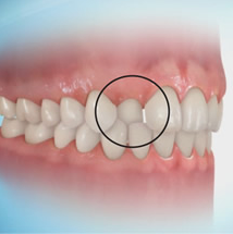
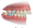
openbite
|
existing space between the mandibular and maxillary teeth can be either anterior or posterior (unilateral or bilateral)
|
|
|
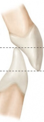
overbite
|
deep vertical overlap of the maxillary teeth onto the mandibular teeth that exceeds the normal, or 1/3 the depth of the mandibular incisors
|
|
|
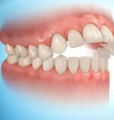
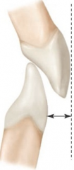
overjet
|
horizontal overlap creating a protrusion or space between the labial surface of the mandibular incisors and the lingual surface of the maxillary incisors
|
|
|
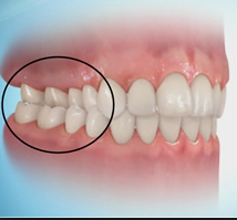
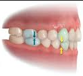
crossbite
|
a facially positioned mandibular tooth or teeth which can be either anterior or posterior (buccal/lingual)
|
|
|
edge to edge (end to end)
|
a contacting of the incisal edges or cusp tips rather than an interdigitation of cusp and fossae (actually a crossbite)
|
|
|
underjet
|
horizontal relationship where the maxillary anteriors are lingual to the mandibular anteriors
|
|
|
occlusal deviations involving individual teeth are?
|
labioversion/ buccoversion
linguoversion infraversion supraversion torsoversion |
|
|
labioversion/ buccoversion
|
tooth positioned more facially than normal
|
|
|
linguoversion
|
tooth positioned more lingually than normal
|
|
|
infraversion
|
tooth positioned below the plane of occlusion
|
|
|
supraversion
|
tooth positioned above the plane of occlusion
|
|
|
torsoversion
|
rotated tooth
|
|
|
neutrocclusion
|
class 1- normal
mesognathic deviations of single or several anterior teeth overjet, overbite w/ molars in ideal positioning |
|
|
distocclusion (division 1)
|
class 2
retrognathic maxillary teeth in labioversion, protrusion of incisors (over jet, overbite, crowding, labial incline of max incisors) |
|
|
distocclusion (division 2)
|
class 2
linguoversion of mandibular teeth protrusion of maxillary lateral incisors, retrusion of the maxillary central incisors can occur on only 1 side of arch |
|
|
mesiocclusion
|
class 3
prognathic mandibular teeth mesial to normal position |
|
|
mesognathic
|
normal profile
|
|
|
prognathic
|
mandible protrudes
|
|
|
retrognathic
|
mandible retrudes
|
|
|
centric occlusion
|
relationship of the occlusal surfaces of 1 arch to those in opposing arch
post teeth closed, ant teeth has light to no contact |
|
|
centric relation
|
most retruded position of the condyle in the mandibular fossa
|
|
|
functional malocclusion
|
occlusal deviation created by habits or muscular dysfunctions (thumbsucking may cause)
|
|
|
what determines the mesial step of primary second molars
|
if the 2nd molars are in class 1 position the permanent teeth will usually be guided into a class 1 position
|
|
|
terminal plane
|
often primary molars will show a cusp to cusp (end to end) relationship
|
|
|
the maxillary teeth in contact with the mandibular teeth in any functional relationship describes
|
occlusion
|
|
|
when the permanent mandibular 1st molar and canine are more mesial (or ant.) than normal it is referred to as what class?
|
class 3 mesiocclusion
|
|
|
a rotated tooth is
|
torsoversion
|
|
|
a horizontal overlap of the maxillary teeth creating protrusion is a
|
overjet
|
|
|
when the mesiobuccal cusp of the maxillary 1st molar rests in the mesiobuccal groove of the mandibular molar the angle classification is
|
class 1 neutrocclusion
|
|
|
anterior crossbite
|
|
|
post crossbite
|
|
|
edge-to-edge bite (ant teeth)
(end-to-end: post teeth) |
|
|
open bite
|
|
|
overjet (vertical overlap)
|
|
|
overjet (horizontal overlap)
|
|
|
neutrocclusion- class 1
|
|
|
distocclusion- class 2
|
|
|
mesiocclusion- class 3
|