Reading...
![]()
Play button
![]()
Play button
![]()
Use LEFT and RIGHT arrow keys to navigate between flashcards;
Use UP and DOWN arrow keys to flip the card;
H to show hint;
A reads text to speech;
102 Cards in this Set
- Front
- Back
|
Intercellular Communication is accomplished chemically via 3 molecules:
|
1) Neurotransmitters---Close distances and fast-acting
2) Local Mediators---immediate around cell 3) Hormones---Far distances and slow-acting |
|
|
Local Mediators
|
May be proteins, amino acid derivatives, or fatty acids (prostaglandins)
-Part of Paracrine System |
|
|
Parts of nervous system include:
|
Brain, spinal cord, nerves, neural support cells, and sense organs (eye, ear, etc)
|
|
|
Neuron
|
Functional unit of nervous system. Highly specialized; lost capacity to divide.
|
|
|
What do neurons depend entirely on for energy?
|
Uses facilitated diffusion to import glucose without the need of insulin.
Requires effective aerobic respiration. |
|
|
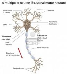
Dendrites
|
Protrude and act as antennae, sensors that receive stimuli as individual units and collectively transmit to cell body (soma).
|
|
|
Axon Hillock
|
Connects soma to axon and is the location where action potential is initiated in all directions including down the axon.
Contains large amounts of VG-Na+ channels |
|
|
Resting Potential
|
Uneven distribution of cations across the membrane mediated by the Na/K-pump (~ -70mv) creating a polarized membrane (negative inside) when the cell is at rest.
|
|
|
Na/K-pump
|
A integral membrane protein-pump using 1 ATP to actively transport 3 Na+ ions out of the cell and 2 K+ ions into the cell.
|
|
|
Initiation of Action Potential
|
Influx of Na+ ions that raises the electrical potential above the "critical threshold" in an all-or-nothing event.
|
|
|
Action potential is defined by
|

Depolarization: Period when VG-Na+ channels open to allow influx of Na+ ions to depolarize the cell to peak voltage.
Repolarization: Outflow of K+ ions through slow-acting VG-K+ channels. Occurs a little before peak voltage. |
|
|
Hyperpolarization
|
Period after repolarization that allows the outflow of many K+ ions to cause the membrane potential to drop below resting potential.
|
|
|
Absolute Refractory Period
|
Period when no stimulus can initiate another action potential. Begins when VG-Na+ channels inactivate at peak voltage and continues as the cell is repolarizing.
Na+ and K+ ions are on opposite sides!!! |
|
|
Relative Refractory Period
|
Period when cell is susceptible to another action potential but requires an abnormally large stimulus.
|
|
|
Accommodation
|
An action potential that may not occur due to a threshold stimulus happening too slowly.
|
|
|
Schwann Cells
|
Encase long, discrete sections of the axons of neurons in the PNS by wrapping layers of their plasma membranes around the axon, creating myelin sheaths.
|
|
|
Oligodendrocytes
|
Wraps layers around the axon, creating myelin sheaths in the CNS.
|
|
|
Nodes of Ranvier
|
Small, unmyelinated areas of the axon that occur in regular intervals down the axon's length and permit saltatory conduction.
|
|
|
What is the significance Myelin Sheaths and Saltatory Conduction?
|
Myelin insulation significantly accelerates the transmission of impulse as depolarization jumps from one node of Ranvier to the next, effectively skipping across long, insulated portions of the axon.
|
|
|
Nervous tissue support cells
|
Glial or Neuroglia Cells: Outnumber neurons 10 to 1. Capable of dividing (eg. brain injury)
|
|
|
6 types of Glial cells and function
|
1) Microglia
2) Ependymal Cells 3) Satellite Cells 4) Astrocytes 5) Schwann Cells 6) Oligodendrocytes |
|
|
Microglia
|
Arise from monocytes (WBCs) and phagocytize microbes and cellular debris in CNS.
|
|
|
Ependymal Cells
|
Epithelial cells that line space containing cerebrospinal fluid and use cilia to circulate the cerebrospinal fluid.
|
|
|
Satellite Cells
|
Support ganglia (PNS).
|
|
|
Ganglia
|
Groups of cell bodies in the PNS.
|
|
|
Astrocytes
|
Star-shaped neuroglia in the CNS that give physical support to neurons, and help maintain mineral and nutrient balance in the interstitial space.
|
|
|
White Matter
|
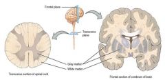
The appearance of myelinated axons.
|
|
|
Gray Matter
|

The appearance of neuronal cell bodies (soma).
Cerebrum has a gray appearance. |
|
|
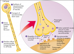
VG-Ca2+ Channels
|
Open when action potential impulse reaches the synapse allowing the influx of Ca2+ ions that bind to regulatory proteins and causes exocytosis of NT vesicles.
|
|
|
Structure of Synapse (3):
|
1) Presynaptic Neuron- contains and releases neurotransmitters through the presynaptic membrane.
2) Synaptic Cleft- space between the presynaptic membrane and the postsynaptic membrane. 3) Postsynaptic Membrane- contain ligand-gated ion channels; located on neurons, muscles, glands, or organs. |
|
|
Chemical Synapse or (Motor-Plate end; neuron to muscle)
|

Terminal end of the axon containing neurotransmitters just inside the presynaptic membrane; unidirectional.
|
|
|
Electrical Synapse
|
Uncommon and bidirectional synapse. Composed of gap junctions between cells that signals much faster than chemical synapses. (eg. cardiac muscle, visceral smooth muscle, escape reflexes, retina of vertebrates)
|
|
|
Ligand-gated ion channels
|
Postsynaptic receptors which bind specific neurotransmitters serving as ligands that induce an open conformational change to allow the influx of ions.
|
|
|
What happens to neurotransmitters after they bind and quickly release from post-synaptic membrane? (3)
|

1) Slowly diffuse out of synaptic cleft
2) Degraded by enzymes 3) Relocate back to presynaptic neuron via active transport |
|
|
Acetylcholine esterase
|
Enzyme that degrades acetylcholine in synaptic cleft to keep neurons from actively firing.
|
|
|
MAO and COMT
(Monoamine Oxidase and cate-chol-O-methy transferase) |
Metabolites that render epinephrine inactive by oxidizing and methylating, respectively. (2)
|
|
|
Exponential decline
|
The declination of stimulus with distance from synapse after EPSP or IPSP
|
|
|
EPSP
(Excitatory Postsynaptic Potential) |
Combination of firing synapses creating a excitatory change where Na+ gates open to become depolarized ----> action potential continues (~40-80 synapses)
|
|
|
IPSP
(Inhibitory Postsynaptic Potential) |
K+ gates open (out) and further polarizes postsynaptic neuron---> difficult to generate action potential
|
|
|
G-Protein
|
Common receptor protein along the inside of a post-synaptic membrane that initiates a second-messenger system.
|
|
|
The _____-subunit of the G-protein breaks free upon receptor-binding and activates....(4)
|
-alpha-subunit
1) activates separate specific ion channels 2) activates a second messenger (i.e. cAMP, cGMP) 3) activates intracellular enzymes 4) activates gene txn |
|
|
Acetylcholine
|
Neurotransmitter:
excitatory response @ neuromuscular junctions inhibitory response @ other junctions (vagus nerve) |
|
|
GABA
|
Inhibitory NT among neurons in the brain
|
|
|
Somatic Nervous System
|
-Controls voluntary actions.
-Stimulus from external environment and innervates skeletal muscle; uses ACh on nicotinic receptors. -1 sensory neuron (synapse in CNS) and 1 motor neuron |
|
|
ANS (Autonomic Nervous System)
|
-Controls involuntary action.
-Receives signals from visceral organs and innervates smooth/cardiac muscle, and glands. -Divided into Sympathetic and Parasympathetic Nervous Systems. |
|
|
How many motor neurons does the ANS use?
|
2!
1) Presynaptic neuron= CNS, uses ACh 2) Postsynaptic ganglion= PNS, uses ACh or adrenaline |
|
|
Sympathetic Nervous System
|
"Flight or Flight"
-Cell bodies lie far from effectors. -Signals come from spinal cord (ventral area). -Use adrenic receptors on effectors |
|
|
Parasympathetic Nervous System
|
"Rest and Digest"
-Cell bodies lie inside or close to effectors. -Signals come from lower spinal cord and brain ('para') -Use nicotinic receptors (ganglion) and muscarinic receptors (effectors) |
|
|
What and where are muscarinic receptors found?
|
-Type of cholinergic receptor (nicotinic is other)
-Found on effectors of parasympathetic nervous system |
|
|
CNS (Central Nervous System)
|
-Brain and spinal cord.
-Composed of interneurons |
|
|
PNS (Peripheral Nervous System)
|
-Conveys information to and from nervous system.
-Divided into ANS and somatic nervous system |
|
|
What are the 5 types of stimuli humans respond to?
|
1) tactile=touch
2) olfactory=smell 3) gustatory=taste 4) auditory 5) visual |
|
|
What is the function of sensory receptors?
|
Register a given stimulus and gather information
|
|
|
What are the 5 main sensory receptors?
|
1) MECHANO= stretch, tactile, proprioceptors, auditory
2) CHEMO= olfactory, gustatory 3) THERMO 4) ELECTROMAGNETIC= photoreceptors 5) NOCIECEPTORS= pain |
|
|
Sensory Neurons
|
Afferent Neurons.
-Carry information from sensory receptors to CNS. |
|
|
Interneurons
|
Associative Neurons.
-Relay and process info between neurons in CNS |
|
|
Motor Neuron
|
Efferent neuron or effector.
-Convey signals from CNS to target (muscle, gland, or organ) |
|
|
What does the neural tube give rise to?
|
-Brain (anterior)
-Spinal Cord (posterior) |
|
|
What 3 components protect the CNS?
|
1) Meninges; connective tissue
2) Bone; spinal cord and skull 3) Cerebrospinal Fluid (CSF); liquid shock absorber |
|
|
What does the hindbrain become?
|

-Cerebellum
-The Pons -Medulla Obligata |
|
|
What is the function of the midbrain?
|
-Integration of visual and audio info
|
|
|
What 2 dinosaurs arise from the forebrain?
|
-diencephalon
-telecephalon |
|
|
What 3 structures is the diencephalon composed of?
|
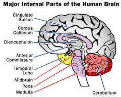
-Hypothalamus
-Thalamus -Pituitary Gland |
|
|
What 3 structures is the telecephalon composed of?
|

-Basal Nuclei
-Limbic System -Cerebral Cortex or Cerebrum |
|
|
Cerebral Cortex or Cerbrum splits into these lobes...
|
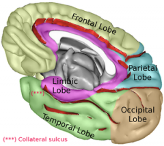
Frontal
Parietal Occipital Temporal |
|
|
Function of Spinal Cord
|
-simple spinal reflexes
-control of primitive processes i.e. walking, urination, function of sex organs |
|
|
Medulla: location and function?
|
-Hindbrain
-Controls autonomic processes (i.e. blood pressure, heart rate, respiratory rate, reflexes like sneezing+vomiting) |
|
|
The Pons: location and function?
|
-Some autonomic processes
-anti-gravity and posture balance |
|
|
Cerebellum: location and function?
|
-Integration center
-Coordination of refined and complex movement, balance, and posture (i.e. dancing, sewing, piano) |
|
|
Thalamus: location and function?
|
-Forebrain diencephalon
-Somatic conscious sensation |
|
|
Hypothalamus: location and function?
|
-Forebrain diencephalon
-Homeostasis (i.e. body temperature) -Primitive emotions (i.e. appetite, rage, sex drive) |
|
|
Pituitary gland: location, regulation, and function?
|
-Forebrain diencephalon
-Regulated by the hypothalamus -Homeostasis via hormone release -Anterior Pituitary releases 6 peptide hormones -Posterior Pituitary releases 2 peptide hormones |
|
|
Basal Nuclei: location and function?
|
-Forebrain telecephalon
-regulate body movement |
|
|
Limbic System: location and function?
|
-Forebrain telecephalon
-emotion |
|
|
Cerebral Cortex or Cerebrum: location and function?
|
-Forebrain telecephalon
-Higher thought processes: cognition, attention, memories, intelligence, language/communication, abstract thought, reading |
|
|
What does the corpus callosum do?
|
Thick bundle of axons connecting the right and left hemispheres of cerebrum.
|
|
|
Simple Reflex Arc
|
-Rapid, monosynaptic reflex involving 1 sensory and 1 motor neuron
-Belongs to PNS |
|
|
Function of Vestibular and Auditory Systems
|
1) maintenance of postural equilibrium and balance
2) reception of sound |
|
|
What are the 3 parts of the ear?
|

-outer ear
-middle ear -inner ear |
|
|
Outer Ear
|

-pinna (ear flap)
-auditory canal |
|
|
Middle Ear
|
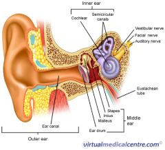
-tympanic membrane or eardrum
-malleus, incus, stapes |
|
|
Inner Ear
|

-Oval window (small entry)
-Cochlea -Hair cells in Organ of Corti -Vestibular apparatus: Semicircular canals |
|
|
Movement of Sound through Ear
|
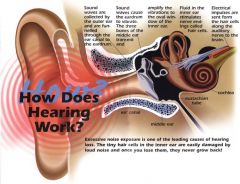
1. Sound enters pinna and is directed through ear canal (outer ear)
2. Produces vibrations at tympanic membrane which travel through malleus, incus, and stapes which act as a lever system, ↑F (middle ear) 3. -Goes through small oval window (↑P), spirals in cochlea and causes movement of hair cells (cilia) at the Organ of Corti that tranduces into neuronal signals. |
|
|
Travel of Light through Eye
|
1. Light refracts of cornea -->transverses through aqueous humor
2. Passes through pupil and lens 3. Proceeds through vitreous humor until it reaches photoreceptors at the retina |
|
|
Cornea
|

Transparent layer which refracts light at front of eye through the aqueous humor
|
|
|
What are the 2 liquid-gel cavities of the eye?
|

-the aqueous humor (anterior)
-vitreous humor (posterior) |
|
|
Iris
|

-Colored portion of eye
-Muscular tissue that constricts and dilates to regulate amount of light through pupil |
|
|
Lens
|
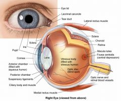
Converging lens that can:
-flatten (farther focal point)....eyes wide open -contract (closer focal point)...eyes squinting |
|
|
Ciliary Body
|

Circular muscular tissue surrounding the lens that controls focal length by contracting and relaxing
|
|
|
Retina
|

Back portion of eye containing photoreceptors (rods and cones)
|
|
|
Rods
|
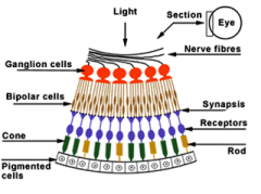
-Registers black/white and dim light
Dark: Depolarized---->inactive rhodopsin Dim Light: Hyperpolarized-----> active rhodopsin |
|
|
Cones
|
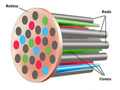
Register bright light and colors
-uses the protein opsin (similar to rhodopsin) |
|
|
What is the largest organ in the human body?
|
Skin
|
|
|
3 main functions of the skin?
|
1. Maintain body temperature
2. Environment sensory input 3. Protection barrier |
|
|
Other 4 functions of the skin?
|
4. Excretion
5. Immunity 6. Blood reservoir 7. Vitamin D synthesis |
|
|
3 main layers of the skin?
|

-epidermis
-dermis -subcutaneous layer |
|
|
Epidermis
|

Avascular, stratified squamous epithelium that consists of 5 separate layers
|
|
|
Stratum Corneum
|
-Most outer layer of epidermis
-Many layers of dead cells and keratin protein -Forms waterproof-like barrier |
|
|
Stratum Germinativum
|
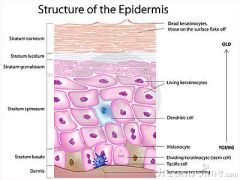
-inner epidermis layer
-skin cells differentiate -where keratin is produced (loses nuclei) |
|
|
Dermis
|
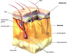
-Thickest layer of skin below epidermis
-Derived from mesodermal tissue |
|
|
Where would you find blood vessels, nerves, sebaceous (oil) and sweat glands???
|
Dermis
|
|
|
Subcutaneous Layer
|
-Most bottom layer of skin
-Composed of adipose tissue--->insulation & protection |