![]()
![]()
![]()
Use LEFT and RIGHT arrow keys to navigate between flashcards;
Use UP and DOWN arrow keys to flip the card;
H to show hint;
A reads text to speech;
89 Cards in this Set
- Front
- Back
|
Reporting Yeast |
Document how many (budding vs. non), bacteria observed (type and amount), and cells (blood vs. epithelial) form the entire slide; Don't forget which ear |
|
|
Reporting on Otododectes |
Count the number of mites and eggs you see per slide; Don't forget which ear |
|
|
Lyme Disease |
Caused by the bacteria: Borrelia burgdorferi; Spread by hard ticks (Ixodes scapularis); When removing a tick, ensure you remove the head because it may cause an abscess |
|
|
Volume of Ejaculate |
Measured with a volumetric flask; Total ejaculate for a bull is usually 5mls (male cats= 0.04mls); Ejaculate volume is larger than spermatozoa and varies amongst collection methods; Three components: sperm free fraction, sperm rich fraction, and sperm poor fraction |
|
|
Gross Appearance |
Opacity (reflects spermatozoa concentration); Words to describe: Thick, creamy, opaque, milky opaque, opalescent milky, watery and white (normal bulls- opaque/creamy/white) and discoloration due to contamination |
|
|
Sperm Motility |
If correlated with fertility!; Improper specimen handling adversely affects the motility; 2 assessments: wave motion, and motility |
|
|
Wave Motion |
Gross motility based on swirling activity observed in a drop of semen on a microscope slide at low magnification (4X); Very good= vigorous swirling; Good= moderate slow swirling; Fair= barely swirling; Poor= no swirling; Bulls have high sperm density so should show vigorous swirling; Wave motion is decreased if sperm concentration is decreased |
|
|
Motility |
Diluted drop of semen with warm physiological saline with a coverslip and observed at 10X; 1 drop of semen on slide, dilute with saline to produce a monolayer, place coverslip; Subjective: Very good= rapid linear activity (80-100%); Good= moderate linear activity (60-80%); Fair= slow linear or erratic activity (40-60%); Poor= very slow, erratic activity (20-40%); A satisfactory sample should have at least 60% |
|
|
Sperm Concentration |
Diluent is formalin, Sodium bicarb and distilled water; Take 0.1mls semen and add 9.9mls diluent; Gently mix sample until homogeneous; Place semen on a Hemacytometer and allow to sit for 2-3 minutes; Count both grid areas at the 10X (withing 10%?- take the average); Multiply the number b 1000000 and this will result in a # sperm per ml of the diluted semen sample; |
|
|
Normal Sperm Concentration for a Bull |
1200 million/mL (range 300-2500 million/mL) |
|
|
Live:Dead Sperm Ratio Procedure |
Have microscope slides and stain prewarmed to body temperature;
Pipette a drop of stain (negrosin/eosin mixture) onto the end of a slide, then pipette a small droplet of semen next to the stain; Rock that slide back and forth a few times to mix the sperm and stain; Smear like a blood smear; Dry the slide rapidly; Examine using 100X objective lens |
|
|
Live:Dead Sperm Ration Count |
Observe at 100X and count 200 cells; result expressed as a percent of live sperm; Live sperm resist staining, dead sperm pick up the eosin and appear pink, the background should appear blue |
|
|
Sperm Morphology |
Observe 100-500 cells; Abnormalities are divided into: Head, midpiece, and tail; Generally 20% of sperm are abnormal |
|
|
Sperm Morphology: Primary |
Serious abnormalities; Occur during production; Head too big/small, doubled, oddly shaped; Midpiece swollen, kinked, double, twisted, essectrically attached to head; Tails that are coiled |
|
|
Sperm Morphology: Secondary |
Storage in the epididymus to the smear; Tailless heads; Bent or broken tails; Protoplasmic droplets on midpiece |
|

|
Misshapen Head |
|

|
Elongated Head |
|

|
Double head |
|

|
Double Tail |
|
|
Other cells in sperm... |
Normal semen has few: WBCs, RBCs, and epithelial cells; NEVER has: bacteria and yeast |
|
|
Formal Evaluation |
Signalment; Volume; Gross appearance; Wave motion; Motility; Concentration; Live:Dead Sperm Ratio; Morphology |
|
|
Normal Total Sperm Ejaculate for a Bull |
4-5 billion |
|
|
Normal Morphology for a Bull |
Greater than 70% normal |
|

|
Anestrus: Non-cornified squammous epithelial cells, may contain some neutrophils, no RBCs |
|

|
Proestrus: Cornified squammous epithelial cells replace non cornified, neutrophils decrease, and RBC's increase |
|

|
Estrus: Cornified squammous epithelial cells, no neutrophils, RBCs present in small numbers, neutrophils increase as diestrus approaches |
|

|
Diestrus: Cornified epithelial cells are replaced by non cornified, abundant cytological debris, neutrophils increase in number by the 3rd day of diestrus and decrease to few by about the 10th day, no RBCs |
|
|
Cytology Report: Lower Power (10X) |
Scan for large objects, cell clusters, parasites, fungal hyphae and crystals |
|
|
Cytology Report: High power (40X) |
Determine prominent cell type |
|
|
Cytology Report: Oil immersion (100X) |
Describe cellular characteristics |
|
|
Imprints |
For Muscle Tissue Samples: Done on external lesions or from removed tissues; Easy to collect; Collects fewer cells; Greater contamination; Disadvantages: few cells collected, cells obtained from the surface of a lesion may not be representative of the whole lesion; |
|
|
Procedure for making Imprints |
Imprint dirty then clean; Make several dirty imprints; Blot tissue on clean, wet (saline), absorbed material to remove fluid; Dry lesion; Make several clean imprints (do not drag); Dry the slide quickly; Stain with dirty diff quick; Observe under 40X and oil;
|
|
|
FNA- Adipose Tissue (small mass) |
21g needle and a 12cc syringe; Alcohol skin prep; Mass held securely; Needle introduced in center of mass; Strong negative pressure applied (3/4 of syringe); Sample several areas (avoid surrounding tissue); Relieve negative pressure before removing; Several squash preps are made; Dry quickly and stain with dirty diff quick; Observe under 40X and oil |
|
|
FNA- Adipose Tissue (large mass) |
21g needle and a 12cc syringe; Alcohol skin prep; Needle stays within mass; Redirect to other areas; Negative pressure relieved during redirection and removal; Several squash preps are made; Dry quickly and stain with dirty diff quick; Observe under 40X and oil |
|
|
Non-Aspirate- Bone Marrow |
Mass held firmly; Needle with syringe attached is introduced in mass (syringe pre-filled with air); Needle is moved rapidly back and forth through the mass five to six times along the same tract; Cells are collected by shearing and capillary action; Needle removed from mass, material is expelled by depressing plunger; Repeat in more than one site and create several slide; Squash prep and air dry quickly; Stain with dirty diff quick; Observe under 40X and oil |
|
|
Hair Pluck |
Grasp hair close to the base and remove in the direction of the follicle; Place on microscope slide and confirm you did not break the hair and the follicle is intact; Positive for ringworm- fungal spores attached to the hair shaft (40% accurate) |
|
|
Culture Swabs |
Use to isolate bacteria and viruses; Areas that can be swabbed include: Mucus membranes of the urethra, cervix, rectum, eye, throat, vagina, and penis; Fluid collected from thoracic or abdominal cavity; And non purulent wounds; It is important to follow asepsis in order not to contaminate the sample and to ensure you have the appropriate aerobic/anaerobic medium for the bacteria being tested |
|
|
Procedure for Culture |
Ensure appropriate sampling (aerobic vs. anearobic or both); Aseptically handle all materials; Peel back plastic wrap exposing the caps; Open tube and discard plug; Take sample with swab; Insert swab into tube and secure cap |
|
|
Post Sampling for Culture |
Refrigerate labeled (Animal ID, site, date and time cultured on each sample) transport media vials containing swabs (do not freeze); Send with refrigerated transport packs in a styrofoam shipper so that culture is set-up within 48hrs of taking the swab- older samples will be rejected as they are unfit; Cultures generally take 7 days fom the time of placing the material on the selective culture media; Negative status cannot be confirmed before 7 days; Identification of fungal or bacterial contamination which would preclude reading of the test, will be done as early as possible so that re-sampling and resubmission can occur |
|
|
Canine Heartworm Antigen Test Kit |
Snap test used to test for the presence of adult D. Immitis antigens in dog or cat blood; |
|
|
ERD Health Screen Feline Urine Test |
Detects level of albumin in feline urine; A test used to determine if a cat has damaged nephrons due to a variety of reasons |
|
|
Canine Pregnancy Test Kit |
Determines pregnancy by measuring relaxin levels in plasma or serum; Valid 20-31 days post LH surge; |
|

|
Hymenolepis spp.; Tapeworm |
|

|
Syphacia spp. Pin worm; Inhabit the cecum and colon of rats and mice; Nonpathogenic in immunocompeten rodents; In labs, infections may have effects on behaviour, growth, intestinal physiology, and immunology; Effective pinworm surveillance and eradication vital in research animal facilities; Diagnosed by the use of perianal tape impression |
|

|
Aspiculuris spp. Pin worm; Found in mice and rats; Located in the colon and cecum |
|

|
Cryptosporidium spp. Protozoa; Causes diarrhea; Fatal in immunocompromised; Thick fecal smear |
|

|
Eimeria spp. Live in the small intestines; Diagnosed by fecal float; Can be sporulated or unsporulated |
|
|
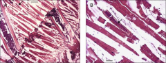
Trichosomoides spp. Larvae develop in the striated muscle; Migrate to he nasal mucosa; Worms can be found between the incisors in the mucosa; Under natural conditions, the inflammation of the nasal mucosa that is induced by the parasites might reduce the competitiveness of infected rodents when foraging or looking for potential mates |
|

|
Spironucleus spp. Small symmetrical, flagellated protozoa; Found in the intestinal tract; Most common in fish |
|

|
Dentostomella spp. Pin worm of gerbils; Causes diarrhea; Fecal float |
|

|
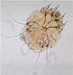
Notoedres spp. Scabby, scaly facial disease known as head mange; Highly contagious; Similar to sarcoptes but smaller and rounder |
|
|
Tixacarus cavia; Guinea pigs; Affects the head, shoulders, back, and flanks of the guinea pig, may spread; Secondary bacterial infections can occur |
|

|
Psoroptes spp. Rodent and rabbits; Scraping or exudate from ears |
|

|
Cheyletiella spp. Rabbits; "Walking dandruff" Direct observation, mites usually direction surface of skin; Excessive dandruff |
|
|
What sample do you require for a Canine Pregnancy Test? |
Serum or Plasma collected in an anti-coagulated with EDTA or Sodium Citrate |
|
|
How many times should the Canine Pregnancy Test be performed and in what time interval, to confirm a negative result? |
The test should be performed twice, one week apart in order to confirm the status |
|
|
Describe a negative and positive result of the Canine Pregnancy Test |
Positive= one pink/purple band in both window 2 & 3; Negative= No band reading in the #2 window but there will be a band in #3 |
|
|
What does the Heartworm Antigen Test Diagnose? |
Dirofilaria immitis antigen |
|
|
Why would we perform the Heartworm test? |
Routine testing |
|
|
What would a negative test result look like for Heartworm? |
Negative= only the positive control spot develops |
|
|
What would a positive test result look like for Heartworm? |
Any color development in the sample spots as well as the positive control |
|
|
When would you deem the test invalid for Heartworm? |
If there is background color, and if the positive control spot does not appear |
|
|
What does the ERD Health Screen diagnose? |
Detection of microalbuminuria in cats |
|
|
When would you test a patient with the ERD Health Screen? |
Health screen and evaluation for early renal damage is indicated |
|
|
What test do you perform before proceeding with the ERD? Why? |
Prior to performing the ERD, you must obtain the urine specific gravity. The result will determine where to place/add urine in the test and whether to dilute the sample. |
|
|
Name 4 diseases that damage nephrons allowing albumin to leak into the urine |
Inflammatory disease; Infectious disease; Metabolic disease; Neoplasia |
|
|
What type of sample do you require for a Canine Parvo Antigen Test and how would you collect it? |
You need a fecal sample collected directly from the anus |
|
|
What form of CPV is prevalent in puppies? Why not the other form? |
Enteritis is prevalent in puppies because the myocardial form is rare |
|
|
Once the pouch is open, the CPV test should be used within how many minutes? |
The test should be completed within 10 minutes |
|
|
What does a positive result look like for CPV? |
Once pink/purple band in window #2 and window #3; |
|
|
What would an invalid result for CPV look like? |
There will be no pink/purple band in window #3 |
|
|
Transudates |
Fluid that passes through a membrane which filters out much of the protein and cellular elements to yield a watery solution; Due to increased pressure in the veins and capillaries pressure forcing fluid through the vessel walls or low levels of protein the blood serum - a filtrate of blood; causes edema; SG: <1.017; TP: <3g/dL |
|
|
Transudates commonly found in... |
Acites or pleural effusions found with heart failure or other circulatory disorders |
|
|
Modified Transudates |
Transudate with additional protein and/or cells; May be in a transitional stage progressing to an exudate; Moderate cellularity; TP: 2.5-7.5 g/dl; SG: 1.017 or less than 1.025; Sediment shows mesothelial cells and macrophages; Amber or pink and turbid |
|
|
Exudates |
Fluid rich in protein and cellular elements that oozes out of blood vessels due to inflammation and is deposited nearby; Altered permeability of blood vessels permits the passage of large molecules and solid matter through their walls; Increased cellularity; TP: >3g/dl; SG: >1.025; Sediment shows neutrophils, lymphocytes, and small number of macrophages; Higher cell count and protein value are indicative of inflammation or injury; ex: Perspiration, pus, and serum; Further classified into : septic or non septic and degenerative or non-degenerative |
|

|
Septic exudate; Neutrophils with intracellular bacteria |
|
|
Non septic Exudate |
Sediment does no show intracellular bacteria |
|
|
Degenerative Exudate |
Sediment shows nuclear changes such as pyknosis, karyorrhexis, and karyolysis |
|
|
Non-degenerative Exudate |
sediment does not show nuclear changes |
|
|
Modified Centrifuged Buffy Coat Smear |
Used to concentrate WBCs and platelets when looking for microfilaria, amastogotes (leishmania), erhlichia, and other blood parasites and mast cells |
|
|
Blood Typing |
Determine the patient's blood type before a transfusion; Canine tests for DEA and cats for A & B blood groups |
|
|
Canine Blood type |
DEA 1.1 positive= RBCs carry that antigen; Donors are usually DEA 1.1 negative or match the recipient; If DEA 1.1 negative recipient is given DEA 1.1 positive blood, the recipient may develop antibodies and destroy the RBCs during the second transfusion |
|
|
Feline Blood type |
No universal donor among cats as they have natural antibodies against the blood antigen that they lack; Cats are typed to match the donor and recipient; Even in the first incompatible transfusion with cats, there is a transfusion reaction; 90% of cats are type A |
|
|
Crossmatching |
Always done prior to transfusion to assess donor and patient serologic compatibility; If not compatible and blood is administered, the patient will have a hemolytic transfusion reaction (rapid destruction of donor red cells); Crossmatch does not check for incompatible WBC or platelet antigen so reactions are still a risk with any transfusion |
|
|
Major Crossmatch |
This compares donor erythrocytes to recipient plasma to test for antibodies; You are checking for preformed (aquired or naturally occurring) antibodies in recipient plasma against donor erythrocytes |
|
|
Minor Crossmatch |
This compares donor plasma to recipient erythrocytes and checks for preformed antibodies in donor plasma that could hemolyze recipient red cells; Usually the donor plasma is markedly diluted after transfusion and is unlikely to produce a significant transfusion reaction, most often done in patients who have already had a transfusion |
|
|
Occult Blood Test on Fecal Sample |
Reagent tablets are used for detection of RBCs and/or hemoglobin in feces; The procedure relies on the peroxidase-like activity of hemoglobin to enhance the oxidation of a chromagen in the tablet to orthotoluidine, which has a blue colour; Small quantity of fresh feces is smeared onto a filter paper, regeat tablet is placed on top, wait 5-10 seconds, if blue = positive; The test reagent reacts with myoglobin of muscle, carnivores should be fed a diet free of raw meat at least 3 days before testing so this doesn't result in a false positive |
|
|
Fecal wet mounts identify... |
protozoas (Giardia + Spirochetes), and purposful movement |
|
|
Fecal Smears identify... |
Clostridium; Spirochetes; Campylobacter- confirm with a culture; Cryptosporidium - diagnose with acid-fast stain |