![]()
![]()
![]()
Use LEFT and RIGHT arrow keys to navigate between flashcards;
Use UP and DOWN arrow keys to flip the card;
H to show hint;
A reads text to speech;
319 Cards in this Set
- Front
- Back
|
___________ circulation brings the nutrients and O2 to the tissues and returns deoxygenated blood back to the heart. |
Systemic circulation brings the nutrients and O2 to the tissues and returns deoxygenated blood back to the heart. |
|
|
___________ circulation takes the deoxygenated blood entering the heart, oxygenates it and returns it back to the heart. |
Pulmonary circulation takes the deoxygenated blood entering the heart, oxygenates it and returns it back to the heart. |
|
|
Blood vessels that carry blood away from the heart are __________. |
Blood vessels that carry blood away from the heart are arteries.
|
|
|
Blood vessels that carry blood towards the heart are __________. |
Blood vessels that carry blood towards the heart are veins. |
|
|
_________ valve is present between the right atrium and right ventricle. |
The tricuspid valve (right AV valve) is present between the right atrium and right ventricle.
|
|
|
The____________valve is present between the left atrium and left ventricle.
|
The Mitral valve (bicuspid/ Left AV valve) is present between the left atrium and left ventricle.
|
|
|
The____________valve is present between the right ventricle and pulmonary artery. |
The pulmonary valve (Semilunar valve) is present between the right ventricle and pulmonary artery.
|
|
|
The____________valve is present between the left ventricle and aorta. |
The aortic valve is present between the left ventricle and aorta.
|
|
|
Weight of heart |
250g-350g |
|
|
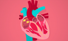
Pericardium? Enlist its layers |
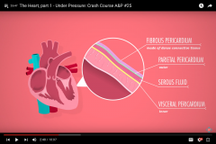
The human heart is situated in a protective sac called pericardium
|
|
|
Fibrous Pericardium |
Fibrous Pericardium - Outermost layer of pericardium -Tough and consists of fibrous proteins -Attaches the heart to the rest of the body -Protects the heart from physical damage |
|
|
Serous layer of pericardium |
Serous layer of pericardium Contains parietal and visceral layer separated by fluid called pericardial fluid which lubricates the heart. |
|
|
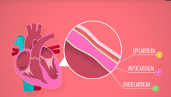
Layers of heart |
|
|
|
Epicardium |
Epicardium: -Outermost layer of heart -Fused with visceral layer of pericardium -Lubricates the heart -Prevents damage due to friction during contraction. |
|
|
Myocardium |
Myocardium:
-Middle layer of the heart -Consists of cardiac muscle responsible for contracting the heart |
|
|
Endocardium |
Endocardium -Innermost layer of the heart -Consists of simple squamous endothelial cells -Direct contact with the blood in the heart |
|
|
Chordae tendineae |
The chordae tendineae (tendinous chords), or heart strings, are cord-like tendons that connect the papillary muscles to the tricuspid valve and the mitral valve in the heart.
|
|
|
Papillary muscles
|
The papillary muscles are muscles located in the ventricles of the heart.
They attach to the cusps of the atrioventricular valves (also known as the mitral and tricuspid valves) via the chordae tendineae FUNCTION:They contract to prevent inversion or prolapse of these valves on systole (or ventricular contraction). |
|
|
Muscles of ______(atria/ventricles) are stronger/thicker than that of _______(atria/ventricles) . |
Muscles of ventricles are stronger/thicker than that of atria .
|
|
|
Parts of interventricular septum |
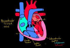
Parts of interventricular septum:
i)Membranous (thinner) ii)Muscular (thicker) |
|
|
In which part of the interventricular septum are birth defects more common? |
Membranous part |
|
|
When can the 1st heart sound be heard? What is it also called? |
LUB=1st heart sound=S1 -It can be heard when the mitral and tricuspid valves snap shut |
|
|
When can the 2nd heart sound be heard?What is it also called? |
DUB=2nd heart sound=S2 -It can be heard when the pulmonary and aortic valves snap shut. |
|
|
Systole |
Systole is the contraction phase of the cardiac cycle (contrast with diastole) that results in the ejection of blood into an adjacent chamber or vessel i.e. emptying of atria and ventricles |
|
|
When does systole occur? |
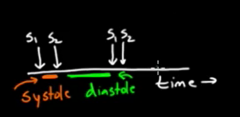
Time between S1 and S2. |
|
|
Diastole |
Diastole is the part of the cardiac cycle when the heart refills with blood following systole (contraction).
|
|
|
When does diastole occur? |

Between s2 and next s1 |
|
|
Which vessels have a high volume of blood (arteries vs veins) |
Veins have high volume of blood |
|
|
Which vessels have a high pressure?(arteries vs veins) |
Arteries have high pressure |
|
|
Which vessels have valves? |
Veins have valves |
|
|
We spend __ time in systole and ___of the time in diastole. |
We spend 1/3rd of the time in systole and 2/3rd of the time in diastole |
|
|
While reading/ recording blood pressure the highest value represents______ phase of the cardiac cycle while the lowest value represents the _______. |
While reading/ recording blood pressure the highest value represents systole (eg: 120) phase of the cardiac cycle while the lowest value represents the diastole (eg:80).
|
|
|
Which of the following arteries accompanies the great cardiac vein?
• circumflex artery • anterior interventricular artery • posterior interventricular artery • right marginal artery |
• Anterior interventricular artery(left anterior descending artery), accompanies the great cardiac vein.
NOTE: It is a branch of left coronary artery |
|
|
When viewed from the back (posterior view), the most obvious structure lying in the coronarysulcus is the ____________.
|
When viewed from the back (posterior view), the most obvious structure lying in the coronarysulcus is the coronary sinus.
|
|
|
Coronary sinus receives most of the ________blood from the heart andempties into the __________.
|
Coronary sinus receives most of the venous blood from the heart and empties into the right atrium.
|
|
|
Name the tributaries of Coronary sinus |
![Tributaries of coronary sinus are:[SMG]-Small cardiac vein,
-Middle cardiac vein, and
-Great cardiac vein.](https://images.cram.com/images/upload-flashcards/86/48/05/17864805_m.jpg)
Tributaries of coronary sinus are:[SMG]
-Small cardiac vein, -Middle cardiac vein, and -Great cardiac vein. |
|
|
__________ vein is a remnant ofthe embryonic left superior vena cava.
State its location |
The Oblique vein is a remnant of the embryonic left superior vena cava. LOCATION: It is a small vein that arises along the left side of the left atrium just beneath the lower left pulmonary artery (called the oblique vein). |
|
|
.With respect to the great cardiac vein answer the following: -Where does it open into the coronary sinus -Where does it receive blood from? |
reat cardiac vein: opens into the left extremity of the coronary sinus.
This vein receives tributaries from the left atrium and both ventricles. NOTE: The left marginal vein, is of considerable size, and ascends along the left margin of the heart. |
|
|
.With respect to the Small cardiac vein answer the following: -Where does it open into the coronary sinus -Where does it receive blood from? |
Small cardiac vein: -Opens into the right extremity of the coronary sinus. -This vein receives blood from the back of the right atrium and ventricle -The right marginal vein ascends along the right margin of the heart and joins the small cardiac vein in the coronary sinus, or opens directly into the right atrium. |
|
|
• The middle cardiac vein: ends in the coronary sinus near its______extremity.
|
The middle cardiac vein: ends in the coronary sinus near its right extremity.
|
|
|
• The oblique vein: ends in the coronary sinus near its_______ extremity; this vein is continuous above with the ligament of __________.
|
• The oblique vein: ends in the coronary sinus near its left extremity; this vein is continuous above with the ligament of left vena cava |
|
|
Name the cardiac veins that DO NOT end in the coronary sinus |
The following cardiac veins do not end in the coronary sinus:
• The anterior cardiac veins • The smallest cardiac veins (thebesian veins) NOTE: The right marginal vein frequently opens into the right atrium, and is therefore sometimes regarded as belonging to this group. |
|
|
Where do the anterior cardiac veins collect blood from? Where do they open into? |
• The anterior cardiac veins: comprising three or four small vessels which collect blood fromthe front of the right ventricle and open into the right atrium;
|
|
|
Which vessel accompanies the middle cardiac vein? which vessel is it a branch of?? |
The posterior (or descending) interventricularartery, a branch of the right coronary artery, accompanies the middle cardiac vein
|
|
|
Sympathetic stimulation will have which direct effect on the heart?
• decreased automaticity • AV block • increased vagal response • bradycardia • increased stroke volume |
• increased stroke volume |
|
|
The strength and frequency of the heart beat are controlled by the______ nervous system
|
The strength and frequency of the heart beat are controlled by the autonomic nervous system
|
|
|
Part of autonomic nervous system involved in control of heart? |
Bothparasympathetic and sympathetic parts of the autonomic nervous system are involved in the control ofthe heart.
|
|
|
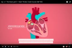
The heart has an internal nervous system made up of the ______ and the _____ nodes.
|
The heart also has an internal nervous system made up of the SA (sinoatrial node) and the AV (atrioventricular)node.
|
|
|
The AV bundle is also called ______. |
The AV bundle is also called bundle of his. |
|
|
The AV bundle (His) leaves the AV node near the lower part of the ______ septum and splits over the upper part of the_____ septum into a left bundle branch and a right bundle branch.
|
The AV bundle (His) leaves the AV node near the lower part of the interatrial septum andsplits over the upper part of the interventricular septum into a left bundle branch and a right bundle branch.
|
|
|
________ is known as the pacemaker of the heart |
SA node is known as the pacemaker |
|
|
AV node
|
AV node is an area of specialized tissue between the atria and the ventricles of the heart, specifically in the posteroinferior region of the interatrial septum near the opening of the coronary sinus, which conducts the normal electrical impulse from the atria to the ventricles.
|
|
|
The sympathetic fibers arise from segments _____of the spinal cord.
|
The sympathetic fibers arise from segments T2-T4 of the spinal cord and are distributed through the middle cervical and cervico-thoracic (or stellate) ganglia and the first four ganglia of the thoracic sympathetic chain
|
|
|
The effect of the sympathetic nerves at the SA node is an _________ in heart rate.
|
The effect of the sympathetic nerves at the SA node is an increase in heart rate.
|
|
|
The ________ nerve provides parasympathetic control to the heart.
|
The vagus nerve provides parasympathetic control to the heart.
|
|
|
The effect of the vagus nerve at the SAnode is it _______ the heart rate
|
The effect of the vagus nerve at the SAnode is the opposite of the sympathetic nerves; it decreases the heart rate.
|
|
|
The vagus nerve also _________the excitability of the junctional tissue around the AV node, and this results in _________ transmission.
|
The vagus nerve also decreases the excitability of the junctional tissue around the AV node, and this results in slower transmission.
|
|
|
_________stimulation in AV node may produce an AV block.
|
Strong vagal stimulation here may produce an AV block.
|
|
|
Which of the following is the correct conduction pathway through the heart?
• SA node- ventricular muscle- AV node- His bundle- bundle branches- Purkinje fibers -atri al muscle • SA node- atrial muscle- AV node- bundle branches- His bundle- Purkinje fibersventricular muscle • SA node- atrial muscle- AV node- His bundle- bundle branches- Purkinje fibersventricular muscle • SA node- Purkinje fibers- AV node- His bundle- bundle branches- atrial muscleventricular muscle |
SA node - atrial muscle-AV node- His bundle -bundle branches- Purkinjefibers - ventricular muscle
|
|
|
The SA node is in the wall of the _______, near the entrance of the_______.
|
The SA node is in the wall of the right atrium, near the entrance of the superior vena cava.
|
|
|
The SAnode typically depolarizes spontaneously at the rate of____to ________ times per minute
|
The SAnode typically depolarizes spontaneously at the rate of 60 to 100 times per minute,causing the atriato contract.
|
|
|
Describe the various components of ECG complex during sinus rhythm |
In sinus rhythm, every P-wave is followed by a QRS complex, the R-R interval is regular,and the P-R interval is less than 0.2 seconds.
|
|
|
P wave |
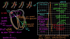
The P wave is the electrical recording from the body surface of atrial depolarizationand precedes atrial contraction. |
|
|
QRS |
Ventricular depolarization |
|
|
PR |
Impulse between SAand AV node |
|
|
T wave |
Ventricular repolarization |
|
|
QT |
Ejection of blood |
|
|
A fast sinus rhythm, faster than 100 beats a minute, is known as ________.
|
A fast sinus rhythm, faster than 100 beatsa minute, is known as sinus tachycardia
|
|
|
Slow rhythm, slower than 60 beats a minute, is known as _________.
|
Slow rhythm, slower than 60 beats a minute, is known as sinus bradycardia.
|
|
|
The apex of the heart is located at the level of the:
• third left intercostal space • fourth left intercostal space • fifth left intercostal space • sixth left intercostal space |
• fifth left intercostal space
|
|
|
The apex of the heart is formed by the tip of the left ventricle and is located at the level of the ___________.
|
The apex of the heart is formed by the tip of the left ventricle and is located at the level of the fifth left intercostal space
|
|
|
Location of SA node |
The sinuatrial node is located at the junction of the superior vena cava and the rightauricle
|
|
|
________ is the most rapidly depolarizing cardiac muscle tissue of the heart. |
SA node is the most rapidly depolarizing cardiac muscle tissue of the heart. |
|
|
The apex of the heart is located at the level of the: • third left intercostal space • fourth left intercostal space • fifth left intercostal space • sixth left intercostal space |
• fifth left intercostal space |
|
|
The apex of the heart is formed by the tip of the _____.
|
The apex of the heart is formed by the tip of the left ventricle. |
|
|
The (atria/ventricles) are larger and thicker walled than the (atria/ventricles) . |
The ventricles are larger and thicker walled than the atria. |
|
|
The_______ ventricle is larger and thicker walled than the right.
|
The left ventricle is larger and thicker walled than the right; since it pumps blood through all other vessels of the body
|
|
|
Resistance to pulmonary blood flow in the lungs causes a strain on the right ventricle and results in _________.
|
Resistance to pulmonary blood flow in the lungs causes a strain on the right ventricle and results in ventricular hypertrophy.
|
|
|
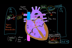
The ______ and ________ veins, carry oxygenated blood to the heart.
|
The pulmonary and umbilical veins, both of carry oxygenated blood to the heart.
|
|
|
Which of the following describes the function of the ductus arteriosus in thefetus?
• it shunts blood from the aorta to the pulmonary artery • it shunts blood from the pulmonary artery to the aorta • it shunts blood from the right atrium to the left atrium • it shunts blood from the umbilical vein to the inferior vena cava |
• it shunts blood from the pulmonary artery to the aorta
|
|
|
After birth, the ductus arteriosus becomes the ________, which connects the ________ to the _____. |
After birth, the ductus arteriosus becomes the ligamentum arteriosum, which connects the arch of the aorta to the left pulmonary artery. |
|
|
Factors that cause the ductus arteriosus to close |
-Inc. O2 levels -Dec. prostaglandins |
|
|
Whenit does not close, it is termed as ___________.
|
Whenit does not close, it is termed a patent ductus arteriosus
|
|
|
The __________ is a depression in the right atrium of the heart, the remnant of a thin fibrous sheet that covered the foramen ovale during fetal development. |
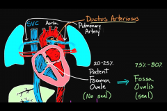
The fossa ovalis is a depression in the right atrium of the heart, the remnant of a thin fibrous sheet that covered the foramen ovale during fetal development. |
|
|
During fetal development, the______allows blood to passfrom the right atrium to the left atrium, bypassing the nonfunctional fetal lungs whilethe fetus obtains its oxygen from the placenta.
|

During fetal development, the foramen ovale allows blood to passfrom the right atrium to the left atrium, bypassing the nonfunctional fetal lungs whilethe fetus obtains its oxygen from the placenta.
|
|
|
Upon birth, the foramen ovale is initially closed by the ___________as pressure in the left atrium exceeds that in the right atrium.
|
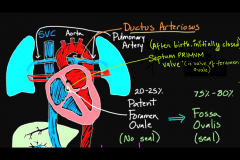
Upon birth, the foramen ovale is initially closed by the septum primum (valve of foramen ovale) as pressure in the left atrium exceeds that in the right atrium.
|
|
|
A worker in the meat-processing industry comes down with an illness,presenting with symptoms of fever, headache, and sore throat. A few dayslater, he feels chest pain and has pink, frothy sputum. His physician statesthat the worker has a viral infection caused by coxsackie B. This patient has inflammationof which layer of the heart?
• epicardium • myocardium • endocardium • pericardium |
• myocardium Myocarditis: is the inflammation of the muscular layer of the heart (myocardium) |
|
|
Arrangement of cardiac muscle cells in the myocardium |
The cardiac muscle cells are arranged in a spiral configuration. This spiral arrangement allows the heart to"wring"the blood from the ventricles toward the aortic and pulmonary semilunar valves. |
|
|
The major sensory nerve tothe parietal pericardium is from branches of the _______nerve.
|
The major sensory nerve tothe parietal pericardium is from branches of the phrenic nerve (C3-CS).
|
|
|
Contents of the middle mediastinum |
The middle mediastinum containsthe pericardium and the heart and the immediately adjacent parts of the great arteries,phrenic nerves, main bronchi, and other structures in the root of the lungs. |
|
|
The left atrium and left ventricle receive their major arterial supply from which artery?
• anterior interventricular branch of the left coronary artery • circumflex branch of the left coronary artery • marginal branch of the right coronary artery • posterior interventri cular branch of the right coronary artery |
• circumflex branch of the left coronary artery
|
|
|
The coronary arteries and their major branches lie within _________ tissue.
|
The coronary arteries and their major branches lie within subepicardial connective tissue.
|
|
|
The right and left coronary arteries, arise from the ascending aorta immediately above the aortic valve. |
The right and left coronary arteries, arise from the ascending aorta immediately above the aortic valve. |
|
|
The right coronary artery (RCA)arises from the __________.
|
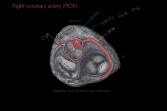
The right coronary artery (RCA)arises from the anterior aortic sinus of the ascending aorta
|
|
|
The right coronary artery(RCA) gives rise to __________,immediately after leaving the ascending aorta.
|
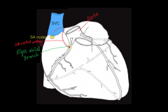
The right coronary artery (RCA)gives rise to anterior right atrial branch, immediately after leaving the ascending aorta.
|
|
|
The anterior right atrial branch gives rise to the ___,which supplies the______. |
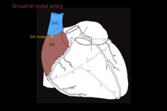
The anterior right atrial branch gives rise to the sinoatrial nodal artery ,which supplies the SA node (the pacemaker of the heart). |
|
|
Name the branch of the right coronary artery which supplies the right ventricle. |
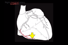
The marginal branch of the right coronary artery supplies the right ventricle. |
|
|
What is the marginal branch of the right coronary artery also known as? |
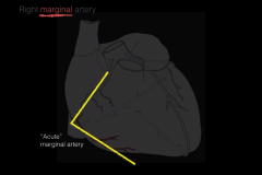
The right marginal artery is also known as the acute marginal artery. |
|
|
Name the branch of the right coronary artery which supplies both the ventricles. |
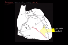
The posterior descending artery supplies both the ventricles. NOTE: It then anastomoses with the circumflex artery from the left coronary artery. |
|
|
Another name for the posterior descending artery (branch of right coronary artery) |
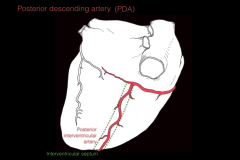
Another name for the posterior descending artery (PDA)=Posterior interventricular artery
|
|
|
What is the dominance of the coronary arterial system? |
The dominance of the coronary arterial system is defined by which artery gives rise to the PDA |
|
|
Right Dominant heart |
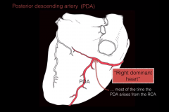
Most of the time the PDA arises from the RCA which implies that the heart is "RIGHT DOMINANT". |
|
|
Left Dominant heart
|
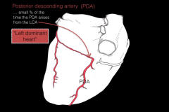
A small % of the time the PDA arises from the LCA which implies that the heart is "LEFT DOMINANT" |
|
|
Co-Dominant heart
|
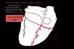
A VERY small % of the time the PDA arises from the both the RCA and LCA which implies that the heart is "CO-DOMINANT" |
|
|
Location of AV nodal artery |
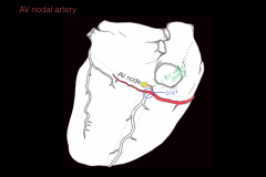
-AV nodal artery arises at the crux, which is the intersection of the RCA and the PDA. -It supplies the AV node |
|
|
Which among the two is larger? -RCA -LCA |
The left coronary artery is usually larger than the right coronary artery,
|
|
|
Origin of LCA |
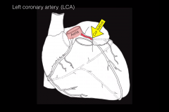
The left coronary artery arises from the left posterior aortic sinus of the ascending aorta and passes forward between the pulmonary trunk and the left auricle.
|
|
|
Structures supplied by LCA |
LCA supplies the major part of the heart, including the greater part of the left atrium, left ventricle, and ventricular septum.
|
|
|
Branches of LCA |
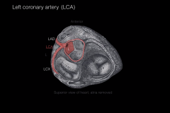
Branches of LCA : i)Anterior interventricularbranch (descending branch) and ii)Circumflex branch. |
|
|
In which phase of the cardiac cycle do the coronary arteries receive most of their blood? |
Coronary arteries receive the majority of their blood flow during ventricular relaxation, or diastole, when the left ventricle is filling with blood
|
|
|
The ___________r artery is the one most often involved in coronary occlusionsand is often the one that is bypassed in bypass cardiac surgery.
|
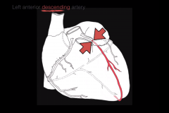
The anteriorinterventricular artery is the one most often involved in coronary occlusionsand is often the one that is bypassed in bypass cardiac surgery.
|
|
|
What is the left anterior descending artery also known as? |
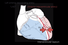
Left anterior descending artery is also known as Anterior interventricular artery |
|
|
Which of the following does NOT empty directly into the right atrium? • azygous vein • inferior vena cava • superior vena cava • coronary sinus |
• azygous vein |
|
|
A patient with a "heart-valve problem" comes into the dental clinic for periodontal therapy. She says that her old periodontist always gave her antibiotics before treatment, and insisting that the dentist hear the problem, she places the stethoscope in the left fifth intercostal space medial to the nipple line. Which heart valve is best heard over the apex of the heart? • tricuspid valve
• mitral valve • pulmonary valve • aortic valve |
• mitral valve (bicuspid valve)
|
|
|
What is the left marginal artery also known as? |
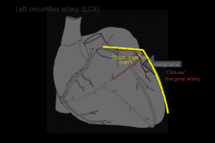
Left marginal artery=Obtuse marginal artery |
|
|
Where does the coronary sinus open? |
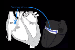
Right atrium |
|
|
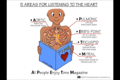
Enlist the areas where we auscultate heart sounds |
|
|
|
Third heart sound is produced by___ |
Third heart sound (S3) is produced by Rapid ventricular filling
|
|
|
Fourth heart sound is produced by___
|
Fourth heart sound is produced by Atrial contraction.
|
|
|
A 1 0-year-old girl comes into the physician suffering from rheumatic fever.She is presenting with aortic valve stenosis, which is causing her dizzinessand syncopal episodes. In the healthy heart, after ventricular systole, the aorticvalve:
• prevents reflux of blood into the right ventricle • prevents reflux of blood into the right atrium• prevents reflux of blood into the left atrium • prevents reflux of blood into the left ventricle |
prevents reflux of blood into the left ventricle
|
|
|
Overdistension of the valves of the atrioventricular orifices of the heart is prevented by the _______ and the ___________ . |
Overdistension of the valves of the atrioventricular orifices of the heart is prevented by the papillary muscles and the trabeculae carneae (muscle ridges and bulges lining the right ventricle of the heart). |
|
|
Papillary muscles are found only in the ______of the heart.
|
Papillary muscles are found only in the ventricles of the heart.
|
|
|
Major Jones Criteria for Diagnosing Rheumatic Fever
|
Major Jones Criteria for Diagnosing Rheumatic Fever:
• Migratory polyarthritis: a temporary migrating inflammation of the large joints • Carditis: inflammation of heart muscle (myocarditis) and may affect endocardium and pericardium too • Subcutaneous nodules: containing Aschoff bodies • Sydenham chorea: involuntary rapid movements of the extremities • Erythema marginatum: a long standing reddish rash distributes in a"bathing suit" pattern |
|
|
The papillary muscles _____(help/do not help) the valves to close.
|
The papillary muscles DO NOT help the valves to close. Instead, these muscles prevent the cusps from being everted (or being blown out) back into the atrium during ventricular contraction.
|
|
|
The sulcus terminalis helps sepearate the _________ from ______. |
The sulcus terminalis helps separate the right pectinate muscles from sinus venarum. Note: The sulcus terminalis externally marks the divisions of the right atrium |
|
|
Pectinate muscles
|
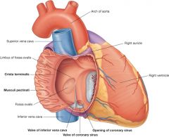
The pectinate muscles are prominent ridges of atrial myocardium located on theinner surfaces of much of the right atrium and of both auricles (which are smallconical pouches projecting from the upper anterior portion of each atrium).
|
|
|
Crista terminalis
|
The crista terminalis is a vertical muscular ridge that runs along the right atrial wall from the opening of the superior vena cava to the inferior vena cava.
|
|
|
__________ provides the origin for the pectinate muscles.
|
The crista terminalis provides the origin for the pectinate muscles.
|
|
|
The _________ represents the junction between the sinus venosus and the heart in the developing embryo.
|
The crista terminalis represents the junction between the sinus venosus and the heart in the developing embryo.
|
|
|
Sinus venarum
|
The smooth muscle wall of the right atrium= sinus venarum |
|
|
Sinus venarum develops from the _________ of the embryonic heart. |
Sinus venarum develops from the sinus venosus of the embryonic heart. |
|
|
The ________ is located in the right atrium at the junction of the crista terminalis near the opening of the superior vena cava. |
The SA node is located in the right atrium at the junction of the crista terminalis near the opening of the superior vena cava. |
|
|
The diaphragmatic surface of the heart is formed by:
• right atrium and right ventricle • right atrium and both ventricles •left ventricle only • right ventricle only • both ventricles |
The left and right ventricles make up the diaphragmatic surface of the heart.
Note:This partrests on the fibrous part of the diaphragm. |
|
|
About _________ of the heart's mass is to the left of the body midline.
|
About two-thirds of the heart's mass is to the left of the body midline.
|
|
|
The anterior surface of the heart is also known as the________surface.
|
The anterior surface of the heart is also known as the sternocostal surface.
|
|
|
Borders of the heart |
Three borders of the heart: • Right border: made up of the right atrium • Inferior border: made up of right atrium, right ventricle, and left ventricle • Left border: made up of the left ventricle |
|
|
The __________ makes up the so -called base of the heart.
|
The left atrium makes up the so -called base of the heart.
|
|
|
When the body is in the supine position, the heart rests on its ______, and the _______of the heart projects up and to the left and fits into a depression on the diaphragm.
|
When the body is in the supine position (lying on its back), the heart rests on its base, and the apex of the heart (the tip of the left ventricle) projects up and to the left and fits into a depression on the diaphragm.
|
|
|
Cardiac muscle has a shortened action potential compared to skeletal muscle. In cardiac muscle, the action potential is caused by opening of two types of channels.
• both statements are true • both statements are false • the first statement is true, the second is false • the first statement is false, the second is true |
• the first statement is false, the second is true
|
|
|
The action potential in a ventricular muscle fiber, averages about 105 millivolts, which means that the intracellular potential rises from a very negative value, about -85 millivolts, between beats to a slightly positive value, about +20 millivolts, during each beat
|
The action potential in a ventricular muscle fiber, averages about 105 millivolts, which means that the intracellular potential rises from a very negative value, about -85 millivolts, between beats to a slightly positive value, about +20 millivolts, during each beat.
|
|
|
The presence of the _______ in the action potential causes ventricular contraction to last as much as 15 times as long in cardiac muscle as in skeletal muscle.
|
The presence of the plateau in the action potential causes ventricular contraction to last as much as 15 times as long in cardiac muscle as in skeletal muscle.
|
|
|
Two major differences between the membrane properties of cardiac and skeletal muscle account for the prolonged action potential and the plateau in cardiac muscle:
|
1)Skeletal muscle:-fast sodium channels Cardiac muscle:fast sodium channels+slow calcium channels 2) Immediately after the onset of the action potential, the permeability of the cardiac muscle membrane for the potassium ions decreases about five-fold, an effect that does not occur in skeletal muscle. |
|
|
The strength of cardiac muscle contraction is directly proportional to _________ concentration.
|
The strength of cardiac muscle contraction is directly proportional to intracellular Ca2+ concentration.
|
|
|
The refractory period of ______(atrial/ventricular) muscles is much shorter than that for the ______(atria/ventricles). |
The refractory period of atrial muscle is much shorter than that for the ventricles (about 0.15 second for the atria compared with 0.25 to 0.30 second for the ventricles).
Therefore,the rhythmical rate of contraction of the atria can be much faster than t hat of the ventricles. |
|
|
________muscle cells have a short refractory period that allows them to be stimulatedto contract a second time before they have relaxed from an initial contraction.
|
Skeletal muscle cells have a short refractory period that allows them to be stimulatedto contract a second time before they have relaxed from an initial contraction.
|
|
|
Which valve is unique in having a different number of cusps than the others?
• mitral valve • tricuspid valve • pulmonary semilunar valve • aortic semilunar valve |
• mitral valve
|
|
|
AV valves are ______during diastole. |
AV valves are open during ventricular diastole. |
|
|
______ valves are openduring ventricular systole. |
Pulmonary and aortic valves are openduring ventricular systole. |
|
|
An electrocardiogram is a graphic illustration of the:
• cardiac cycle • cardiac conduction system • cardiac output • systemic and pulmonary circuits |
• cardiac conduction system
An electrocardiogram (ECG or EKG) is a test that measures the electrical activity of theheart. |
|
|
Why is there no distinctly visible wave representing atrial repolarization in the ECG?
|
There is no distinctly visible wave representing atrial repolarization in the ECG because atrial repolarization occurs during ventricular depolarization and is thus obscured.
|
|
|
An ECG that shows extra P waves before each QRS complex indicates____________.
|
An ECG that shows extra P waves before each QRS complex indicates partial heart block (or second-degree block).
|
|
|
An ECG that shows the P wave and the QRS complex being dissociated is indicative of ______.
|
An ECG that shows the P wave and the QRS complex being dissociated is indicative of complete heart block.
|
|
|
Association between P wave and QRS-T complex |
There is no correlation betweenthe P wave and the QRS-T complex on the ECG.
|
|
|
Cardiac function is the volume of blood pumped each minute, and isexpressed by which equation?
• CO = SV- HR • CO = SV + HR • CO = SVx HR • CO = SV I HR |
Cardiac function is the volume of blood pumped each minute, and is expressed by: |
|
|
_________is perhaps the single most important factor that is used in relation to the circulation, for it is responsible for transport of substances to and from the tissues
|
Cardiac output (CO) is perhaps the single most important factor that is used in relation to the circulation, for it is responsible for transport of substances to and from the tissues
|
|
|
The average resting cardiac output is about _____liters per minute for men and 10% to 20% ______(less/more) forwomen.
|
The average resting cardiac output is about 5.6liters per minute for men and 10% to 20% less forwomen. |
|
|
Heart Rate (HR) is ____(directly/inversely) proportional to cardiac output |
Heart Rate (HR) is directly proportional to cardiac output. |
|
|
In an adult HR is normally ______ beats per minute (bpm).
|
In an adult HR is normally 80-100 beats perminute (bpm).
|
|
|
Heart rate is an intrinsic factor of the______node in the heart and is modified by_____ ,________and ________ factors.
|
Heart rate is an intrinsic factor of the SA (pacemaker) node in the heart and is modified by autonomic, humoral and local factors.
|
|
|
Name the factors which determine the stroke Volume
|
Stroke Volume (SV) is determined by three factors: [SV=3PAC]
Preload, Afterload and Contractility |
|
|
The______ gives the volume of blood that the ventricle has available to pump, as well as the end diastolic length of the muscle.
|
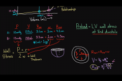
The preload gives the volume of blood that the ventricle has available to pump, as well as the end diastolic length of the muscle (increased preload increases stroke volume).
|
|
|
The ________ is the force that the musclecan create at the given length.
|
The contractility is the force that the musclecan create at the given length.
|
|
|
Increased contractility_________ (increases/decreases) stroke volume |
Increased contractility increases stroke volume |
|
|
______ is thearterial pressure against which the muscle will contract .
|
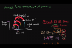
Afterload is thearterial pressure against which the muscle will contract
|
|
|
Increased afterload _______ stroke volume
|
Increased afterload decreases stroke volume.
|
|
|
___________= End Diastolic Volume - End Systolic Volume
|
Stroke Volume (SV)= End Diastolic Volume - End Systolic Volume
|
|
|
The average SV is _____ to_____ ml |
The average SV is 70 to 80 ml |
|
|
Cardiac output on the left side of the heart is _________(>,<,=) the cardiac output on the right ride of the heart. |
Cardiac output on the left side of the heart is = the cardiac output on the right side of the heart. |
|
|
BP = ______ x _______.
|
BP(Blood pressure) = CO (Cardiac output)x TPR (Total peripheral resistance).
|
|
|
The Bainbridge Reflex is a positive feedback mechanism in which there is a compensatory increase in heart rate, due to a rise in right atrial pressure.
It is commonly referred to as an Atrial Reflex. • both statements are true • both statements are false • the first statement is true, the second is false • the first statement is false, the second is true |
• both statements are true
The Bainbridge Reflex is a positive feedback mechanism in which there is a compensatoryincrease in heart rate, due to a rise in right atrial pressure. It is commonly referredto as an Atrial Reflex. |
|
|
The stretch receptors of the atria that elicit the Bainbridge Reflex transmit their afferent signals through the ______nerves to the_____of the brain.
|
The stretch receptors of the atria that elicit the Bainbridge Reflex transmit their afferent signals through the vagus nerves to the medulla of the brain.
Then efferent signals are transmitted back through vagal and sympathetic nerves to increase heart rate and strength of heart contraction. |
|
|
What does the Bainbridge Reflex help prevent? |
Bainbridge Reflex helps prevent damming of blood in the veins, atria, and pulmonary circulation.
|
|
|
Location of baroreceptors? |
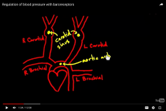
Baroreceptors are located on internal carotid arteries and aortic arch. |
|
|
A ________in arterial pressure stretches these receptors to initiate a reflex that inhibits the ________ center and induces vasodilation.
|
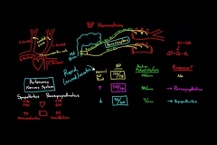
An increase inarterial pressure stretches these receptors to initiate a reflex that inhibits the medullaryvasoconstrictor center and induces vasodilation.
|
|
|
The baroreceptors in the ________predominate over those in the_____ and they respond more vigorously to changes in pressure (stretch) than they do to elevated or reduced nonpulsatile pressure.
|
The baroreceptors in the internal carotid arteries predominate over those in the aorta and they respond more vigorously to changes in pressure (stretch) than they do to elevated or reduced nonpulsatile pressure.
|
|
|
____ and ______in the medulla oblongata are stimulated by a decrease in blood P02 and by anincrease in blood PC02. |
Peripheral chemoreceptors (carotid and aortic bodies) and central chemoreceptorsin the medulla oblongata are stimulated by a decrease in blood P02 and by anincrease in blood PC02.
|
|
|
Stimulation of chemoreceptors in the medulla ___(increases/decreases) the heart rate and depth of respiration, but it also produces peripheral _______(vasoconstriction/vasodilation). |
Stimulation of chemoreceptors in the medulla increases the heart rate and depth of respiration, but it also produces peripheral vasoconstriction. |
|
|
Cardiopulmonary baroreceptors present in the cardiac chambers and large pulmonary vessels have less influence on blood ______but participate in regulation of blood_______.
|
Cardiopulmonary baroreceptors present in the cardiac chambers and large pulmonary vessels have less influence on blood pressure but participate in regulation of blood volume.
|
|
|
Your patient has a defective mitral valve, allowing backflow. Which of the following cardiac phases will be least affected by this defect?
• isovolumetric contraction • filling phase • isovolumetric relaxation • ejection phase |
Filling phase (because the mitral valve is open through this phase normally)
|
|
|
Normally, which phase would have the highest ventricular pressure?
• isovolumetric contraction • filling phase • isovolumetric relaxation • ejection phase |
• ejection phase (isovolumetric contraction would have an increasing pressure right up until the ejection where the pressure would be the highest)
|
|
|
The spontaneous generation of an action potential within the SA node initiates a sequence of eventsknown as the __________.
|
The spontaneous generation of an action potential within the SA node initiates a sequence of eventsknown as the cardiac cycle.
|
|
|
Each cardiac cycle lasts approximately ______ seconds and spans the intervalfrom the end of one heart contraction to the end of the subsequent heart contraction.
|
Each cardiac cycle lasts approximately 0.8 seconds and spans the intervalfrom the end of one heart contraction to the end of the subsequent heart contraction.
|
|
|
Summary of the events that occur during the diastole
|
summary of the events that occur during the diastole phase:
• Atrioventricular valves are open • The sinoatrial node, which starts cardiac conduction, contracts causing atrial contraction • The atria empty blood into the ventricles • Semilunar valves close, preventing backflow into the ventricles |
|
|
Summary of the events that occur during the systole
|
Summary of the events that occur during the systole phase: • The ventricles contract • Atrioventricular valves close and semilunar valves open • Blood flows to either the pulmonary artery or aorta
|
|
|
Blood flow to the coronary arteries would be greatest during _________ in a resting individual.
|
Blood flow to the coronary arteries would be greatest during ventricular relaxationin a resting individual.
|
|
|
Ventricular volume is greatest following_______.
|
Ventricular volume is greatest following atrial systole.
|
|
|
Ventricular pressure is greatest during _______.
|
Ventricular pressure is greatest during ventricular ejection.
|
|
|
Increased ventricular volume increases ______fiber length.
|
Increased ventricular volume increases end-diastolic fiber length.
Note: This is why an increased filling of the ventricle during diastole causes a more forceful heartbeat. |
|
|
______ begins with the second heart sound.
|
Diastole begins with the second heart sound.
|
|
|
The _____ valve closes before the_______valve; this causes"splitting" of the second heart sound.
|
The aortic valve closes before the pulmonary valve; this causes"splitting" of the second heart sound.
|
|
|
Which portion of the cardiac conduction system that is most likely malfunctioning in the following: Higher than normal heart rate (tachycardia). |
Sinoatrial node (the pacemaker of the heart) |
|
|
Which portion of the cardiac conduction system that is most likely malfunctioning in the following: Ventricles contract nearly simultaneously with the atria. |
Atrioventricular node (the portion responsible for delaying impulses asthey pass from the atria to the ventricles) |
|
|
Which portion of the cardiac conduction system that is most likely malfunctioning in the following: Right ventricle does not contract on the lateral side. |
Purkinje fibers (they are not transmitting impulses to the lateral side ofthe right ventricle) |
|
|
Which portion of the cardiac conduction system that is most likely malfunctioning in the following: Entire left ventricle does not contract. |
Atrioventricular bundle (the AV bundle is divided, and the left bundle is not transmitting impulses).
Note: Technically he could have a problem with all Purkinje fibers on the left side of heart, but the most likely problem would be at the source of the split (the AV bundle) |
|
|
An impulse is delayed in the AVnode for about ________ seconds to allow the atrial blood to empty into the ventricles beforeventricular contraction occurs.
|
An impulse is delayed in the AV node for about 0.13 seconds to allow the atrial blood to empty into the ventricles before ventricular contraction occurs.
|
|
|
AV bundle (bundle of His) originates in the AV node, passes subendocardially down the right side of the interventricular septum for about ______cm and then divides into the right and left bundle branches.
|
AV bundle (bundle of His) originates in the AV node, passes subendocardially down the right side of the interventricular septum for about 1cm and then divides into the right and left bundle branches.
|
|
|
The wave of depolarization travels extremely _________through the bundle branches and purkinje fibers (total elapsed time of _____seconds).
|
The wave of depolarization travels extremely fast through the bundle branches and purkinje fibers (total elapsed time of 0.03 seconds).
|
|
|
The ventricles are completely depolarized during which isoelectricportion of the ECG?
|
S-T segment
|
|
|
This portion of the ECG represents atrial depolarization.
|
• Pwave
|
|
|
This portion of the ECG represents the segment between depolarization ofthe atria and depolarization of the ventricle.
|
P-R interval
|
|
|
P-R interval is approximately ______ seconds
|
P-R interval is approximately 0.16 seconds
|
|
|
Q-T interval is approximately ______ seconds.
|
Q-T interval is approximately 0.35 seconds.
|
|
|
When is the ECG isoelectric? |
The ECG is isoelectric between: i) T and P waves (the ventricle is at resting membrane potential). This period of ventricular diastole, when the ventricle is filling with blood, greatly diminishes at high heart rates. ii)S-T segment: represents the period when the ventri cles are depolarized |
|
|
Venous return (VR) is the flow of blood back to the heart. Under steady-state conditions, venous return must equal__________ when averaged over time because the cardiovascular system is essentially a closed loop .
|
Venous return (VR) is the flow of blood back to the heart. Under steady-state conditions, venous return must equal CO (cardiac output) when averaged over time because the cardiovascular system is essentially a closed loop .
Note:Otherwise, blood would accumulate in either the systemic or pulmonary circulations. |
|
|
Frank-Starling relationship
|
An increasein myocardial fiber length, as occurs with augmented ventricular filling (preload)during diastole, produces a more forceful ventricular contraction. This relationship between fiber length and strength of contraction is known as the Frank-Starling relationship or Starling's law of the heart.
|
|
|
Preload value is related to ______ pressure.
|
Preload value is related to right atrial pressure.
|
|
|
The most important determining factorfor preload is ________.
|
The most important determining factor for preload is venous return.
|
|
|
Afterload for the left ventricle is determined by _________;
|
Afterload for the left ventricle is determined by aortic pressure;
|
|
|
Afterload for the right ventricle is determined by ____ pressure
|
Afterload for the right ventricle is determined by pulmonary artery pressure
|
|
|
lncrease in heart rate will also increase cardiac output, EXCEPT_________?
|
An increase in heart rate will also increase cardiac output, EXCEPT at very high heart rates where there will be less time for filling.
|
|
|
The myocardium functions only ________________ (aerobically/anaerobically) and in general, it uses substrates inproportion to their arterial concentration.
|
The myocardium functions only aerobically and in general, it uses substrates inproportion to their arterial concentration.
|
|
|
Which of the following equations correctly defines blood flow through the vasculature?
· flow = (initial pressure - final pressure) x resistance • flow = (initial pressure - final pressure ) I resistance • flow = resistance I (initial pressure - final pressure) |
• flow= (initial pressure- final pressure) / resistance
|
|
|
Blood flow is _________proportional to the pressure difference between the two ends of the vessel (the pressure gradient) and is ______proportional to the resistance of the vessel.
|
Blood flow is directly proportional to the pressure difference between the two ends ofthe vessel (the pressure gradient) and is inversely proportional to the resistance of thevessel.
|
|
|
The larger the vessel, the ______ the resistance
|
The larger the vessel, the less the resistance
|
|
|
Resistance = __________ x _________/[_________]
|
Resistance = viscosity (of blood) x length (of vessel) /[(radius)^4]
Note: It is the fourth power of the radius. This means that if the radius is doubled the resistance will decrease by a factor of 16. |
|
|
The major physiologicalregulation of blood flow is via the activation of _______.
|
The major physiological regulation of blood flow is via the activation of vascular smooth muscle (vasoconstriction).
Note: This fact explains why arterioles, with their ability to quickly constrict or dilate, are the most critical factor in controlling blood flow to peripheral tissues. |
|
|
When blood vessels are connected, the blood flows from the higher pressure site to the lower pressure site and the rate of flow is proportional to the _________.
|
When blood vessels are connected,the blood flows from the higher pressure site to the lower pressure site and therate of flow is proportional to the pressure difference.
|
|
|
The overall pressuredifference is between the ascending aorta and the entrance to the right atrium(the circulatory pressure [about 100 mmHg]).
|
The overall pressure difference is between the ascending aorta and the entrance to the right atrium (the circulatory pressure [about ________mmHg]).
|
|
|
Stretch receptors are located in ___________ |
Stretch receptors are located in carotid sinus. Respond to actual stretching, not chemicals |
|
|
High BP activates the ______ nerve |
High BP activates the Carotid Sinus nerve |
|
|
Activation of ___________ nervous system and inhibition of ___________ to drops BP |
Activation of parasympathetic nervous system and inhibition of sympathetic to drops BP |
|
|
Stimulation of Carotid Sinusleads to __________heart rate, __________ arterial blood pressure and __________ venous return |
Stimulation of Carotid Sinusleads to decreased heart rate, decreased arterial blood pressure and decreased venous return |
|
|
Increased pressure in the carotid sinus increases the discharge of efferent fibers that travel in the _____ CN |
Increased pressure in the carotid sinus increases the discharge of efferent fibers that travel in the 9th CN |
|
|
Stretch receptors in the atria elicit _______ reflex |
Stretch receptors in the atria elicit Bainbridge reflex --- Think Bridging the gap from the heart to the Lungs |
|
|
Bainbridge reflex |
Bainbridge reflex • Initiated by excess blood volume in R atrium • Sensitive to both Pressure and stretch • Goal is to get heart to pump harder to transfer excess blood from pulm circ to systemic circ •• A sudden increased in flow into the R atrium will cause an increased blood flow to the lungs in 2-3 heart beats |
|
|
Stimulation of the heart by the________ nerve keeps the HR down |
Stimulation of the heart by the vagus nerve keeps the HR down |
|
|
_______ (Increased/Decreased) vagal activity results in decreased cardiac oxygen consumption |
Increased vagal activity also results in decreased cardiac oxygen consumption |
|
|
Increased vagal activity ___________(does /doesn't) result in “decreased transit time through the AV node |
Increased vagal activity does not result in “decreased transit time through the AV node |
|
|
Then efferent sympathetic to the heart increase the __________ and______________ |
Then efferent sympathetic increases the HR and strength of contractions |
|
|
When PCO2 goes up, also increases_______ |
When PCO2 goes up, also increases H+ because CO2 + H2O forms carbonic acid, which then disassociates to H+ and bicarbonate |
|
|
Breathing a gas mixture with 5% CO2 ultimately leads to a stimulation of ______________ |
Breathing a gas mixture with 5% CO2 ultimately leads to a stimulation of central chemoreceptors CO2 affects central chemoreceptor the most |
|
|
central chemoreceptors are located in the |
central chemoreceptor are located in the Medulla • MAJOR regulators of ventilation • central receptors are sensitive to low pH ♦ Detect H+ changes in CSF (cerebrospinal fluid)♦ NO detection of PO2 ♦ Detect increase in PCO2 |
|
|
Peripheral chemoreceptors are located in ________ and ________ |
Peripheral chemoreceptors are located in carotid and aortic bodies |
|
|
Peripheral chemoreceptors detect change in blood _____________________ |
Peripheral chemoreceptors detect change in blood PO2 (if <60mmHg), PCO2, and H+ (or pH) ion concentrations |
|
|
A marked fall in the oxygen tension in arterial blood would stimulate the receptors in the ___________ |
A marked fall in the oxygen tension in arterial blood would stimulate the receptors in the aortic & carotid bodies |
|
|
• Normal Hemoglobin concentration is about _________ blood |
• Normal Hemoglobin concentration is about 15gm/dl blood |
|
|
Normal arterial oxygen content is about _____________. |
Normal arterial oxygen content is about 20ml/dl blood. |
|
|
An anemic individual breathing room air with a hemoglobin concentration of 10 gm/dl blood is expected to have normal _________________ and reduced _____________. |
An anemic individual breathing room air with a hemoglobin concentration of 10 gm/dl blood is expected to have -normal arterial oxygen tension and -reduced arterial oxygen content |
|
|
Administration of 90:10 N20 to O2 will (inc/dec)_______ RESPIRATION and it will result in ___________ |
Administration of 90:10 N20 to O2 will DEPRESS RESPIRATION and it will result in Respiratory Acidosis |
|
|
Subanesthetic doses of nitrous (20-40% N2O) cause ________ |
♦ NOTE Subanesthetic doses of nitrous (20-40% N2O) cause increased respiration |
|
|
______ has the largest share of systemic CO |
Liverhas the largest share of systemic CO |
|
|
_________ has the highest blood flow per gram of tissue |
Kidneyhas the highest blood flow per gram of tissue especially the adrenals |
|
|
Increased O2 demand of heart is met by increased ___________, NOT by increased extraction of O2 |
Increased O2 demand of heart is met by increased coronary blood flow, NOT by increased extraction of O2 |
|
|
Total Peripheral Resistance (TPR): -Regulates the flow of blood from________ -Caused by accumulation of_______ |
Total Peripheral Resistance (TPR): Regulates the flow of blood from the sys. arterial to venus circulation Resistance to blood flow from the entire circulatory system Caused by accumulation of vasodilator local metabolites (lactate, K+ ions, & adenosine) |
|
|
Factors determining Autoregulation of blood flow Heart |
Factors determining Autoregulation of blood flow Heart: O2, adenosine, NO (Unique in that when hypoxic causes vasoconstriction) |
|
|
Factors determining Autoregulation of blood flow Brain |
Factors determining Autoregulation of blood flow Brain: CO2, pH |
|
|
Factors determining Autoregulation of blood flow Kidneys |
Factors determining Autoregulation of blood flow Kidneys: Myogenic and tubuloglomerular feedback |
|
|
Factors determining Autoregulation of blood flow Lungs |
Factors determining Autoregulation of blood flow Lungs: Hypoxia causes vasoconstriction (* Unique like the heart) |
|
|
Factors determining Autoregulation of blood flow Skeletal Muscle |
Factors determining Autoregulation of blood flow Skeletal Muscle: Local metabolites, Lactate, Adenosine, K+ (vasodilators) NOTE: Adenosine causes vascular smooth muscle to relax |
|
|
Posseult’s Law |
Posseult’s Law = Viscosity (of blood) x length (of vessel) / (Radius)4 |
|
|
Factors affecting blood resistance |
Factors affecting blood resistance: ♦ Vessel Radius: Larger the vessel, the less resistance ♦ Viscosity: Directly proportional to resistance ♦ Vessel Length: Directly proportional to resistance |
|
|
___________ contains the highest viscosity blood in the body |
The vena cava contains the highest viscosity blood in the body |
|
|
Viscosity increases with ________, ____________ &__________. |
Viscosity increases with polycythemia, Hyperproteinemic states (Multiple Myeloma), and Hereditary Spherocytosis May vary with hematocrit |
|
|
Increasing the radius produces the greatest __________(increase/decrease) in resistance in a single artery |
Increasing the radius produces the greatest decrease in resistance in a single artery |
|
|
CO (formulae) |
CO = HR x SV CO = BP/TPR (BP) Pressure = Flow (CO) x Resistance (TPR) |
|
|
During exercise, CO ________ initially as a result of an increase in ___________ |
During exercise, CO increases initially as a result of an increase in SV |
|
|
After prolonged exercise, CO ________ as a result of an increase in ________ |
After prolonged exercise, CO increases as a result of an increase in HR |
|
|
Effect of very high HR on CO |
If HR is too high, diastolic filling is incomplete and CO drops (i.e. ventricular tachycardia, pulse >200) |
|
|
Factors affecting SV |
Factors affecting SV:[CAP] -Contractility (increases SV) ♦ Afterload (decreases SV) ♦ Preload (increases SV) |
|
|
Factors that increase Contractility |
ContractilityIncreased with [CID] Catecholamines (increased activity of Ca2+ pump in sarcoplasmic reticulum) • Sympathetic action of increasing HR, conduction velocity, and contractiility Increased intracellular Ca2+ Decreased Extracellular Ca2+ Digitalis (increased intracellular Na+, resulting in increased Ca2+) |
|
|
Factors that decrease Contractility |
ContractilityDecreased with [BAHH]: B1 Blockers Acidosis Heart Failure Hypoxia/Hypercapnea |
|
|
Vasodilators ______(increase/decrease) afterload |
Vasodilators decrease afterload (i.e. hydralazine) |
|
|
Venous Dilators______(increase/decrease) preload |
Venous Dilators decrease preload |
|
|
A drop in BP results from ______(increase/decrease) in venous return |
A drop in BP results from decreased venous return |
|
|
(Increase/Decrease) ___________of thoracic/abdominal pressure will decrease VR |
Increase of thoracic/abdominal pressure will decrease VR |
|
|
In the completely isolated, blood perfused mammalian heart, adjustments to increase workload appear to be mediated primarily by mechanisms associated w/ __________________ |
In the completely isolated, blood perfused mammalian heart, adjustments to increase workload appear to be mediated primarily by mechanisms associated w/ increased diastolic volume |
|
|
A fall in BP causes ______(increased/decreased) activity of the vasoconstrictor center and ______(increased/decreased) activity of the cardioinhibitory center |
A fall in BP causes increased activity of the vasoconstrictor center and decreased activity of the cardioinhibitory center |
|
|
The characteristic of the aorta most responsible for the maintenance of diastolic BP is ______________ |
The characteristic of the aorta most responsible for the maintenance of diastolic BP is elastic distensibility |
|
|
Systolic BP may be abnormally high when arterial compliance is ______(increased/decreased) |
Systolic BP may be abnormally high when arterial compliance is decreased (think arteriosclerosis) |
|
|
What are the main factors directly involved in the maintenance of systemic arterial blood pressure ? |
The main factors directly involved in the maintenance of systemic arterial blood pressure are cardiac output, blood volume, blood viscosity and peripheral resistance (not heart rate) |
|
|
A drop in BP results from ______(increased/decreased) CO |
A drop in BP results from decreased CO |
|
|
The arterial blood pressure might be abnormally high in a ______________ accident |
The arterial blood pressure might be abnormally high in a cerebrovascular accident (NOT cardiac shock, heart failure, anaphylactic shock, or ventricular fibrillation) |
|
|
In the presence of a constant HR, changes in BP may be attributed mainly to alterations in ____________ |
In the presence of a constant HR, changes in BP may be attributed mainly to alterations in resistance in arterioles |
|
|
Postural hypotension is compensated by_________ (constriction/dilation) of systemic arterioles |
Postural hypotension is compensated by constriction of systemic arterioles |
|
|
The most likely cause of hypertension is generalized_________ (constriction/dilation)of arterioles |
The most likely cause of hypertension is generalized constriction of arterioles |
|
|
The amt of blood passing through the capillaries of the systemic circulation/min = amt of blood passing through the ________/min |
The amt of blood passing through the capillaries of the systemic circulation/min = amt of blood passing through the aorta/min |
|
|
If two vessels are connected in parallel, their total resistance to blood flow is _________(greater/less) than the resistance of either vessel alone |
If two vessels are connected in parallel, their total resistance to blood flow is less than the resistance of either vessel alone |
|
|
What would the primary effect of substituting a rigid arterial system for a compliant arterial system be? |
The primary effect of substituting a rigid arterial system for a compliant arterial system would be that continuous flow in the capillaries would change into pulsatile flow |
|
|
Plasma colloid osmotic pressure acts in the same direction as___________pressure |
Plasma colloid osmotic pressure acts in the same direction as tissue pressure |
|
|
Interstitial colloid osmotic pressure acts in the same direction as ________ pressure |
Interstitial colloid osmotic pressure acts in the same direction ascapillary fluid pressure |
|
|
Why is colloid osmotic pressure of blood imp? |
Colloid osmotic pressure of the blood is important because it prevents excess loss of fluid from capillaries |
|
|
Albumin is produced in the _____ |
Albumin is produced in the liver Aldosterone and Vasopressin are NOT |
|
|
________ is main organ responsible for reg of osmotic pressure with reg of water resorption |
Kidney is main organ responsible for reg of osmotic pressure with reg of water resorption via ADH When balance is off, edema occurs and lymphatics pick up the extra fluid left in the interstitium |
|
|
BP is much _______(lower/higher) in Pulmonary Circuitthan systemic circuit |
BP is much lower in Pulmonary Circuit than systemic circuit |
|
|
Compared with systemic circulation under normal conditions, pulmonary circulation is characterized by ________, _________and___________ |
Compared with systemic circulation under normal conditions, pulmonary circulation is characterized by low pressure, equal flow and low resistance |
|
|
Hematocrit |
Hematocrit • Proportion of RBCs in a sample of blood |
|
|
Hematocrit values for males and females |
Hematocrit : ♦ 46.2% -- Males ♦ 40.6% -- Females |
|
|
______ excretion is a good indicator of RBC destruction per day |
• Bile excretion is a good indicator of RBC destruction per day |
|
|
Some parts of a hemoglobin molecule |
Some parts of a hemoglobin molecule – iron, protein, histidine, pyrrole ring (not magnesium) |
|
|
Hb synthesis involves _______ & _________ (metals) |
Hb synthesis involves iron & copper |
|
|
Heme synthesis occurs in the ___________ |
Heme synthesis occurs in the liver and bone marrow |
|
|
Name the two forms in which the Globin portion of Hb exits |
Globin portion of Hb exists in 2 Forms: 1♦ Taut (T) has low affinity for Oxygen 2♦ Relaxed (R) has high affinity for Oxygen (300x) |
|
|
Hb is important due to its ability to combine reversibly with O2 at the ________ heme prosthetic group |
Hb is important due to its ability to combine reversibly with O2 at the ferrous heme prosthetic group (not ferric heme group) |
|
|
MetHemoglobinhas its iron in__________state |
MetHemoglobin has its iron in Fe3+ (ferric) state and cannot function (NOT reduced hemoglobin) |
|
|
Dominant factor controlling GI absorption of iron is _____________ |
Dominant factor controlling GI absorption of iron is saturation of mucosal cells |
|
|
Hb Types: HbA |
Hb Types: Hb A – Normal type |
|
|
Hb C |
Hb C – Abnormal in which Lysine has replace glutamic acid causing reduced plastiCity of the RBCs |
|
|
Hb H |
Hb H – Abnormal composed of 4 beta chains (resulting in alpHa-tHalassemia) |
|
|
Hb M |
Hb M – Abnormal Hbs in which single amino acid substitution favors the formation of Methemoglobin |
|
|
• Hb A1C |
• Hb A1C – Hemoglobin of a diabetic pt |
|
|
Hb S |
• Hb S – Sickle Cell anemia ♦ Abnormal Hb in which valine replaced glutamic acid in the beta chain This causes Hb to become less soluble under low O2 tension& to polymerize into crystals that distort the RBCs into a sickle shape (sickle-cell anemia) |
|
|
Major effect of sickle cell anemia is from ___________ |
Major effect of sickle cell anemia is from decreased solubility of the deoxy form of hemoglobin. NOTE: This would cause an increase of the isoelectric pH |
|
|
Sickle-cell anemia is caused by the presence of a ______ substitution for __________ in the sixth amino acid from the N-terminal end of the hemoglobin _____-chain |
Sickle-cell anemia is caused by the presence of a valine substitution for glutamate in the sixth amino acid from the N-terminal end of the hemoglobin B-chain |
|
|
Sickle-cell anemia involves a _________ mutation |
Sickle-cell anemia involves a missense mutation |
|
|
A dental patient presents with a bluish discoloration of the mucous MBs indicative of cyanosis – most likely the result of ____________ |
A dental patient presents with a bluish discoloration of the mucous MBs indicative of cyanosis – most likely the result of increased levels of reduced hemoglobin |
|
|
Among the larger blood vessels, O2 tension is highest in the __________ veins |
Among the larger blood vessels, O2 tension is highest in the pulmonary veins |
|
|
When PO2 is low, Hb has________ (higher/lower) affinity for O2 |
When PO2 is low, (in tissue caps), Hb has lower affinity and unloads O2 |
|
|
Conversion of _______ to ___________ is the most important process in preventing a decrease of >0.2 units in blood pH when CO2 enters |
Conversion of HbO2 to Hb is the most important process in preventing a decrease of >0.2 units in blood pH when CO2 enters |
|
|
Causes of O2 unloading |
Causes of O2 unloading: Low PO2 Bohr effect [Decreased pH = increased arterial [H+] = decreased affinity for O2] Increase in arterial pCO2 Causes oxyhemoglobin to shift to deoxyhemoglobin Increase in Diphosphoglycerate (DPG) Increase in tissue temperature |
|
|
With a decreased affinity for O2 – Curve shift to the Right |
With a decreased affinity for O2 – Curve shift to the Right |
|
|
Increase in arterial pCO2 has the most effect in stimulating________ |
Increase in arterial pCO2 has the most effect in stimulating respiration |
|
|
Oxygen dissociation equation |
Oxygen dissociation equation: ♦ y = (pO2)^n/[ (pO2)^n + (P50)^n] n = 2.8 for hemoglobin |
|
|
_______________ in erythrocytes increases the rate of dissociation of H2CO3 |
Carbonic anhydrase in erythrocytes increases the rate of dissociation of H2CO3. HCO3- reenters the blood plasma for transport to the lungs |
|
|
Wt is Cl- shift |
Because the RBC membrane is not very permeable to cations, the HCO3- efflux is balanced with a Cl- influx, which is known as the Cl- shift Chloride passes from the plasma into the cell as HCO3- passes from cell into plasma |
|
|
_____________ in erythrocytes increases the rate of dissociation of H2CO3 |
carbonic anhydrase in erythrocytes increases the rate of dissociation of H2CO3 |
|
|
Carbonic anhydrase in the kidney tubular cells is associated with______________ |
Carbonic anhydrase in the kidney tubular cells is associated with reabsorption of bicarbonate ion |
|
|
Absence of carbonic anhydrase drastically reduces blood________ carrying capacity |
Absence of carbonic anhydrase drastically reduces blood CO2 carrying capacity |
|
|
Most of the CO2 is transported in the blood as __________ |
Most of the CO2 is transported in the blood as bicarbonate ion (HCO3-) |
|
|
The greatest concentration of carbonic anhydrase is found in __________ |
The greatest concentration of carbonic anhydrase is found in erythrocytes |