![]()
![]()
![]()
Use LEFT and RIGHT arrow keys to navigate between flashcards;
Use UP and DOWN arrow keys to flip the card;
H to show hint;
A reads text to speech;
10 Cards in this Set
- Front
- Back
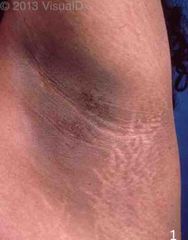
What is this? It is associated with? |
Acanthosis Nigricans -Brown to gray-black papillomatous cutaneous thickening in flexural areas (posterolateral neck- common, axillae, groin, abdominal fold) Associated with: -Obesity -Diabetes mellitus (insulin resistant) -Polycystic ovarian syndrome (PCOS) -Cushing syndrome -Atypical (palmar or mucosal) distributions or acute onset acanthosis nigricans may also be associated with malignancy (usually gastrointestinal adenocarcinoma) |
|
|
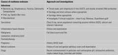
Causes of generalised pruritus |

Back (Definition) |
|
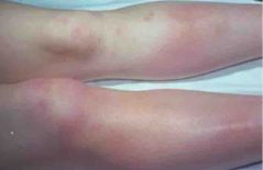
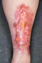
What is this? What causing this condition? |
Erythema nodusum The causes and appropriate investigations are listed in the table. |
|
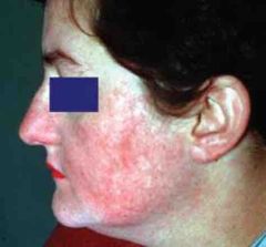
What is this lesion? What symptoms associated with this condition? |

Ans: Malar rash of SLE Associated symptoms as listed in the picture. |
|
|
List DM skin manifestations |
1. Acantosis nigricans 2. Diabetic dermopathy 3. Bullosis diabetocorum 4. Necrobiosis lipoidica 5. Diabetic foot ulcer |
|
What is this lesion? |
Diabetic dermopathy -‘shin spot' or pigmented pretibial papules -Benign asymptomatic red brown macules on shins -Most common skin manifestation of DM |
|
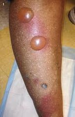
What is this lesion? What is the differential diagnosis? |
Bullosis diabeticorum DDX: 1. Friction bullae 2. Bullae due to burns or edema 3. Bullous fixed drug reaction 4. Bullous pemphigoid 5. Epidermolysis bullosa acquisita |
|
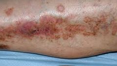
What is this lesion? Describe the lesion |
Necrobiosis lipoidica -Sharply demarcated yellow-brown plaques on anterior pretibial region. -Violaceous, irregular border, raised and indurated. -Red-brown papules--> well defined yellow-brown atrophic plaques and telangiectasias--> ulcerations |
|
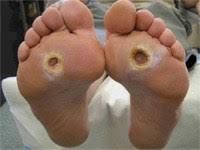
What is this lesion? What classification is used to grade this lesion? |
Diabetic foot ulcer Classified using Wegner ulcer classification |
|
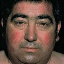
What is this? In which condition can be seen? What other manifestation can be seen in this patient? |
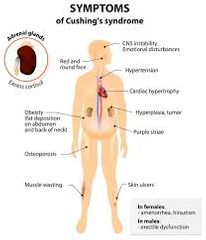
Plethoric moon face can be seen in Cushing's syndrome (erythema and telangiectases of cheeks and forehead) Other manifestation as seen in picture |