Reading...
![]()
Play button
![]()
Play button
![]()
Use LEFT and RIGHT arrow keys to navigate between flashcards;
Use UP and DOWN arrow keys to flip the card;
H to show hint;
A reads text to speech;
101 Cards in this Set
- Front
- Back
|
general functions of cartilage
|
structural support of soft tissues
shock absorption in joints reduce friction between joint sufraces role in growth and development of long bones |
|
|
mature cells in cartilage that secrete extracellular matrix
|
chondrocytes
|
|
|
where chondrocytes are found
|
lacunae (cavities in cartilage)
|
|
|
makes up 95% of cartilage volume
|
ECM
|
|
|
predominant matrix components in cartilage
|
collagen and proteogylcans
|
|
|
how is most cartilage nourished?
|
relies on diffusion from vessels in the adjacent CT (perichondrium)
|
|
|
how is articular cartilage nourished?
|
the synovial fluid provides what?
for this particular type of cartilage? |
|
|
this type of cartilage has no perichondrium
|
articular cartilage
|
|
|
chondroblasts are derived from
|
mesenchymal cells
|
|
|
chondrocytes
|
chondroblasts that have surounded themselves in matrix
|
|
|
lacunae
|
little champers filled with matrix in which chondrocytes reside
|
|
|
perichondreum
|
outer layer surrounding most cartilage
|
|
|
appositional growth of cartilage results from
|
inactive fibroblasts on perichondrium become chondroblasts and add to the surface of the existing cartilage
|
|
|
interstitial growth of cartilage
|
division of chondrocytes and deposition of more matrix
|
|
|
isogenous groups
|
"cell nests" multiple chondrocytes in one lacuna
|
|
|
most common type of cartilage
|
hyaline
|
|
|
principle components hyaline cartilage matrix
|
type II collagen
proteoglycans chondronectin |
|
|
locations where Hyaline cartilage is found
|
articular surfaces of moveable joints
ends of ribs respiratory passages (larynx, trachea, bronchi) |
|
|
forms epiphyseal plates of growing long bones
|
hyaline cartilage
|
|
|
elastic cartilage
|
similar to hyaline cartilage with the addition of elastic fibers
|
|
|
cartilage found in areas requiring flexible support
|
elastic cartilage
|
|
|
areas where elastic cartilage is found
|
auricle of ear, auditory tubes, epiglottis
|
|
|
color of elastic cartilage
|
yellow color due to elastin
|
|
|
fibrocartilage
|
combo of hyalin cartilage and dense CT
|
|
|
primariy fiber type in Fibrocartilage
|
type I collagen
|
|
|
ares where fibrocartilage is found
|
flexible strength, intervertebral discs, symphysis pubis, articular discs, menisci of knee
|
|
|
arrangement of cells in fibrocartilage
|
chondrocytes are in rows, there is no perichondrium
|
|
|
osteoarthritis
|
impaired cartilage function due too poor regenerative capacity
|
|
|
when can cartilage not be regenerated?
|
if injury is not adjacent to the perchondrium
if wound is large |
|
|
what forms scar when cartilage is injured and not regenerated?
|
dense CT forms scar when...
|
|
|
can affect synovial joint function in aged cartilage
|
degenerative calcification
|
|
|
general functions of bone
|
support and attachment sites for soft tissues
protects vital organs site of hematopoieses (formation of blood cells) is bone marrow provides a reservoir of calcium and phosphate |
|
|
classification criteria for mature bone
|
architecture (compact or spongy(cancellous))
shape (long, short, flat, irregular) |
|
|
epiphysis
|
exbanded end of long bone with articular cartilage
|
|
|
diaphysis
|
shaft of long bone
|
|
|
epiphyseal line
|
remnant of the epiphyseal plate (growth area in young bones)
|
|
|
periosteum
|
covering of outer bone surface with outer fibrous layer and inner cellular layer (containing osteoprogenitor cells)
|
|
|
Sharpey's fibers
|
collagen fibers from periosteum that penetrate the mineralized bone matrix, especially in areas where Ligaments and Tendons attach
|
|
|
endosteum
|
line all internal spaces of bone (single layer of bone-lining and osteoprogenitor cells)
|
|
|
osteoblasts
|
found on the surfaces of bone tissue
synthesize organic components of bone matrix differentiate into osteocytes |
|
|
organic components of bone matrix
|
Type I collagen
proteoglycans gylcoproteins |
|
|
osteocytes
|
mature cells found within the lacunae of the bone matrix
have processes that communicated with oterh cells via canaliculi |
|
|
canaliculi
|
channels in bone that allow osteocyte processes to facilitate communication with otehr cells
|
|
|
osteoclasts
|
multinucleated cells
function in bone reapsorption lie within Howship's lacunae |
|
|
Howship's lacunae
|
depressions in which osteoclasts reside also know as "resporption bays"
|
|
|
what are osteoprogenitor cells
where are they primarily found? |
stem cells from which osteoblasts and osteocytes derive
found in periosteum and endosteum |
|
|
Organic Bone Matrix components
|
fibers: Type I collagen
Ground Substance: structural gylcoproteins |
|
|
Inorganic Component of Bone Matrix
|
bone mineral
hydroxyapatite-like material (mostly Ca and Phosphate) |
|
|
mineralized bone matrix only allows what kind of bone growth?
|
appositional bone growth
|
|
|
first bone tissue that is produced (or sometimes as a result of disease)
|
woven (immature, primary bone)
|
|
|
wove bone characteristics
|
non-lamellar, random weave
|
|
|
woven bone is usually replaced by
|
lamellar bone
|
|
|
osteoblasts secrete what?
|
osteoid
|
|
|
osteoid
|
organic component of bone matrix
|
|
|
what triggers the deposition of inorganic matrix on the collage network
|
alkaline phosphatase-rich matrix vesicles
|
|
|
mineralization of matrix may take how long?
|
several months
|
|
|
lamellar bone
|
(mature, secondary) laid out in well-defined layers
|
|
|
lamella of compact bone
|
inner and outer circumferential lamella
concentric lamellae forming the walls of the osteons |
|
|
interstitial lamellae
|
remnants of former osteons, may be found amist current osteons
|
|
|
osteocytes within an osteon are connected to eachother by
|
cellular bridges that run through bony canliculi, which are in continuity with the haversion canal
|
|
|
where the blood supply to the osteon runs
|
haversion canal
|
|
|
volkman's canals
|
lateral connections among the haversion canals
|
|
|
cancellous bone differs from compact in that...
|
osteons are absent in which type of bone?
|
|
|
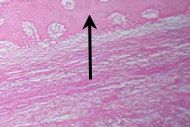
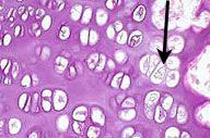
Elastic Cartilage
Type II + elastic fibers (external Ear) |
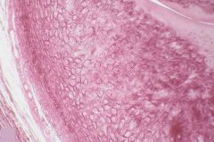
What type of tissue is shown here?
|
|
|
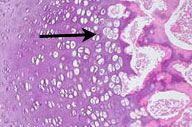
developing hyaline cartilage
|
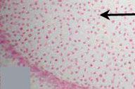
What is this tissue?
|
|
|
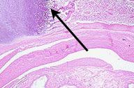
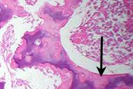
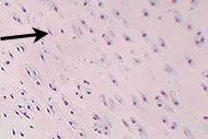
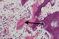
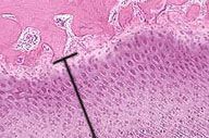
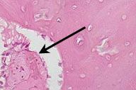
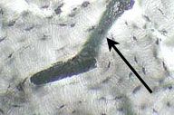
trabeculae, developing bone
|
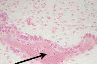
What is the structure indicated by the arrow? What kind of tissue is it made of?
|
|
|
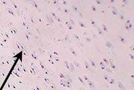
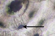
mesenchyme (embryonic CT) surrounding developing bone (intramembranous ossification- because bone is not replacing exiciting cartilage)
|
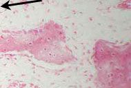
What is lighter staining tissue surrounding the darker staining pink structures?
|
|
|
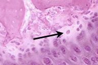
osteoblast
|
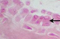
What type of cell is indicated by arrow?
|
|
|
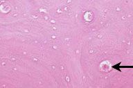
osteocyte
|
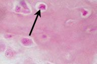
What type of cell is indicated by arrow?
|
|
|
Osteoid- thin layer of unmineralized matrix
|
Non-staining region in upper right corner of micrograph (hint- osteoblasts are lined up just above region)
|
|
|
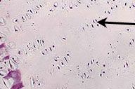
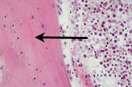
epiphyses- hyaline cartilage
|
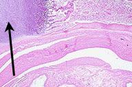
What is the region indicated by the arrow?
|
|
|
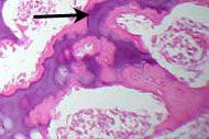
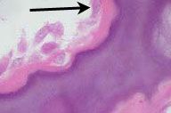
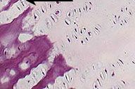
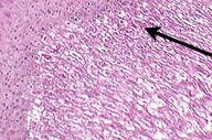
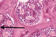
metaphyseal region (or osteogenic zone) of developing bone-
endochondral bone formation is occuring there: lamellae of new bone deposited on remnants of calcified cartilage |
what does the arrow indicate? what is taking place there?
|
|
|
developing compact bone (taken from a picture of the cortex of a diaphysis)
|
What type of tissue is in the top part of this slide?
|
|
|
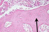
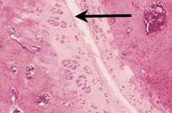
metaphyseal region marked by hypertrophy of the chondrocytes , the calcification of their matrix (evidenced by the more deeply basophilic staining), and the deposition of eosinophilic bone material over the remnants of calcified cartilage matrix (towards right)
|
what region is indicated by arrow?
what is happening? |
|
|
calcified cartilage matrix
|
what is the purple tissue indicated by arrow?
|
|
|
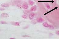
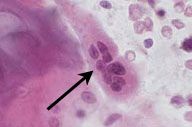
chndrocytes
hypertrophy and death |
What type of cells are these? What is happening?
|
|
|
bone material, deposited on purple calcified cartilage matrix
|
What is the pink tissue indicated by arrow?
|
|
|
osteoblast, laying down woven bone on calcified cartilage matrix
|
What is the cell indicated by arrow? What is it doing?
|
|
|
woven bone
|
what type of tissue predominates (and is indicated by arrow)
|
|
|
Resting Zone (reserve cartilage zone)
|
What is the zone indicated by arrow?
|
|
|
Zone of proliferation
|
What zone is shown in lower left part of slide?
|
|
|
the zone of maturing cartilage where cell size increases and the cells begin to arrange themselves in columns
|
What zone is shown here?
|
|
|
zone of hypertrophy and calcification in which the chondrocytes grow greatly in size, die, and their surrounding matrix becomes calcified (deeply basophilic)
|
What zone is shown in the bottom left region?
|
|
|
Metaphyseal (Osteogenic) Zone- where bone tissue is laid down upon spicules of calcified cartilage matrix.
|
What zone is shown here?
|
|
|
Woven bone with osteoblasts forming edosteum on the right
|
what is the tissue indicated by the arrow? what is the region to the right?
|
|
|
Periosteum (woven bone to right)
|
What is the arrow point to? What is to the right?
|
|
|
osteoclast is a large, multinucleated cell with noticeably eosinophilic cytoplasm. Often times, but not apparent in this image, they may be found in a depression in the bone surface known as a resorption bay or Howship's lacunae. The remnants of calcified cartilage matrix seen here are a sure indication that the bone seen in this image if forming by endochondral ossification.
|
What type of cell is indicated by the arrow?
|
|
|
2ndary ossificaiton center, developing in the epiphysis- invasion of the epiphyseal cartilage by blood vessels and perivascular connective tissue elements containing osteoprogenitor and other cells is the first step in the development of the secondary ossification center.
|
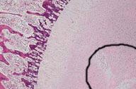
What is developing in the circle region? Where is this region located?
|
|
|
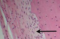
endosteum along edge of woven (immature, primary) bone
|
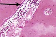
What is indicated by the arrow?
|
|
|
lammelar (mature, secondary) bone
|
what is shown in running horiz/diag in this slide
|
|
|
metaphyseal bone, epiphyseal cartilage visible in upper left, epiphyseal bone (NOT SHOWN) would be exterior to that
|
what tissue predominates in slide? What is the tissue visible in upper left?
|
|
|
epiphyseal bone, epiphyseal cartilage (growth plate), and beginning of transition into developing metaphyseal bone
|
What are the 3 layers here from top left to bottom right?
|
|
|
the very thin zone of reserve cartilage. When this population of cells is exhausted and replaced by bone tissue, the bone can no longer growth in length at this site. This is known as closure of the epiphysis.
|
what zone of cartilage is shown here? why is it significant?
|
|
|
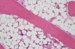
marrow within spaces of immature cancellous bone
|
what is shown in the circular spaces? what are the surrounding structures?
|
|
|
a portion of the zygapophyseal joint between successive vertebra, with the articular surfaces being covered by hyaline cartilage.
|
the space indicated is part of what?
|
|
|
the resorption cavity as evidence of bone remodeling.
|
What does this space tell you about the surrounding tissue?
|
|
|
Haversion canal
|
What is indicated by arrow?
|
|
|
osteon
|
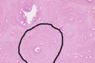
what is the structure demarcated by the circle?
|
|
|
lacunae housing the shrunken remains of osteocytes.
|
arrow indicates what?
|
|
|
Volksman's Canal- connects Haversion systems
|
What is this?
|
|
|
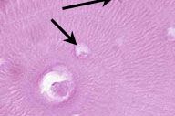
lacunae with canaliculi (which in life would contain cellular processes interconnecting neighboring osteocytes, are visualized here as the black 'tentacles' radiating from the lacunar spaces)
|
What is shown here?
|