Reading...
![]()
Play button
![]()
Play button
![]()
Use LEFT and RIGHT arrow keys to navigate between flashcards;
Use UP and DOWN arrow keys to flip the card;
H to show hint;
A reads text to speech;
31 Cards in this Set
- Front
- Back
|
where is CSF secreted from?
|
the brain ventricles
|
|
|
what are the functions and effects of CSF?
|
buoyancy of brain, physical support of brain, transfer of neuromodulators, acts as lymphatic system for drainage of fluid in CNS
|
|
|
1. interventricular foramen (of monroe)
2. anterior horn 3. body 4. trigone 5. posterior horn 6. cerebral aqueduct 7. 4th ventricle 8. median aperture 9. lateral aperture 10. inferior horn 11. 3rd ventricle |
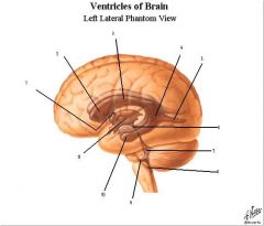
label the parts of the ventricles?
|
|
|
what ventricles and associated parts are bilateral?
|
the lateral ventricles and lateral apertures
|
|
|
ventricles are lined with what kind of cells? what is special about the area superficial to the cell layer?
|
simple cuboidal layer called ependymal cells and neural stem cells maybe in the subependymal region in adult brains.
|
|
|
what cells produce CSF and where are they located in the ventricles?
|
the choroid plexus produces them. They are located on the superior aspect of the 3rd ventricle, the interventricular foramen to the posterior body of the lateral ventricles around the anterior trigone to the superior aspect (called the glomus which is often calcified in adults) of the inferior horn of the lateral ventricles. A seperate area is on the posterior wall of the 4th ventricle down the median aperture
|
|
|
what arteries supply the choroid plexus?
|
the anterior and posterior choriodal arteries supply the choroid cells in the lateral and 3rd ventricles while the anterior and posterior inferior cerebellar arteries supply the choroid cells i nthe 4th ventricle area.
|
|
|
what is the flow of CSF?
|
the choroid cells on the choroid villi get blood and secrete CSF in the ventricles which goes into subarachnoid space from 3rd and 4th ventricles. It circulates around spinal cord and brain and then the arachnoid villi drain it into the superior sagittal sinus.
|
|
|
how are the villi of the choroid plexus formed?
|
a choroid capillary covered in pia mater protrudes forming the villi
|
|
|
ependymal cells become these on the tips of choroid villi and what is their function?
|
epithelium cells which secrete CSF
|
|
|
what flows in a choroid capillary?
|
CSF
|
|
|
1. optic
2. infundibular 3. pineal 4. lateral |
label the ventricular recesses
|
|
|
1. prepontine
2. chiasmatic 3. cistern of lamina terminalis 4. superior 5. cisterna magna |
label the cisterns of the brain
|
|
|
what do cisterns contain?
|
CSF
|
|
|
the lumbar cistern is found on what vertebral levels?
|
between L2 and S2
|
|
|
conus medularis ends at what vertebral body?
|
L2
|
|
|
what is defined by enlargement of the lateral ventricles with increased intracranial pressure?
|
hydrocephalus
|
|
|
what is a common site of blockage in obstructive hydrocephalus (non communicating)?
|
cerebral aqueduct
|
|
|
why do babies with hydrocephalus have huge heads? what can be done to treat them?
|
the skull has not fused, so CSF leaks... Can put shunt in right hemisphere that drains to abdomen.
|
|
|
what can be the causes of communicating hydrocephalus?
|
subarachnoid obstruction (block the arachnoid villi) via bacterial meningitis, blood clots from subarachnoid hemorrhage, or protein in CSF (Guillain barre can do this).
|
|
|
This condition is characterized by dementia and parkinson's like symptoms in the elderly along with increased intracranial pressure, enlarging the ventricles. Pressure eventually returns to normal and the etiology is unknown.
|
normal pressure hydrocephalus
|
|
|
this condition is characterized by increased CSF pressure with headache, etc. Usually effects young obese females, pregnancy, estrogen or cortisone treatment, adrenal and parathyroid abnormalities, drugs (tetracycline).
|
pseudotumor cerebri
|
|
|
where are good spots for lumbar puncture?
|
between L3 and L4 or between L4 and L5
|
|
|
what is the composition of the CSF?
|
clear, colorless, little protein, 1-5 WBC's per ml, increased Na and Cl compared to plasma, and decreased K and glucose compared to plasma
|
|
|
what is the composition of CSF in diseased state (bacterial meningitis, viral meningitis, multiple sclerosis)?
|
1000-10000 neutrophil count, decreased glucose, increased protein, 200 plus lymphocytes per ml, IgG, increased oligoclonal bands, and myelin basic protein
|
|
|
how must materials from blood travel to the CSF compartment and why?
|
through receptor mediated transport because the choroid epithelial cells that surround the capillary have tight junctions and will not allow blood to pass into the CSF.
|
|
|
Where can brain and CSF exchange fluid?
|
Between the ependymal cells that do not have the tight junctions
|
|
|
describe the blood brain barrier
|
it is a non fenestrated endothelium with tight junctions and selective transporters. Most large molecules cannot pass through
|
|
|
what is the role of the astrocytes? what is NOT the role of astrocytes?
|
they may induce the endothelium to form tight junctions and prevent cellular invasion to the CNS, but they are not a barrier to transport of any molecule
|
|
|
what kind of transport can fenestrated peripheral capilaries do that CNS caps cannot? what can they both do?
|
CNS cannot do pinocytosis, but they both can have lipid soluble transport (EtOH!!)
|
|
|
what areas of the brain do not have blood brain barrier, thus they respond to levels of metabolites in the blood?
|
choroid plexus, pineal gland, neurohypophysis, area postrema (responds to toxins in blood and triggers vomitting)
|