Reading...
![]()
Play button
![]()
Play button
![]()
Use LEFT and RIGHT arrow keys to navigate between flashcards;
Use UP and DOWN arrow keys to flip the card;
H to show hint;
A reads text to speech;
17 Cards in this Set
- Front
- Back
|
Organization of the Muscular System
|
There are about 600 human skeletal muscles
Muscles are organized and named by: shapes of muscles actions of muscles innervation of muscles |
|
|
Muscle Names
|
Nomina Anatomica is the officially recognized system of Latin names used by anatomists.
Some English names for muscles are slight modifications of the Latin names. |
|
|
Functions of Muscles
|
Movement, Control, Stability, Communication, Heat Production (85%)
|
|
|
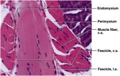
Connective Tissues of Muscle
|
Epimysium - covers the whole muscle, blends into tendons and CT sheets called the deep fasciae that surrounds and separates muscles, Deep fascia grades into the superficial fascia (also called the hypodermis which contains various amounts of adipose between muscles and skin) contains blood vessels and nerves
Perimysium-layer of CT surrounding a fascicle (bundle of muscle cells), contains blood vessels and nerves Endomysium- thin layer of areolar CT surrounding each muscle cell (muscle cell = muscle fiber), contains capillaries and nerves |
|
|
Superficial Fascia and Deep Fascia
|
Superficial Fascia (hypodermis): found bw skin and muscles adipose tissue with vessels and nerves
Deep Fascia (epimysium): found around and between muscles contains neurovascular bundles |
|

|
red = individual muscle w/ fascicles
yellow outer= superficial fascia (hypodermis) yellow w/ red & blue circle = neurovascular bundle white = deep fascia (epimysium) |
|
|
slide 9 lecture 7
know: endomysium (thin white collagen fibers) perimysium Facicles |
|
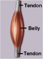
General Parts of a Skeletal Muscle
|
Origin: attach to the more stationary end of a muscle
Belly: thick, middle region of a muscle Insertion: attachment to the more mobile end of muscle Example: biceps brachii origin is scapula and insertion is radius |
|
|
Muscle Attachments
|
Connective tissue fibers of a muscle merge with the connective tissue of bone (periosteum).
Tendons & Aponeuroses |
|
|
Tendons
|
merge with the bone periosteum and they are connected by extremely strong Perforating Fibers (Sharpey’s fibers). Stress will tear the tendon before pulling the tendon loose from either muscle or bone
|
|
|
Aponeuroses
|
are flat sheet-like tendons as under the scalp, palm, foot and other areas that attach muscle to the collagen fibers of the dermis.
example: palmaris longus tendon fans out to become the palmar aponeurosis |
|
|
Skeletal Muscle Shapes
|
Fusiform Muscles: thick in middle and tapered at ends, biceps brachii m.
Convergent Muscle: broad at origin and tapering to a narrower insertion, pectoralis major m. Parallel Muscles: long, uniform,parallel fascicles shorten more than other muscles, rectus abdominis m. Circular muscles: act as sphincters, ring around body openings, orbicularis oris m. and orbicularis occuli m. Pennate muscles: fascicles insert obliquely on a tendon unipennate, bipennate or multipennate, palmar interosseus m., rectus femoris m., deltoid m. |
|
|
Coordinated Muscle Actions
|
Agonist: the Prime Mover produces most of the force
Synergist: aids the agonist stabilizes the nearby joint modifies the direction of movement that occurs Antagonist: opposes the prime mover prevents excessive movement and injury Fixator: prevents movement of the bone that the prime mover is attached to |
|
|
Muscle Actions during Elbow Flexion
|
Agonist = biceps brachii m.
Synergist = brachialis m. Antagonist = triceps brachii m. Fixator = muscle that holds the origin bone firmly in place such as rhomboideus major m. holds the scapula |
|
|
Intrinsic and Extrinsic Muscles
|
Intrinsic muscles are entirely contained (both origin and insertion) within a region such as the hand like the palmar interosseous m.
Extrinsic muscles move the fingers but origin is outside of the hand like the flexor digitorum profundus m. |
|
|
Skeletal Muscle Innervation
|
Skeletal muscles are activated by motor neurons
Motor Neurons branch out of the central nervous system from the skull (cranial nerves) and the spine (spinal nerves) Cranial nerves arising from the brain exit the skull through foramina and are numbered I to XII Spinal nerves arising from the spinal cord exit the vertebral column through intervertebral foramina |
|
|
Afferent Innervation
Efferent Innervation |
Afferent: sensory info from senses to brain
Efferent: motor somatic, outgoing to skeletal muscle *autonomic, outgoing to glands, cardiac muscle, and smooth |