Reading...
![]()
Play button
![]()
Play button
![]()
Use LEFT and RIGHT arrow keys to navigate between flashcards;
Use UP and DOWN arrow keys to flip the card;
H to show hint;
A reads text to speech;
124 Cards in this Set
- Front
- Back
|
What cranial nerves exit through the jugular foramen?
|
Cranial nerves IX, X, and XI.
|
|
|
TRUE OR FALSE: CN III divides into its superior and inferior divisions BEFORE entering the superior orbital fissure.
|
FALSE: enters the orbit via the SUPERIOR ORBITAL FISSURE; Divides here into the superior and inferior divisions
|
|
|
The ophthalmic division of trigeminal nerve exits the skull through the _____________, and supplies sensation to skin of the _________, while the maxillary division exits the skull through the _____________, and supplies the ____________.:
A)foramen cecum, eyelid; jugular foramen, larynx B) foramen ovale, stomach; orbital canal, maxillary teeth C)superior orbital fissure, forehead; foramen rotundum, upper lip D)superior orbital fissure, upper lip; foramen rotundum, eyeball |
C
|
|
|
TRUE OR FALSE: The mandibular division of CN V supplies both motor and sensory innervation to structures within the head and neck including the tongue.
True False |
TRUE
|
|
|
Inability to completely abduct the eye could be the result of an injury to:
CN III CN XIII CN VI CN X CN V |
CN VI
|
|
|
TRUE OR FALSE: Glossopharyngeal nerve (CN IX) is responsible for parasympathetic innervation to the parotid gland, sensation to the auditory tube and taste to the anterior one third of the tongue.
|
FALSE:POSTERIOR 1/3 of tongue
|
|
|
The _______ division of CN ___ is responsible for our perception of balance and equilibrium.
|
vestibular, VIII
|
|
|
Taste perceived from the anterior two-thirds of the tongue is due to the __________, a branch of CN __________.:
and ____________, a branch of CN____________ |
chorda tympani, VII; anterior two thirds of the tongue.
CN IX, anterior one third of the tongue. |
|
|
The vagus nerve innervates all of the following structures EXCEPT:
abdominal viscera soft palate heart nasal cavity inferior pharynx |
Nasal Cavity
|
|
|
A patient presents with an inability to shrug her shoulders. This is due to a paralysis of the ______ muscle, which is innervated by CN ____.
serratus posterior, XI trapezius, XI latissimus dorsi, XI sternocleidomastoid, XI dorsal scapular, XI |
trapezius, XI
|
|
|
HYPOGLOSSAL NERVE- CN XII
|
• A purely motor nerve from the medulla
• Leaves the skull through the HYPOGLOSSAL CANAL. • Joined temporarily by fibers from spinal nerves C1 and C2, which form the superior limb of the ansa cervicalis. • Supplies motor innervation to all of the muscles of the tongue except palatoglossus (supplied by vagus). |
|
|
CN I
|
• The Olfactory Nerve
• FUNCTION: special sensory (or visceral afferent - smell) • fibers pass through foramina in the CRIBRIFORM PLATE of the ethmoid bone, pierce the dura and arachnoid of the brain and enter the olfactory bulb. • Induces visceral responses via the autonomic nervous system (i.e., salivation in response to the aroma of food) |
|
|
CN II
|
OPTIC NERVE
• FUNCTION: special sensory (somatic afferent - vision) • Formed by retinal ganglion axons that converge at the optic disc at the back of the eye. • Surrounded by cranial meninges • subarachnoid space is filled with CSF. • travels through the OPTIC CANAL from the eye socket to the middle cranial fossa where it forms the optic chiasm. |
|
|
CN III
|
OCULOMOTOR NERVE
FUNCTIONS: motor to 4 of the 6 extraocular muscles; parasympathetic motor to ciliary muscle and sphincter pupillae. • Chief motor nerve to the ocular and extraocular muscles. • Leaves the cranial cavity and enters the orbit via the SUPERIOR ORBITAL FISSURE. o Divides here into the superior and inferior divisions o The inferior division also carries presynaptic autonomic fibers to the CILIARY GANGLION |
|
|
CN IV
|
TROCHLEAR Nerve
• FUNCTION: motor to the superior oblique extraocular muscle • Passes through the SUPERIOR ORBITAL FISSURE where it supplies the superior oblique muscle of the eye. • Injury inhibits the eyeball from turning out and down (inferolaterally), seen clinically as DIPLOPIA (double vision). • Only cranial nerve to emerge dorsally from the brainstem. |
|
|
CN V
|
• TRIGEMINAL NERVE
• Sensory and motor innervation to various parts of the head and neck; first two divisions are purely sensory |
|
|
CN V(1)
|
OPHTHALMIC division of TRIGEMINAL NERVE:
o Sensory nerve that passes through the SUPERIOR ORBITAL FISSURE o Supplies the eyeball, conjunctiva, lacrimal gland and sac, nasal mucosa, frontal sinus, external nose, upper eyelid, forehead and scalp. |
|
|
CN V(2)
|
MAXILLARY division of TRIGEMINAL NERVE:
o Sensory nerve that passes through the FORAMEN ROTUNDUM. o Relays sensation from the skin of the face over the maxilla including the upper lip, maxillary teeth, nasal mucosa, maxillary sinuses and palate. |
|
|
CN V(3)
|
MANDIBULAR division of TRIGEMINAL NERVE:
o Both a motor and sensory nerve. o Passes through the FORAMEN OVALE that is both a sensory and motor nerve. Motor component: supplies the MUSCLES OF MASTICATION, mylohyoid, anterior belly of the digastric, tensor veli palatini and tensor tympani. Sensation from the skin over the mandible, including the lower lip and side of the head, mandibular teeth, mucosa of the mouth and the anterior two thirds of the tongue |
|
|
CN VI
|
ABDUCENT NERVE
• FUNCTION: motor to the lateral rectus extraocular muscle. • Arises between the pons and the medulla on the brain and will travel through the SUPERIOR ORBITAL FISSURE to innervate the lateral rectus muscle. • The action of this muscle is to ABDUCT the eye. |
|
|
CN VII
|
FACIAL NERVE
• FUNCTIONS: motor to muscles of facial expression, parasympathetic to submandibular, sublingual, and lacrimal glands, and special sensory (taste). • Emerges between the pons and medulla on the brain and exits the skull through the internal acoustic meatus, facial canal and finally the STYLOMASTOID FORAMEN. • 5 TERMINAL BRANCHES: o Temporal o Zygomatic o Buccal o Mandibular o Cervical • Branches supply: o the MUSCLES OF FACIAL EXPRESSION o Muscles of auricle and occipitalis muscle (posterior auricular) o Taste to the anterior two thirds of the tongue (chorda tympani), o Parasympathetic innervation to the submandibular and sublingual glands (chorda tympani) o Lacrimal glands and mucous glands of the soft palate, nose, and paranasal sinuses (greater petrosal nerve) |
|
|
CN VIII
|
VESTIBULOCOCHLEAR NERVE
• FUNCTION: special sensory (somatic afferent - hearing & equilibrium) • Vestibulocochlear nerve enters the INTERNAL ACOUSTIC MEATUS where it then divides into vestibular (equilibrium) and cochlear (hearing) divisions. |
|
|
CN IX
|
GLOSSOPHARYNGEAL NERVE
• FUNCTIONS: special sensory (taste), sensory, motor and parasympathetic • Conveying taste from the posterior one third of the tongue. • Sensory innervation to the pharyngeal mucosa, palatine tonsil, posterior one third of the tongue, auditory tube, middle ear, carotid sinus and carotid body. • The only muscle innervated by CN IX is the stylopharyngeus muscle. • The glossopharyngeal nerve supplies parasympathetic innervation to the parotid gland. • passes through the JUGULAR FORAMEN to exit the skull. |
|
|
CN X
|
VAGUS NERVE
• FUNCTIONS: sensory, motor, parasympathetic and some special sensory (taste) • Extensive innervation throughout the body; responsible for sensation from the inferior pharynx, larynx and thoracic & abdominal viscera. • Taste from the root of the tongue and the taste buds located on the epiglottis is also carried by CN X. • The vagus nerve innervates the muscles of the soft palate, pharynx, the intrinsic laryngeal muscles and the palatoglossus muscle of the tongue. • Parasympathetic innervation of the thoracic and abdominal viscera down to the splenic flexure is also a function of CN X. • The vagus nerve will exit the skull with the glossopharyngeal nerve (CN IX) at the JUGULAR FORAMEN. |
|
|
CN XI
|
ACCESORY NERVE
• FUNCTION: motor • Leaves the skull at the JUGULAR FORAMEN and is responsible for the motor innervation of the sternocleidomastoid and trapezius muscles. • Injury to the accessory nerve presents clinically as an inability to shrug one's shoulders, due to the paralysis of the trapezius muscle. |
|
|
CN XII
|
HYPOGLOSSAL NERVE
• A purely motor nerve from the medulla • Leaves the skull through the HYPOGLOSSAL CANAL. • Joined temporarily by fibers from spinal nerves C1 and C2, which form the superior limb of the ansa cervicalis. • Supplies motor innervation to all of the muscles of the tongue except palatoglossus (supplied by vagus). |
|
|
Smallest CN and only one to emerge from dorsal aspect of brainstem?
|
Trochlear Nerve (CN IV)
|
|
|
Divisions of Trigeminal Nerve
|
Ophthalmic, Maxillary & Mandibular
|
|
|
Olfactory Bulb
|
location of synapse for the 20 bundles of unmyelinated special visceral afferent fibers arising from olfactory neurons in the olfactory are (in upper 1/3 of nasal mucosa)
|
|
|
Optic nerve (CN II) formed from?
|
Axons of ganglion cells of the retina which converge at the optic disk
|
|
|
Optic Chiasma
|
(Chiasma="cross") When the optic nerve leaves the orbit throu the optic canal, the fibers from the nasal side of either retina cross over one another on the opposite side of the brain
|
|
|
nerve that enters thru the tendinous ring of the superior orbital fissure
|
CN III, Oculomotor Nerve
|
|
|
Nerve that passes thru the lateral wall of cavernous sinus during its course
|
CN IV, Trochlear Nerve
|
|
|
First branchiomeric nerve? Supplies what? Type of ganglion?
|
Trigeminal Nerve (CN V); supplies 1st branchial arch; sensory (general somatic afferent & ganglion (semilunar or trigeminal) consisting of cell bodies of GSA fibers
|
|
|
Mediates afferent limb of corneal reflex? Nerve type?
|
CN V, br. 1 Trigeminal Nerve, Opthalmic branch; sensory nerve
|
|
|
Mediates afferent limb of sneeze reflex? Cell bodies where? Snesory where?
|
CN V, br. 2, Trigeminal Nerve, Maxillary Branch; cell bodies in trigeminal ganglion; sensory to midface (below eye, but above upper lip), palate, paranasal sinuses, maxillary teeth
|
|
|
Which nerve passes through the Foramen Ovale? Type of fibers and to where?
|
CN V, br. 3, Trigeminal Nerve, Mandibular br.;
SVE fibers to tensor veli palatini, tensor tympani, muscles of mastication & ant. belly of digastric & mylohyoid ms.; sensory to lower part of face |
|
|
CN that provides sensory to lower part of face (name spec. where) and mediates afferent limb of jaw jerk reflex?
|
Trigeminal Nerve, Manibular br., CN V, br. 3;
BELOW lowr lip and mouth, scalp, jaw, mandibular teeth, ant 2/3 of tongue |
|
|
Lateral Rectus muscle innervated by? What type of nerve fibers? Where does it travel tru to get there?
|
CN VI, Abducens Nerve; GSE fibers; pierces dura on dorsum sellae of sphenoid bone; passes thru cavernous sinus, enters orbit thru superior oribital fissure and then goes on to muscle
|
|
|
Somatic Motor Cranial Nerves are what type of fiber and innervate what structure?
|
GSE; Striated muscle derived from somites; CN III, VI, & XII
|
|
|
Visceral motor Cranial Nerves are what type of fiber and innervate what structure?
|
GVE; Smooth muscle of blood vessels, glands, and organs;
CN III,VII, IX, X |
|
|
Branchial Motor Cranial Nerves are what type of fiber and innervate what structure?
|
SVE; striated muscle derived from branchial arteries;
CN V, VII, X, XI |
|
|
General Sensory Cranial Nerves are what type of fiber and innervate what structure?
|
GSA;Mechanical, pain/temperature, propprioception of skin & mucous membranes;
CN V, VII, IX, X |
|
|
Visceral Sensory Cranial Nerves are what type of fiber and innervate what structure?
|
GVA;Mechanical, pain/temp, proprioception of oral cavity, pharynx and larynx;
CN IX, X |
|
|
Special Somatic Sensory Cranial Nerves are what type of fiber and innervate what structure?
|
SSA; Vision, audition & balance;
CN II, VIII |
|
|
Special Visceral Sensory Cranial Nerves are what type of fiber and innervate what structure?
|
SVA; Olfaction, taste;
CN I, VII, IX, X |
|
|
Which cranial nerve is actually a CNS tract rather than a true cranial nerve? Why?
|
The Optic Nerve, CN II; the retina and the optic nerve develop from the optic vesicle, an outgrowth of the brain
|
|
|
Which CN is the shortest and least "nerve-like"? Why?
|
CN I, the Olfactory Nerve; it consists primarily of sensory neurons in nasal mucousa that enter cranium (via cribriform plate of ethmoid bone) & synapse in the OLFACTORY BULB! axons from bulb project to higher processing centers and look like a CN, but is NOT!
|
|
|
Each eye has its own?
|
Monocular crescent
|
|
|
What happens in the optic chiasm? What does this do to visual field?
|
Some fibers cross the ipsilateral tract, and others cross the contralateral side; partial decussation produces homologous representation of visual field in each tract
|
|
|
Structures where optic tract synapses
|
thalamus, hypothalamus, midbrain
|
|
|
Which CN exits brain laterally at ponto-medullary junction? What does it do next?
|
CN IX, the Glossopharyngeal Nerve; exits cranium at the jugular foramen, where 2 sensory ganglia are located just distal, called the superior & inferior glossopharyngeal ganglia
|
|
|
What nerve innverates the Pharynx and posterior 1/3 of tongue? Innervation provides what?
|
Glossopharyngeal, CN IX; general sensation to pharynx & post 1/3 of tongue; taste to post 1/3 tongue
|
|
|
What Brachiomeric muscle is innervated by CNIX?
|
The stylopharyngeus
|
|
|
Where does CNIX (glossoph.) re-enter cranium? why?
|
distal to the inferior glossoph. ganglion; crosses thru middle ear to form tympanic plexus
|
|
|
What is the Lesser Petrosal Nerve?
|
br of CN IX (Glossoph.) that is preganglionic parasympathetic & courses thru middle cranial fossa, exits thru the foramen ovale & synapses in the OTIC GANGLION
|
|
|
Where do POST-GANGLIONIC fibers of CNIX (Glossophl)go?
|
They follow the auriculotemporal nerve (CN V3) to the parotid gland
|
|
|
What gives rise to the carotid nerve? What does it do?
|
CN IX (Glossphl); small visceral sensory nerve of two sensory organs at the bifurcationof the common carotid body, to monitor blood CO2 levels (carotid body) and arterial BP (carotid sinus)
|
|
|
Where does the Vestibulocochlear Nerve (CNVIII) arise from? exit brain from? What nerve does it accompany and where does it go?
|
Two sensory organs in inner ear; border btwn pons & medulla just lateral to the facial nerve; accompanies facial nerve into internal acoustic meatus
|
|
|
CNVIII fibers separate where? then what?
|
The cochlear and vestibular fibers separate out and innervate their respective targets
|
|
|
Hearing is mediated by CN???
|
VIII, Vestibulocochlear; cell bodies of primary sensory neurons are located in the spiral ganglion in the cochlea
|
|
|
What CN mediates balance? where? how?
|
CN VIII (VSTBLCR; cell bodies of primary sensory neurons mediating blance (position & acceleration) are located in the vestibular ganglion in bony labyrinth
|
|
|
Where does CN VI originate from and where does it go?
|
Motor neurons originating on ventral surface of brain at junction of pons & meedulla; pierces dura immediately & courses anteriorly beneath
|
|
|
Which CN enters orbit thru the Superior Orbital fissure? Does what?
|
CNVI, the abducens nerve; to innervate lateral rectus m.
|
|
|
Where does CNVII originate? what next?
|
CNII, the FACIAL NERVE, orig. laterally at junction btwn pons & medulla; travels to enter internal acoustic meatus of temporal bone; shortly after, major trunk makes sharp angle, called GENU
|
|
|
What is the GENU?
|
it is where the geniculate ganglion located, which contains cell bodies of general sensory neurons innervating skin behind ears as well as special sensory neurons innervating the anterior 2/3 of tongue
|
|
|
Where does the main trunk of CN VII descend? What next?
|
Post & Infer to exit thru stylomastoid foramen;
|
|
|
Branchial motor axons form characteristic branching pattern from what nerve? What next?
|
CNVII, the FACIAL NERVE; innervation of facial expression muscles & others: stapedius, stylohyoid, post. belly of diagastric
|
|
|
Name the branches of CNVII?
|
Facial Nerve Branches:
-temporal -zygomatic -buccal -mandibular -cervical |
|
|
How do general sensory neurons course in CNVII?
|
In a posterior auricular branch to innervate the post. surface of auricle & ext. surface of tympanic membrane
|
|
|
What is the route of Great Petrosal Nerve?
|
Collection of parasympathetic preganglionic fibers from CNVII(visceral motor)that projects from GENU to middle cranial fossa thru a sm. opening, the hiatus for Greater Petrosal Nerve; then courses ant. & medially; then at the foramem lacerum, it dives thru a sm canal & exits the skull
|
|
|
What does the Great Petrosal Nerve join with? then what?
|
Deep Petrosal Nerve; enters the pterygoid canal & courses into the pterygopalatine fossa & runs into pterygopalatine ganglion--> Post Ganglionic fibers distributed via CN V to glands of Nasal and Oral cavities & lacrimal glands
|
|
|
CN V and CN VII (Facial) work together how?
|
Post-ganglionic fibers of CNVII (facial) are distributed via CN V, to target tissues, including glands of Nasal and Oral cavities & lacrimal glands
|
|
|
Facial Nerve (CNVII) gives off what branch as it descends towards the STYLOMASTOID FORAMEN? Then what?
|
CHORDA TYMPANI; nerve contains axons of SPECIAL SENSORY neurons for taste as well as pregang & postgang. parasymp neurons
|
|
|
Describe route/function of CHORDA TYMPANI
|
passes thru middle ear cavity & exits cranium at petrotympanic fissure to enter infratemporal fossa; joins lingual nerve (which carries special sensory axons to ant. 2/3 of tongue)
|
|
|
Explain Lingual Nerve Route/Function
|
Carries special sensory axons to ant. 2/3 of tongue; visceral motor fibers carried to submandibular ganglion and then postgangnlionic fibers are carried to submandibular and sublingual glands
|
|
|
Identify the branches of the facial nerve in the face
|
Branches of the facial nerve include:
--temporal, --zygomatic, --buccal, --marginal mandibular --cervical The temporal branch of the facial nerve emerges from the superior border of the parotid gland and crosses the zygomatic arch. The zygomatic branches pass via two or three branches to the eye to innervate the inferior part of the orbicularis oculi and other facial muscles inferior to the orbit. Buccal branches pass external to the buccinator to innervate this muscle and muscles of the upper lip. The (marginal) mandibular branch innervates the muscles of the lower lip and chin. It emerges from the inferior border of the parotid gland and crosses the inferior border of the mandible deep to the platysma to reach the face. The cervical branch passes from the inferior border of the parotid gland to the mandible to innervate the platysma. |
|
|
CNVII originates? then what is the pathway until its first division?
|
THE FACIAL NERVE
• Originates laterally at junction between pons and medulla • Enters internal acoustic meatus of temporal bone o Quickly makes sharp angle in petrous bone (genu) – where geniculate ganglion is at • Main trunk of CN VII → goes posterior and inferior, exits thru stylomastoid foramen • Main branch divides |
|
|
Main trunk of CNVII divides into?
|
FACIAL NERVE: CNVII
--Temporofacial -- Cervicofacial |
|
|
Motor innervations of CNVII?
|
FACIAL NERVE, CNVII, MOTOR:
o Stapedius o Stylohyoid o Posterior belly of digastrics o Muscles of facial expression o Secretomotor : • lacrimal • Submandibular • Sublingual • mucous glands of nasal & oral cavities |
|
|
Sensory innervations of CNVII?
|
Taste (SVA) from anterior 2/3rds of tongue; part of skin of external auditory meatus
|
|
|
Motor Innervation of Auriculotemporal? Branch of what?
|
secretomotor to parotid gland: postganglionic parasympathetic from communicating br. of otic ganglion; preganglionic parasympathetic from lesser petrosal br. of glossopharyngeal n. (IX)
---BR of CNV3, TRIGEMINAL, MANDIBULAR BR |
|
|
Name 2 Buccal Nerves; review differences
|
1)Buccal Nerve, of Mandibular div. of Trigeminal (CN V3)
-not a motor nerve -sensory of cheek, oral mucosa 2) Buccal Br of Facial Nerve (CNVII) -not a sensory nerve - has temporo-&cervico-facial divisions - motor nerve to facial expression muscles |
|
|
Petrossal Nerves: name them and describe differences
|
1) Lesser Petrosal nerve:
from tympanic nerve, branch of Glossopharyngeal Nerve, CNIX; otic ganglion (synapses); secretomotor (preganglionic parasympathetic) for parotid gland; axons join auriculotemporal nerve 2) Greater Petrossal Nerve; from Facial (VII), no branches; secretomotor (preganglionic, parasympathetic), goes to lacrimal gland and mucous glands of lower nasal cavity, maxillary sinus & palate; joins w/ deep petrosal |
|
|
Auricotemporal Nerve from CN?? joins with ?? nerve from CN?? where?
|
Mandibular div of Trigeminal n (V3) axons from the lesser petrosal nerve (after synapsing in otic ganglion) from the tympanic nerve from Glossopharyngeal nerve (CNIX)
|
|
|
This nerve, from CN?? lets you wiggle your ears
|
Auricular, from the Auricular branch of CNVII (facial)
--Anterior and Superior Ear: Temporal Branches of CNVII --Posterior Ear: Posterior Branch of Auricular br of CNVII |
|
|
The Facial Nerve emerges from the ? of the skull
|
stylomastoid foramen
|
|
|
The genticulate ganglion lies in the ____? gives off ???
|
The Genu;
1) Greater Petrossal Nerve, which carries parasymp fibers to the pterygopalatine ganglion 2) Branch to Tympanic Plexus 3) Branch to Sympathetic Plexus on middle meningeal artery |
|
|
Preganglionic PARAsympathetic fibers from the pterygopalatine ganglion go to what glands?
|
lacrimal, nasal, palatine
|
|
|
Nerves of the orbit? (in general)
|
---3 motor nn. to its muscles (CN III, IV, VI) [LR6, SO4, AO3]
---sensory ophthalmic division of CN V ---optic nerve arises in the retina of the eye, the other nn. enter the orbit through the sup. orbital fissure |
|
|
The Greater petrosal nerve meets up with the Deep pretrosal nerve in the ___________; it has fibers from the ____________ and supplies____________
|
ptergopalatine ganglion; superior cervical ganglion;
|
|
|
Name two CNs that have parasympathetic/sympathetic associations, but no ganglia; explain
|
VAGUS (X):
never has any named, discrete ganglion associated with it, only diffuse ganglia in organ walls. TRIGEMINAL (V): Branches of trigeminal nerve (V) carry pre- and postganglionic fibers around the head, but trigeminal nerve does not have any preganglionic nuclei of its own. |
|
|
Name the 4 pairs of parasympathetic ganglia in the head & their associated CNs
|
1)ciliary ganglion - oculomotor nerve (III)
2)pterygopalatine ganglion - facial nerve (VII) 3)submandibular ganglion - facial nerve (VII) 4)otic ganglion - glossopharyngeal nerve (IX) |
|
|
PRESYNAPSE:
--CN III nerve carries preganglionic parasympathetic fibers into the orbit;fibers follow the infer div of oculomotor nerve, and branch anterosuperiorly as a short (2-4 mm) motor root of the ciliary ganglion, on the lateral side of the optic nerve, near apex of orbit. --A sensory root passes from the nasociliary nerve (from V1) to the ciliary ganglion, but its fibers pass right through. --A sympathetic root passes from the cavernous sinus to the ganglion, and it also passes through (all sympathetics past the superior cervical ganglion are postsynaptic) |
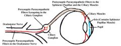
Explain the CILIARY GANGLION:
whats goin on up in there, PRESYNAPSE? |
|
|
The motor root of the ciliary ganglion is a branch of the:
A. internal carotid plexus B. external carotid plexus C. inferior division of oculomotor nerve D. superior division of oculomotor nerve E. nasociliary nerve |
C: inferior div of oculomotor nerve (CN III)
|
|
|
What is going on presynapse in the ptergopalatine ganglion?
|
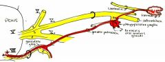
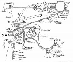
Facial nerve gives off preganglionic fibers in the form of 2 nerves: the greater petrosal and the chorda tympani. Greater petrosal nerve branches from facial at the geniculate ganglion, a sensory ganglion (not to be confused with an autonomic ganglion) along the beginning of the course of the facial nerve within the petrous temporal bone. Greater petrosal exits the anterior slope of the petrous bone to run anteromedially toward the foramen lacerum. It passes through a bit of the cartilage blocking this foramen to enter a canal hidden in its anterior margin, the pterygoid canal (because it lies superior to the pterygoid plates of the sphenoid bone). Greater petrosal is joined by some fibers from the internal carotid plexus (postganglionic sympathetic, of course) called the deep petrosal nerve - they unite to form the nerve of the pterygoid canal. The nerve of the pterygoid canal runs forward to reach the pterygopalatine ganglion, that lies within the pterygopalatine fossa.
|
|
|
What is going on POSTsynapse in the ptergopalatine ganglion?
|

Postganglionic fibers leave the pterygopalatine ganglion within branches of the maxillary division of trigeminal nerve (V2). Some of these branches go to mucous membranes in the nose, paranasal sinuses, palate, and upper pharynx. One branch, the zygomatic nerve, passes through the inferior orbital fissure into the orbit. It runs superiorly up the lateral wall of the orbit, and splits into zygomaticotemporal and zygomaticofacial branches. The postganglionic parasympathetic fibers continue with the zygomaticotemporal nerve, then leave as a communicating branch to reach the lacrimal nerve, a branch of ophthalmic division of trigeminal nerve (V1). Lacrimal nerve carries these secretomotor fibers the last millimeters to reach the lacrimal gland, and then continues on to innervate skin of the upper eyelid laterally.
|
|
|
The _____________ nerve branches from the geniculate ganglion to carry preganglionic parasympathetic fibers to the pterygopalatine ganglion.
A. facial B. greater petrosal C. chorda tympani D. deep petrosal |
B. greater petrosal
|
|
|
The _______________ nerve carries postganglionic parasympathetic fibers into the orbit through the inferior orbital fissure.
PICK ONE zygomatic zygomaticotemporal zygomaticofacial lacrimal |
zygomatic
|
|
|
Tell the submandibular ganglion story - before the synapse
|

Toward the end of its course through the petrous temporal bone, the facial nerve gives off a branch, the chorda tympani nerve, immediately before exiting the skull at the stylomastoid foramen. Chorda tympani recurs back into the middle ear cavity, and then passes anteriorly across the lateral wall of the middle ear, which happens to be the tympanic membrane. It crosses the handle of the malleus, and ultimately leaves the middle ear anteriorly to exit at the base of the skull through the petrotympanic fissure. This leads chorda tympani into the infratemporal fossa, in which it passes anteroinferiorly to join the lingual nerve from behind. It then runs with the lingual nerve to the submandibular ganglion, located in the paralingual space near the deep portion of the submandibular gland.
|
|
|
Preganglionic parasympathetic fibers reach the lingual nerve from the ______ nerve.
PICK ONE: auriculotemporal greater petrosal lesser petrosal chorda tympani |
Chorda Tympani
|
|
|
Explain thesubmandibular ganglion story -POST synapsing, and beyond
|
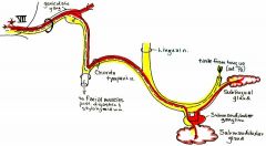
Chorda tympani carries preganglionic parasympathetic fibers that pass to the submandibular ganglion. There, some of these fibers synapse and return to the lingual nerve to travel anteriorly to reach the sublingual gland, to be secretomotor there. Other fibers pass through the submandibular ganglion to reach the submandibular gland, and synapse within the gland on diffuse postsynaptic neurons, to be secretomotor to this gland.
Chorda tympani not only carries preganglionic parasympathetic fibers, but it also carries sensory fibers for taste for the anterior two-thirds of the tongue. These fibers travel into the tongue with the lingual nerve to reach the taste buds on the dorsum and sides of the tongue. |
|
|
Chorda tympani carries what kinds of fibers?
|
preganglionic parasympathetic fibers with secretomotor function (sublingual gland)and also sensory fibers for taste for the anterior two-thirds of the tongue. These fibers travel into the tongue with the lingual nerve to reach the taste buds on the dorsum and sides of the tongue.
|
|
|
Tell the otic ganglion story -until the synapse happens
|
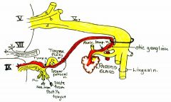
As the glossopharyngeal nerve (IX) passes inferiorly through the jugular foramen, it gives off a tympanic branch, carrying preganglionic parasympathetic fibers, that passes upward through the inferior tympanic canaliculus to pass into the middle ear. There, it forms a tympanic plexus on the promontory, an eminence on the medial wall of the middle ear cavity. Stretching anteromedially from the promontory, and piercing the anterior wall of the middle ear cavity, is the lesser petrosal nerve. Lesser petrosal nerve travels anteromedial on the anterior surface of the petrous temporal bone, just lateral to the greater petrosal nerve. Lesser petrosal passes inferiorly through the skull base, usually through foramen ovale. Immediately below the skull, it passes into the otic ganglion, located on the medial surface of the mandibular division of trigeminal nerve (V3) below its exit through foramen ovale.
|
|
|
The _________ nerve carries preganglionic parasympathetic fibers into the otic ganglion.
|
Lesser Petrosal
|
|
|
The otic ganglion story - after the synapse
|
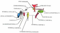
Postganglionic fibers from the otic ganglion pass into the auriculotemporal nerve, a cutaneous sensory branch of V3. The auriculotemporal nerve passes posteriorly on the medial side of the temporomandibular joint capsule, encircles the middle meningeal artery as it ascends to pass through foramen spinosum, and then curves laterally behind the TMJ capsule to reach the parotid gland. The postganglionic parasympathetic fibers distribute to the parotid gland, where they are secretomotor, causing saliva production.
** remember this: glossopharyngeal nerve innervates the posterior one-third of the tongue |
|
|
Postganglionic fibers reach the parotid gland within the ___________ nerve.
PICK ONE: greater petrosal lingual auriculotemporal mandibular division of trigeminal |
auriculotemporal
|
|
|
How does the vagus nerve carry its preganglionic fibers? explain
|
The vagus nerve has NO ganglions.
It carries preganglionic fibers everywhere;In the head, it also delivers skeletal motor fibers to most of the soft palate (except tensor veli palatini muscle), most of the pharynx (except stylopharyngeus muscle), all of the larynx, and the upper esophagus (which is skeletal muscle). These skeletal motor fibers travel in the pharyngeal branches, external branches of superior laryngeal nerve, and the recurrent laryngeal nerves. --Preganglionic parasympathetic fibers accompany the branches of vagus, synapsing as always within the viscera wall, to innervate mucous glands in the lining of the structures innervated - pharynx, larynx, and esophagus. This is most of the mucous membranes in the head and neck, with the exception being the nasal cavity, paranasal sinuses, palate, and upper pharynx. (This region is pterygopalatine ganglion.) --In the neck, the vagus also gives off 2 cervical cardiac nerves that pass inferiorly into the chest, often uniting with the sympathetic cardiac branches on the way. |
|
|
In the head and neck, the primary autonomic function of the vagus nerve is innervation of the:
pick one! laryngeal muscles pharyngeal muscles mucous membranes salivary glands |
Mucous membrane
|
|
|
Summary of ANS in head & neck
|
Sympathetic - internal & external carotid nerves/plexuses - innervate blood vessels, sweat glands, superior tarsal and dilator pupillae muscles.
Parasympathetic - oculomotor n. (III) > inferior division > motor root > ciliary ganglion > short ciliary nn. > sphincter pupillae & ciliary muscles facial n. (VII) > greater petrosal n. > n. of pterygoid canal > pterygopalatine ganglion > zygomatic n. (V2) > zygomaticotemporal n. > lacrimal n. (V1) > lacrimal gland facial n. (VII) > chorda tympani > lingual n. (V3) > submandibular ganglion > submandibular & sublingual glands glossopharyngeal n. (IX) > tympanic n. > tympanic plexus > lesser petrosal n. > otic ganglion > auriculotemporal n. (V3) > parotid gland vagus n. (X) > pharyngeal, superior laryngeal, and recurrent laryngeal nn. > terminal ganglia > mucous glands |
|
|
DESCRIBE PTERYGOPALATINE GANGLION!
|
a PARAsympathetic ganglion; hangs off maxillary division of the trigeminal n. (V2) within the pterygopalatine fossa; preganglionic axons of the greater petrosal n. synapse here; postganglionic sympathetic axons of the deep petrosal n. pass through the otic ganglion without synapsing (they synapse in the superior cervical sympathetic ganglion)
|
|
|
Where do sympathetic axons of the deep petrosal n. pass through without synapsing? Where do they synapse?
|
pass through the otic ganglion without synapsing; synapse in the superior cervical sympathetic ganglion)
|
|
|
What nerves synapse in the ptergopalatine ganglion? Then where do they go?
|
Preganglionic axons of the greater petrosal n. synapse here; joins w/ deep petrosal, forms Vidian Nerve/Nerve of Ptergoid Canal,passes thru otic canal without synapsing; synapses in Superior Cervical Ganglion; gives POSTganglionic SYMPATHETIC innervation to vascular smooth muscles of mucous membranes of lower nasal cavity
|
|
|
Explain what happens in the ciliary ganglion
|
a PARAsympathetic ganglion, located on the lateral side of the optic n. near apex of the orbit; sensory and sympathetic axons pass thru ciliary ganglion without synapse -sensory root is carried via the nasociliary n.and the sympathetic root arrives in the orbit via the internal carotid a.
|
|
|
Explain the NASOCILIARY NERVE
|
ORIGIN: branch of V1
TYPE: GSA BRANCHES: communicating br. to the ciliary ganglion, long ciliary n., anterior and posterior ethmoidal nn., infratrochlear n. INNERVATIONS: nasociliary area, mucous membranes (just sensory!!!) |
|
|
EXPLAIN SHORT CILIARY NERVES
|
ORIGIN:
--SENSORY: ciliary ganglion, from nasociliary (V1) --SYMP:internal carotid plexus --PARASYMP: inf div of CN III TYPE: GSA ACTION: parasymp & symp of sphincter pupillae & ciliary (PS) dilator pupillae (symp) ETC: MIXED! sensory AND ANS; postganglionic parasympathetic neurons whose axons are located in these nerves have their cell bodies located in the ciliary ganglion |
|
|
NASOPALATINE N
|
maxillary division of the trigeminal n. (V2) INNERVATION TO: mucous membrane of the nasal septum; mucous membrane of the anterior portion of the palate nasopalatine n. innervates the mucosa overlying the primary palate (development); it passes through two openings in bone: sphenopalatine foramen and incisive canal
|
|
|
Long ciliary nerves enter/do not enter ciliary ganglion
|
do not enter!
|
|
|
Nerve of Pterygoid Canal/Vidian Nerve
|
--formed by the union of the greater petrosal n. (preganglionic parasympathetic) and the deep petrosal n.(postganglionic sympathetic)
---ends in the pterygopalatine ganglion (parasympathetic) secretomotor (parasympathetic) INNERVATION to: SECRETOMOTOR: --lacrimal gland and mucous glands of nasal cavity and maxillary sinus; --SYMP. innervation to vascular smooth muscle in the same region --- NO SENSORY! --- CONTENTS: preganglionic axons of the greater petrosal n. bound for pterygopalatine ganglion where they will synapse; postganglionic sympathetic axons of the deep petrosal n. which will pass through the pterygopalatine ganglion without synapsing |
|
|
What happens POSTSYNAPSE in the ciliary ganglion?
|
POSTSNYAPSE:
--The presynaptic parasympathetic fibers in the motor root synapse, and then the postsynaptic parasympathetic fibers UNITE with the fibers in the other two roots, sensory and sympathetic, to form short ciliary nerves. --These travel anteriorly to reach the back of the eyeball, pierce it, and pass forward within the walls of the eyeball to reach the smooth muscle of the eyeball. --The parasympathetic fibers innervate 2 muscles: the ciliary muscle and the sphincter pupillae --The sympathetic fibers innervate 2 muscleS: dilator pupilla (dilates pupil) and superior tarsal muscle(holds eyelid up) |
|
|
Explain about lacrimal nerve
|
ORIGINS: ophthalmic division of the trigeminal n. (V1); facial nerve;
INNERVATIONS:carries secretomotor axons to the lacrimal gland skin of the lateral portion of the upper eye lid and its associated conjunctiva lacrimal n. carries the postganglionic parasympathetic axons from the zygomaticotemporal br. of the maxillary n. that originate in the pterygopalatine ganglion |
|
|
How do you test the function of the hypoglossal nereve?
|
Ask patient to stick out tongue, which is done by the genioglossus m (protrudes tongue); if they can't, the nerve is damaged on both sides; if they have a lesion on 1 side, they protruded tongue will deviate towards the side with the nerve lesion
|
|
|
one muscle of tongue that is NOT innervated by the hypoglossal (XII)
|
palatoglossus by Vagus nerve
|
|
|
one muscle motor innervated by glossopharnygeal nerve
|
stylopharyngeus, for elevation of pharynx
|