Reading...
![]()
Play button
![]()
Play button
![]()
Use LEFT and RIGHT arrow keys to navigate between flashcards;
Use UP and DOWN arrow keys to flip the card;
H to show hint;
A reads text to speech;
46 Cards in this Set
- Front
- Back
|
Olfactory Nerve--CN I
Nasal cavity - Cribriform plate - Anterior cranial fossa - Olfactory bulb - Olfactory tract |
|
|
Optic Nerve--CN II
Optic Canal - Middle cranial fossa - Optic Chiasm - Optic Tracts |
|
|
Oculomotor Nerve--CN III
Superior Orbital Fissure - Brain (superior to pons) |
|
|
Trochlear Nerve--CN IV
Superior Orbital Fissure - Lateral aspect of pons |
|
|
Trigeminal Nerve--CN V
V1: Ophthalmic nerve V2: Maxillary nerve V3: Mandibular nerve |
|

|
Ophthalmic Branch of Trigeminal Nerve--CN V1
Superior Orbital Fissure - Lateral Aspect of Pons |
|

|
Maxillary Branch of the Trigeminal Nerve--CN V2
Foramen Rotundum - Lateral Aspect of Pons |
|

|
Mandibular Branch of the Trigeminal Nerve--CN V3
Foramen Ovale - Lateral Aspect of Pons |
|
|
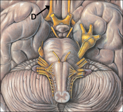
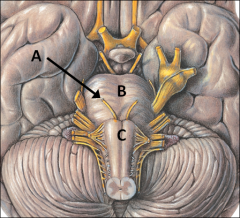
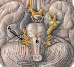
Abducens Nerve--CN VI
Superior Orbital Fissure - Emerges between the pons (B) and medulla (C) near midline |
|
|
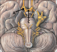
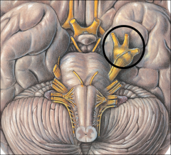
Facial Nerve--CN VII
Emerges between pons (B) and medulla (C), lateral to CN VI. It enters the petrous part of the temporal bone through the internal auditory meatus, passes through the facial canal to the geniculate ganglion, and exits the skull via the stylomastoid foramen. |
|
|
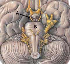
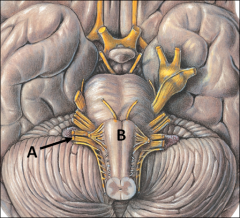
Vestibulocochlear Nerve--CN VIII
Emerges between pons (B) and medulla (C), just inferior to CN VII Enter the petrous part of the temporal bone through the internal auditory meatus. |
|
|
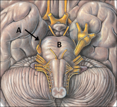
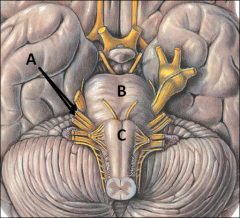
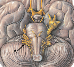
Glossopharyngeal Nerve--CN IX
Emerges on the lateral aspect of the medulla. Exits the cranial cavity, alongside the internal jugular vein, through the jugular foramen. This opening is located just inferior to the petrous part of the temporal bone |
|
|
Vagus Nerve--CN X
Emerges on the lateral aspect of the medulla. Exits the cranial cavity, alongside the internal jugular vein, through the jugular foramen |
|
|
(Spinal) Accessory Nerve--CN XI
Emerges on the lateral aspect of the medulla. (Some fibers originate at the C1-C5 region.) Exits the cranial cavity as CN XI through the jugular foramen. |
|
|
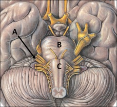
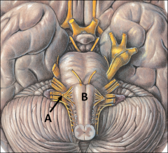
Hypoglossal Nerve--CN XII
Arises off the medulla (B), medial to CN IX, X, and XI. Exits the cranial cavity through the hypoglossal canal (C), an opening located at the base of the occipital condyles |
|
|
Name the structure(s) that pass through the Superior Orbital Fissure.
|
CN III, CN IV, CN V1, CN VI, Ophthalmic veins.
|
|
|
Name the structure(s) that pass through the Optic Canal.
|
CN II, Ophthalmic artery
|
|
|
Name the structure(s) that pass through the Inferior Orbital Fissure.
|
Infraorbital Nerve (branch of CN V2)
|
|
|
Name the structure(s) that pass through the Infraorbital Foramen.
|
Terminal branch of CN V2, Infraorbital nerve, Infraorbital vessels.
|
|
|
Name the structure(s) that pass through the Supraorbital Foramen.
|
Supraorbital nerves (branches of CN V1), Supraorbital vessels
|
|
|
What are the bone(s) that comprise the MEDIAL wall of the orbit?
|
Lacrimal, Maxillary, Ethmoid
|
|
|
What are the bone(s) that comprise the FLOOR of the orbit?
|
Maxillary, Zygomatic
|
|
|
What are the bone(s) that comprise the LATERAL wall of the orbit?
|
Zygomatic, Sphenoid
|
|
|
What are the bone(s) that comprise the ROOF of the orbit?
|
Frontal
|
|
|
Name the structure(s) that pass through the Mental Foramen.
|
Mental nerve
|
|
|
Name the structure(s) that pass through the Mandibular Foramen.
|
Inferior alveolar nerve and vessels
|
|
|
Name the structure(s) that pass through the Mylohyoid Groove.
|
Mylohyoid Nerve
|
|
|
Name the structure(s) that pass through the Foramen Rotundum.
|
CN V2, pterygopalatine ganglion
|
|
|
Name the structure(s) that pass through the Foramen Magnum.
|
Spinal cord, Vertebral Arteries, (spinal) Accessory nerve
|
|
|
Name the structure(s) that pass through the Hypoglossal Canal.
|
CN XII
|
|
|
Name the structure(s) that pass through the Jugular Foramen.
|
Internal Jugular Vein, CN IX, CN X, CN XI
|
|
|
Name the structure(s) that pass through the Stylomastoid Foramen.
|
CN VII
|
|
|
Name the structure(s) that pass through the Carotid Canal.
|
Internal Carotid Artery
|
|
|
Name the structure(s) that pass through the Foramen Lacerum.
|
Nothing. In life it is plugged with cartilage.
|
|
|
Name the structure(s) that pass through the Foramen Spinosum.
|
Middle Meningial Artery (branch of maxillary artery)
|
|
|
Name the structure(s) that pass through the Foramen Ovale.
|
CN V3
|
|
|
Name the structure(s) that pass through the Incisive Foramen.
|
Nasopalatine Nerve, Greater Palatine Nerve
|
|
|
Name the structure(s) that pass through the Internal Auditory Meatus.
|
CN VII, CN VIII
|
|
Name the major branches of the Subclavian A.
|
Internal Thoracic A., Vertebral A., Thyrocervical Trunk, Dorsal Scapular A
|
|

Name the major branches of the Thyrocervical Trunk.
|
Suprascapular A., Inferior Thyroid A.
|
|
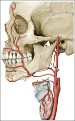
Name the major branches of the External Carotid A.
|
Superior Thyroid A., Lingual A., Facial A., Occipital A., Maxillary A., Superficial Temporal A.
|
|

Outline the flow of blood to the brain beginning with the Internal Carotid A.
|
Internal Carotid A.
Posterior Communicating A. (to Post. Cerebellar A.) Anterior Cerebral A. Anterior Communicating A. Middle Cerebral A. |
|
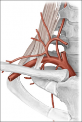
Outline the flow of blood to the brain beginning with the Vertebral A.
|
Vertebral AA.
Posterior Inferior Cerebellar A. Basilar A. Anterior Inferior Cerebellar A. Superior Cerebellar A. Posterior Cerebellar AA. Posterior Communicating A. (to Int. Carotid A.) |
|
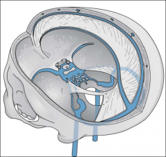
Outline the venous drainage of the head.
|
Superior Sagittal Sinus
Confluence of Sinuses Transverse Sinus Sigmoid Sinus Internal Jugular V. |
|
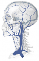
Outline venous drainage from the neck.
|
Superficial Temporal V.
Maxillary V. Retromandibular V. OR External Jugular Facial V. V. alone Internal Jugular V. Subclavian V. Brachiocephalic V. |
|
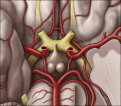
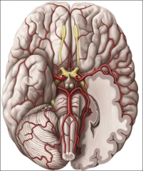
Name the vessels of the Circle of Willis
|
Posterior Cerebral AA.
Posterior Communicating AA. Internal Carotid AA. Anterior Cerebral AA. Anterior Communicating A. |