![]()
![]()
![]()
Use LEFT and RIGHT arrow keys to navigate between flashcards;
Use UP and DOWN arrow keys to flip the card;
H to show hint;
A reads text to speech;
75 Cards in this Set
- Front
- Back
- 3rd side (hint)
|
What are the 4 tissue classifications? ECMN |
Epithelial Connective Muscle Nervous |
|
|
|
What is tissue classification based on? |
It’s based on structure of cells, composition of non cellular extracellular matrix, and cell functions |
|
|
|
What is histology of tissues? What are the 2 histology? |
Microscopic study of tissues 1. Biopsy- removal of tissues for diagnostic purposes 2. Autopsy- examination of organs of a dead body to determine cause of death |
|
|
|
What are 7 characteristics of epithelial tissue? |
Predominantly cells Creates glands Covers body surfaces & forms glands Avascular - means no blood supply - only by diffusion Outside surface of the body Lining of digestive, respiratory and urogenital systems & body cavities Heart & blood vessels |
|
|
|
What does the term mean basement membrane? |
Extracellular- firmed by secretions of both epithelial & connective tissues Acellular glue |
|
|
|
What functions do the Epithelial tissue have? |
Protecting- underlying structures- lining of the mouth Acting- as barriers - skin Permitting- the passage of substances- nephrons in kidneys Secreting- substances- creating - pancreas glands Absorbing - substances- lining of small intestines |
|
|
|
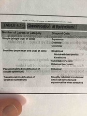
What are the 2 classifications of EPITHELIUM? |
1.Number of layers of cells 2. Shape of cells |
|
|
|
What are the 3 types of layer of cells? Explain |
1. Simple- one layer of cells - each extends from basement membrane to the free surface 2. Stratified- more than one layer- shape of cells of the apical layer 3. Pseudo-stratified- tissue appears to be stratified. But all cells contract basement membrane- simple - transitional |
|
|
|
What are the 4 shapes of epithelium cells? SCCG |
Squamous- flat- scale like Cuboidal- equal in height & width Columnar- taller than wider Goblet- looks like a cup that is a goblet |
|
|
|
What are the functions of the simple cells? |
Allows diffusion of gases, filtration of blood. Secretion & absorption Thin as possible |
|
|
|
What are the functions of the stratified cell? |
More than one layer Protection, particularly against abrasions- eg. Skin |
|
|
|
What are the functions of the Squamous cells? |
Allows diffusion Acts as filter |
|
|
|
What are the functions of the cuboidal & columnar do? |
Secretion Absorption May include goblet cells |
|
|
|
What functions does a goblet cell do? |
Produce mucus Protection |
|
|
|
What are the free surfaces of epithelial tissues? |
Smooth - reduce friction Microvilli- increase surface area for absorption or secretion Stereocilia - elongated microvilli for sensation & absorption Cilia- move materials across the surface |
|
|
|
What are the functions of the simple squamous epithelium? DFPSA |
Diffusion Filtration Protection against friction Secretion Absorption |
|
|
|
What are the locations that simple squamous epithelium is found? 2 |
Lining of the blood & lymphatic vessels Alveoli of the lungs |
|
|
|
What is the structure of simple squamous epithelium? |
Single layer of flat cells |
|
|
|
Where is simple cuboidal epithelium found? |
It’s transitional It’s only found in the lining of the urinary bladder It’s makes & releases substances |
|
|
|
Where is the location of the simple cuboidal epithelium? |
Kidneys tubules Glands & their ducts |
|
|
|
What is the structure of simple cuboidal epithelium? |
Single layer of cube shaped cells - some have microvilli (kidney) or cilia (bronchioles of lungs) |
|
|
|
What is the function of the simple cuboidal epithelium? |
Secretion! In glands & choroid plexus |
|
|
|
Ok now onto simple columnar epithelium . What’s the 1. location 2. Structure 3. Function |
1.glands & some ducts 2. Single layer of tall, narrow cells- cilia (bronchioles of lungs) & microvilli (intestine) 3. Secretion by glands of the stomach & the intestines Absorption by cells of the intestines |
|
|
|
Ok now we discuss stratified squamous epithelium. What’s the location of these? |
Moist— mouth-larynx -esophagus-anus-vagina- inferior urethra& cornea Places that are exposed to the outside world Keratinizated- skin / structure protein & water proof - In keratinized surface cells are dead Layers of FAT CELLS- layers & layers |
|
|
|
What’s the structure of simple squamous epithelium? |
Multiple layers of cells that are cuboidal in the basal layer & progressively flattened towards the surface. |
|
|
|
What is the function of the stratified squamous epithelium? |
Protection against abrasions Caustic chemicals Water loss Infection |
|
|
|
Ok now stratified cuboidal epithelium. What’s the 1. Location 2. Structure 3. Function |
1. Sweat gland ducts 2. Multiple layers of somewhat cube shaped cells 3. Secretion Absorption Protection against infection |
|
|
|
Ok now onto stratified columnar epithelium. What’s the 1. Location 2. Structure 3. Function |
1. Mammary gland ducts 2. Multiple layers of cells with tall thin cells resting on layers of more cuboidal cells (ciliated in the larynx) 3. Protection and secretion |
|
|
|
Ok onto pseudo stratified columnar epithelium. What’s the 1. Location 2. Structure 3. Function |
1. Lining of the nasal cavity. Pharynx. Trachea. Bronchi of lungs 2. All cells reach basement membrane. Appears to be stratified b/c nuclei are at various levels Ciliated & associated with goblet (mucus producing) cells 3. Secretes mucus onto the free surface Move mucus/ fluid that contains foreign particles over the free surface & from passages Interspersed with goblet cells |
|
|
|
Ok now transitional epithelium. What’s the 1. location 2. Structure 3. Function |
1. Lining of the urinary bladder 2. Stratified- cells change shape 3. Accommodates fluctuations in the volume of fluid in an organ or tube. Protection against the caustic effects of urine |
|
|
|
What are glands lined with? |
Lined with epithelium tissue Epithelium with supporting network of C.T. |
|
|
|
What are glands lined with? |
Lined with epithelium tissue Epithelium with supporting network of C.T. |
|
|
|
What are the 2 types of glands that are formed by infolding of epithelium? |
1. Endocrine- makes & secretes hormones- no open contact with exterior. No ducts . Glands in body Stay in bloodstream 2. Exocrine- outside of ur blood stream - OPEN contact maintained with exterior - ducts Not going into blood Secreting something outside the body- sweat / mucus / tears /GI TRACK / mammary glands |
|
|
|
What are exocrine glands classified as? |
By structure By the method of secretion |
|
|
|
What is the classification of structure? |
Unicellular Goblet cells - single cell (mucus) Multicellular - simple & compound (don’t need to go in-depth) |
|
|
|
Explain 5 things about connective tissue. AMDVfOccP? |
Abundant- found in every organ Consists of cells separated by extracellular MATRIX (framework) Many diverse types Performs variety of important functions Tissues that makes organs Lined with cuboidal & columnar Parenchyma?? |
|
|
|
What are the functions of connective tissue ? 7 |
1. Enclose organs as a capsule 2. Connect tissues to one another (tendons & ligaments) 3. Support & movement (bones) 4. Storage(fat) 5. Cushion & insulation (fat) 6. Transport (blood- red & white -immune system) 7. Protect - cells of the immune system |
|
|
|
What specialized cells does the connective tissues produce? |
The extracellular MATRIX |
|
|
|
What is blasts? In connective tissue.. |
Creates the matrix, builds it Eg. Osteoblasts- make bone Eg. Condroblasts - makes cartilage |
|
|
|
What is the term cytes mean with connective tissues? |
Maintain the matrix Eg. Chondrocyte . Keeps the bone the way it is. Just does the function of the cell |
|
|
|
What does the term classy mean in connective tissue? |
Break the matrix down for remodeling Eg. Osteoclasts . Less bone |
|
|
|
What are the 5 types of connective tissue cells? Explain each one |
1. Adipose/fat cells (adipocytes) common is skin-dermis 2. White blood cells (leukocytes) respond to injury/infection- immune for protection 3. Macrophages- fixed (stay in position in CT) & wandering (move by Amoeboid movement through the CT) 4. Platelets- bone marrow - fragments of hematopoietic cells in clotting 5. Undifferentiated Mesenchyme- stem cells could turn into other specific types of cell- into adult cells |

|
|
|
What are the 3 extracellular matrix protein fibres in CT? CRE |
1. Collagen- strong, flexible, inelastic 2. Reticular - fills the spaces & doesn’t stretch 3. Elastic -returns to its original shape after distension or compression Remember like a spring - it stretches than goes back to its shape Note : most tissues are a mixture of all 3 types! |
|
|
|
What is loose(Areolar) CT? |
It’s the filler Superficial fascia Loose packaging material of most organs & tissues. Aka STROMA |
|
|
|
What are the CT with special properties? |
Adipocytes - predominant cells |
|
|
|
What are the 2 cell types in adipocytes? explain YB |
1. Yellow (white) - most abundant type , has a wide distribution. White at birth & yellow with age 2. Brown - found only in specific areas of the body, axillae, neck & near kidneys heat metabolic active Babies done chill cause of Brown fat |
|
|
|
Know: what are the dense regular collagenous CT ? And what are the 2 fibres that resist stretching? TL |
Has abundant COLLAGEN fibres that resist stretching ! 1. Tendons- connect muscles to bones, fibres are not necessarily parallel 2. Ligaments- connect Bones to bones - densely packed fibres |
|
|
|
What are the supporting CT tissue ; Cartilage? |
Chondrocytes- located in the matrix surrounded spaces called Lacunae Type of cartilage determined by components of the matrix Firm consistency Avascular & no nerve supply Heals slowly Cell cartilage No blood supply No nerve supply |

|
|
|
Types of supporting CT : cartilage? HFE |
Hyaline Fibrocartilage Elastic |
|
|
|
What is the Dense Irregular ELASTIC CT? |
Stretches In walls of elastic Big arteries Strong Bundles and sheets of collagenous & elastic fibres oriented in multiple directions |
|
|
|
What are the Dense Irregular Collagenous CT? |
Random oriented network Outside capsule Forms the innermost layer of the dermis of the skin, scars. Capsule of the kidneys and spleen |
|
|
|
What is hyaline cartilage? Smooth |
Large amounts of collagen fibres evenly distributed in PROTEOGYLCAN matrix Smooth surface in the articulation (joint) Areas found in - ribs cage- trachea & bronchi In embryo forms most of skeleton Increase bone length Bone to bone |
|
|
|
What is fibrocartilage? |
Tough Thick collagen fibres distributed in proteoglycan matrix Found in knee. Jaw. Between vertebrae. Found in areas of body where a great deal of pressure is applied to joints |
|
|
|
What are Elastic cartilage? |
More elastin Elastic and collagen fibres embeds in proteoglycans Rigid but elastic properties Locations: external ear & epiglottis |
|
|
|
The supporting CT Bone . Explain |
Hard connective tissues composed of living cells (osteocytes) and mineralization matrix Living CT GIVES STRENGTH & rigidity Allows bones to support & protect other tissues& organs |
|
|
|
What are the organic bone CT |
Collagen fibres |
|
|
|
What are the inorganic bone in CT? |
Hydroxyapatite (ca plus po4) |
|
|
|
What are the 2 types of CT BONE? |
Spongy Compact |

|
|
|
Explain spongy bone? |
Network Hallow Trabeculae of bone with spaces between - looks like sponge- Found inside the bone |

|
|
|
What is compact CT bone? |
Arranged in concentric circle layers around a central canal that contains a blood vessel. Looks like a tree! |
|
|
|
What are the characteristics of muscle tissue? |
Contracts/ shortens with force Moves entire body & pumps blood |
|
|
|
What are 3 types of muscle tissues? SCS |
Skeletal Cardiac Smith |
|
|
|
Define skeletal in muscle tissue?? |
Attached to skeleton / bone Striated & voluntary Conscious control of it |
|
|
|
Define cardiac in muscle tissue.. |
Muscle of heart Striated & involuntary |
|
|
|
Define smooth in muscle tissue |
Associate with tubular structures & with the skin Lines the blood vessels Lines the digestive track Non striated & involuntary |
|
|
|
Which one is voluntary? Skeletal Cardiac Smooth |
Skeletal |
|
|
|
Which ones are involuntary? Skeletal Cardiac Smooth |
Cardiac (Striated) Smooth (non striated) |
|
|
|
What does the tissue membranes do? |
Line surfaces and cavities Protective membrane |
|
|
|
What are the 3 types of tissue membranes? |
Mucous Serous Synovial |
|
|
|
Explain mucous? In tissue membrane |
Lines cavities that open to the outside body Secretes mucus In the respiratory- digestive- urinary and reproductive systems |
|
|
|
Explain serous tissue membrane? |
Simple squamous epithelium called mesothelium Lines cavities not open to exterior Pericardial - pleural - peritoneal Thin layer of loose CT |
|
|
|
What is the synovial tissue membrane? |
Line freely movable joints Produce fluid rich in hyaluronic acid |
|