![]()
![]()
![]()
Use LEFT and RIGHT arrow keys to navigate between flashcards;
Use UP and DOWN arrow keys to flip the card;
H to show hint;
A reads text to speech;
73 Cards in this Set
- Front
- Back
|
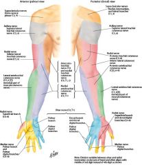
Dermatome - Upper Extrimity |
|
|
|
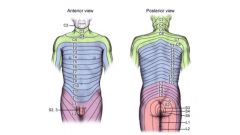
Dermatome - Trunk |
|
|
|
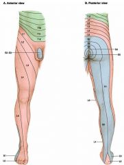
Dermatome - Lower Extremities |
|
|
|
Ulnar Nerve Spinal cord segment Muscle Innervation Sensory Distribution Clinical Motor Features of Paralysis |
Spinal: C8 and T1 Muscles: Flexor Carpi Ulnaris, Flexor Digitorum Profundus (medial half), Interossei, 4th and 5th Lumbricales Sensory: 4th finger (medial portion) & 5th finger Motor: loss of ulnar deviation, weakened wrist and finger flexion, loss of thumb adduction, and loss of most intrinsics ("claw hand") |
|
|
Sciatic Nerve Spinal cord segment Muscle Innervation Sensory Distribution Clinical Motor Features of Paralysis |
Spinal: L4, L5, S1, S2, S3
Muscles: Hamstrings Sensory: None Motor: Weakened hip extension, loss of knee flexion |
|
|
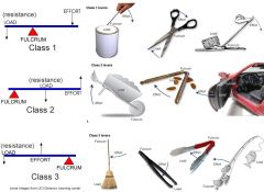
First Class Levers Second Class Levers Third Class Levers |
|
|
|
Mechanics Definition |
The branch of physics dealing with the study of forces and the motion produced by their actions.
|
|
|
Frontal Lobe |
Cognition Problem solving and reasoning Motor skill development Parts of speech Impulse control Spontaneity Regulating emotions Regulating sexual urges Planning |
|
|
Reversal of Muscle Action |
Origin moves toward insertion
|
|
|
Muscle tear (tensile) stretching |
When the muscle is stretched beyond it's normal range
|
|
|
Capsular Pattern |
Inflammation of the joint capsule
When inflammation of a joint is present (known as synovitis or capsulitis), not only does passive stretching of the capsule cause pain but a limitation of range of motion of the involved joint is always found to be in a specific pattern (different for each joint) |
|
|
Which movements are involved in circumduction? |
Flexion, extension, abduction, and adduction |
|
|
What type of joint is the thumb? Movements? |
Saddle Joint Flexion, extension, abduction, adduction (rotation) |
|
|
Passive Insufficiency |
Occurs when a muscle cannot be elongated any farther without damage to its fibers - occurs to the antagonist muscle (the one that's relaxed, not the one actively working)
|
|
|
What causes the patellar reflex? |
"Knee Jerk" Muscle Spindles |
|
|
What does myelin do? |
It increases the speed of conduction |
|
|
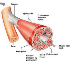
Anatomy of skeletal muscle
|
|
|
|
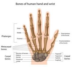
Metacarpals are located where? |
In the palm
|
|
|
Plane and Axis during rotation of head |
Transverse Plane
Vertical Axis (Also supination/pronation, medial/lateral rotation, horizontal abduction/adduction) |
|
|
Definition of Rotary Movement |
(Angular motion) Movement of an object around a fixed point.
|
|
|
Brachial plexus comprised of what 5 nerves |
C5-T1
Axillary Radial Median Ulnar |
|
|
MMT grades |
5 - Normal - Holds against max
4 - Good - Breaks with max 3 - Fair - Breaks with mod 2 - Poor - Gravity Eliminated 1 - Trace - No motion, muscles contract 0 - None |
|
|
Law of acceleration |
The amount of acceleration depends on the strength of the force applied to an object
|
|
|
Periosteum Definition |
The thin fibrous membrane that covers all of the bone except the articular surfaces that are covered with hyaline cartilage
|
|
|
Endosteum Definition |
A membrane that lines the medullary canal - contains osteoclasts which are mainly responsible for bone resorption.
|
|
|
End Feels |
Subjective assessment of the quality of the fell when slight pressure is applied at the end of a joint's PROM.
Bony, Soft Tissue Stretch, Soft Tissue Aproximation, Abnormal Bony, Boggy, Empty, Muscle Spasm, Springy Block, etc. |
|
|
Accessory Movement Definition |
"Joint Play" - not voluntary
|
|
|
What are the antagonists to hip abductors? |
Hip adductors
|
|
|
What position can you test the Brachialis? |
Elbow bent 90 degrees, forearm pronated (Under Biceps Brachii - this muscle is tested with forearm supinated) |
|
|
Deltoid: Origin, Insertion, Action, Innervation |
O: Lateral third of clavicle, Acromion Process, Spine of Scapula I: Deltoid Tuberosity A: Abduction, flexion, medial rotation, horizontal adduction, extension, lateral rotation N: Axillary (C5, C6) |
|
|
Subscapularis: Origin, Insertion, Action, Innervation |
O: Subscapular fossa of the scapula I: Lesser tubercle of the humerus A: Medially rotates N: Upper and Lower subscapular nerve (C5, C6) |
|
|
Pronator Teres: Origin, Insertion, Action, Innervation |
O: Medial Epicondyle of humerus and coronoid process of Ulna
I: Lateral aspect of radius at its midpoint A: Forearm pronation, assists in elbow flexion N: Median Nerve (C6, C7) |
|
|
Radial Nerve Innervates what extensors muscles? |
Wrist, finger, and thumb extensors
|
|
|
Medial Nerve innervates what flexor muscles? |
Wrist and finger flexors on radial side - also most thumb muscles
|
|
|
Where do you place the axis for elbow flexion? |
Lateral Epicondyle
|
|
|
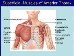
Serratus Anterior: Origin, Insertion, Action, Innervation |
O: Lateral surface of the upper 8 ribs
I: Vertebral border of the scapula, anterior surface A: Scapular protraction and upward rotation N: Long thoracic Nerve (C5, C6, C7) |
|
|
What muscle flexes the mcp/pip |
Flexor Digitorum Superficialis
|
|
|
Muscles that perform ulnar deviation |
Flexor and Extensor Carpi Ulnaris
|
|
|
Bones of the hand |
|
|
|
Joints of the hand |

|
|
|
|
|
|
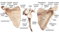
Scapula - Landmarks |
|
|
|
Anterior Tilt |
Occurs when the pelvis tilts forward, moving the ASIS anterior to the pubic symphysis
|
|
|
PCL prevents what movements? |
Anterior displacement of the femur on the tibia
Posterior displacement of the tibia on the femur (Same thing, just changing the perspective of which bone) |
|
|
soleus action |
plantar flexion
|
|
|
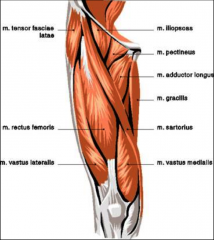
vastus lateralis originates |
linea aspera
|
|
|
action of vastus lateralis |
knee extension
|
|
|
gracialis action |
adduction
|
|
|
insertion iliosoas |
lesser trochanter
|
|
|
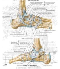
Bones and Joints of the foot |

|
|
|
Tibialis Posterior |

|
|
|
Ligaments of the knee |
|
|
|
Ligaments of the ankle |
|
|
|

|
|
|
|
|
|
|
|
|
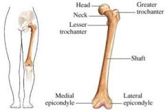
Femur - landmarks |
|
|
|

|
|
|
Plane and axis for abduction and adduction? |
Frontal Plane Sagittal Axis (Also radial/ulnar deviation and eversion/inversion) |
|
|
Plane and axis for flexion and extension |
Sagittal Plane Frontal Axis |
|
|
Posterior or Anterior pelvic tilt with tight hamstrings? |
Posterior |
|
|
Most common ligament injured in ankle sprain |
Lateral ligament |
|
|
Trimalleolar Fractures involve what bones? |
Both malleoli and the posterior lip of the tibia |
|
|
Active Insufficiency |
When a muscle cannot shorten any further (occurs to the agonist) |
|
|
Isometric contraction |
when a muscle contracts without shortening |
|
|
Isotonic contraction |
muscle contracts and changes length |
|
|
Concentric Eccentric |
Against gravity With gravity |
|
|
Cervical Plexus Nerves |
C1-C4 |
|
|
Lumbar plexus |
L1-L4 |
|
|
Sacral Plexus |
L5-S3 |
|
|
Medial Epicondylitis is also known as |
Golfer's Elbow |
|
|
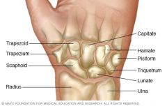
Carpals |
|
|
|
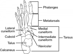
Tarsals |
|