![]()
![]()
![]()
Use LEFT and RIGHT arrow keys to navigate between flashcards;
Use UP and DOWN arrow keys to flip the card;
H to show hint;
A reads text to speech;
94 Cards in this Set
- Front
- Back
|
For the MusculoskeletalSystem what do we Examine and Observe? |
•Examineposture Look for Muscle atrophy–(decrease in size of muscle) Caused by: Lack of nervesupply•Muscle disease•Disuse •Observe Alllimbs/joints make sure the same size •Stands withweight fully on all limbs |
|
|
How would you Observe the MusculoskeletalSystem/ Examination. |
•Watch animalwalk •Hold up ordrag any leg •Bear weightthe same on all legs •Any headbobbing |
|
|
What are the grades of lameness and how many grades are there? |
Grades of lameness: •Grade I – barely perceptible •Grade II –noticeable but weight bearing •Grade III – bearsweight occasionally for balance •Grade IV –completely non-weight bearing |
|
|
What is a Grade 1 Lameness? |
Barely Perceptible |
|
|
What is a Grade 2 Lameness? |
Noticeble but weight bearing |
|
|
What is a Grade 3 Lameness? |
Bears weight occasionally for balance |
|
|
What is a Grade 4 lameness? |
Completely non-weight bearing |
|
|
How do you examine the musculature system through palpation? |
•Developroutine •Palpate andexamine each leg from distal to proximal •Flex andextend each joint individually •Examine alllegs •Examine skinbetween toes and pads •Palpate theneck |
|
|
What are we looking for in Lameness? |
•Acute vsChronic •Trauma? •Signalment •Unilateral vsbilateral •Shifting leglameness •Single limb (Often due tominor strain or sprain, resolves with rest in 1-3 days) |
|
|
How is Lameness Diagnosed? |
•Localizeeffected area by PE •Radiographarea •Further tests: •Joint tap •CT scan •MRI •Serologicaltesting •Exploratorysurgery |
|
|
What is Panosteitis? |
•Self-limitingbenign bone disease |
|
|
Who is prone to getting Panosteitis? |
•Young largeand giant breed dogs, usually in first year of life (6-18 mo, rarely up to5 yrs) •Esp. common inGerman shepherds |
|
|
How is Panosteitis diagnosed and treated? |
Diagnosis: •Exam – palp.mid-shaft of long bones elicits pain•Radiographs (↑intramedullary densities and irregular endosteal surfaces of long bones)
Treatment: –NSAIDS and time |
|
|
What is Osteochondrosis? |
•Degenerationor aseptic necrosis of bone and cartilage followed by reossification •Defect ofendochondral ossification (defect in articular cartilage development) •Hyalinecartilage does not mature into bone, thickens, can not receive nutrients, dies,detaches. |
|
|
What is Osteochondrosis Dissecans? |
•OCD = termused for disease when a flap forms as a result of progression of articularcartilage defect. |
|
|
Where is Osteochondrosis typically seen? |
•Lameness •Seen instifle, hock, elbow; shoulder is most common |
|
|
What breeds and age typically gets Osteochondrosis? |
•Large breeds –Retrievers, Bernese Mt. Dogs, Rottweilers •Young dogs(5-8 months) |
|
|
How do you diagnose and treat Osteochondrosis. |
Diagnosis: •Exam –lameness, pain on flexion of joint•Radiographs –lesion visible•Joint mice –loose cartilage pieces Treatment: •Rest andweight control in early stages•Surgery(remove flap, curettage of lesion) |
|
|
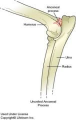
What is Ununited Anconeal Process? |
•Failure of anconeal process tofuse with the olecranon •May bebilateral |
|
|
What kind of animals/age, get Ununited Anconeal? |
•Large breeddogs – German Shepherds•Young dogs(7-8 months) |
|
|
How is Ununited Anconeal Diagnosed and Treated? |
Diagnosis: •Exam –lameness, crepitus on flexion of elbow joint, joint swelling •Radiographs Treatment: Surgicalremoval of anconeal process |
|
|
What is Fragmented Medial Coronoid Process? |
•Coronoidprocess is the cranial medial articular process of the ulna distal to thetrochlear notch•May bebilateral |
|
|
What type of animals get Fragmented Medial Coronoid Process. |
•Large breeddogs – German Shepherds •Young dogs(4-12 months) |
|
|
How is Fragmented Medial Coronoid Process diagnosed and treated? |
Diagnosis: •Exam –Lameness, pain on flexion of elbow and palpation of medial aspect of joint,joint swelling•Radiographs. Treatment: – surgicalremoval of coronoid process. |
|
|
What is Aseptic (Avascular) Femoral Head Necrosis. |
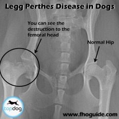
Also called: Legg-Calve-Perthes Disease •Loss of bloodsupply and subsequent degeneration of femoral head. |
|
|
What type of animals get Aseptic Femoral Head Necrosis? |
•Young (4 – 11months) miniature and toy breed dogs •Unilateral orbilateral |
|
|
What are the clinical signs of Aseptic Femoral Head Necrosis? |
Clinicalsigns: irritability and chewing of hip/flank, pain, atrophy of hip muscles,gradual lameness. |
|
|
How do you diagnose Aseptic Femoral Head Necrosis? |
Diagnosis: Radiographs Treatment: Surgery (FHO), neuter due to probable heritable nature of disease. |
|
|
What is Hypertrophic Osteodystrophy (HOD) and who gets it? |
•Developmentaldisorder of metaphysis of long bones of large and giant breed puppies, •Usually at 3 –4 months age (2 – 8 months). |
|
|
Is there a cause of Hypertrophic Osteodystrophy? |
•Cause unknown(diets high in calories, protein, and calcium ?; vit C def ?; CDV ?) |
|
|
Any signs or symptoms or sign of Hypertrophic Osteodystrophy? |
•Bilateral,symmetrical metaphyseal swelling, pain, and lameness •Possiblefever, anorexia, depression •Can be verydebilitating |
|
|
How is Hypertrophic Osteodystrophy diagnosed and treated? |
Diagnosis: –Radiography (metaphyseal lucency and periosteal bone formation) Treatment: -pain relief (NSAIDS), supportive care. |
|
|
What is Hip Dysplasia and who gets it? |
•Abnormaldevelopment or growth of the hip joint – usually bilateral •Congenital,bilateral DJD or hip laxity •Large breeddogs, younger (5-8 months) and older dogs |
|
|
What causes Hip Dysplasia? |
Causes: •Geneticpredisposition •Environmentand dietary factors •Disparitybetween muscle mass and the developing skeletal system |
|
|
What are some Hip Dysplasia Clinical Signs? |
•Difficulty inrising •Stiffness thatdecrease as the animal warms up on exercise •Pain onpalpation of the dorsal pelvic area or over hip joint •Older dogs –waddling gait, +/- of hip muscles•LamenessRestrictedrange of motion of joint |
|
|
How is Hip Dysplasia Diagnosed? |
•Radiographs•OrthopedicFoundation of Animals (OFA) and/or PennHIP positioning to quantitate hip joint laxity•Must be > 24 mo for OFAcertification; will perform preliminary evaluation at > 4 mo & < 24 months age•PennHIP - reliable at16 weeks (4 months) of age – reliability at 6 months of age is somewhatimproved over 4 months of age; reliability at 1 year of age is somewhatimproved over 6 months of age•Severity ofclinical signs may not correspond to degree of radiographic change |
|
|
How is hip dysplaisa treated conservatively? |
•Moderateexercise •Weight control •Antiinflammatory medication •NSAIDs,Tramadol, Prednisone•Chondroprotective agentsAdequan, Cosequin |
|
|
How is hip dysplaisa treated with surgery? |
•severe pain •TPO – triplepelvic osteotomy •Total hipreplacement •FHO – femoralhead and neck osteotomy |
|
|
What is Patellar Luxation and types? Who gets it? |
MedialLuxation: •Toy andminiature breeds Lateralluxation: – less common •Large andmedium breeds •Consideredinherited – usually result of anatomic deformities |
|
|
What are clinical signs of Patellar Luxation? |
•Neonates andyoung puppies – abnormal hind limb fx •Animal skipsor intermittently holds leg |
|
|
How is Patellar Luxation diagnosed and treated? |
Diagnosis: •Palpation toluxate patella •Radiographs Treatment: –None, rest, surgery |
|
|
What is Osteoarthritis? |
•Degenerativejoint disease – DJD •Slowlyprogressive cartilage degeneration |
|
|
What are signs and how diagnose Osteoarthritis? |
Signs: •Pain, lameness•Stiffness –esp. on rising•Crepitus injoint Diagnosis: •Radiographs |
|
|
How is Osteoarthritis treated? |
•Prevention •Rest duringflare-ups only •Diet •Hill’s j/d Canine –omega-3 FA’s, glucosamines, chondroitinsulfate•Medication•Chrondroprotective agents (polysulfated glycosaminoglycans, glucosamines, chondroitinsulfate, hyaluronic acid)•NSAIDSTramadolC |
|
|
What are some Treatments for OA/DJD? |
NSAIDS: •Carprofen - Rimadyl •Meloxicam -Metacam•Deracoxib - Deramaxx •Firocoxib - Previcox •Tepoxalin - Zubrin. Nutraceuticals(cartilage building blocks): •Glucosamineand chondroitin sulfate - Cosequin (PO)•Chondroprotective agents•HyaluronicAcid - Hyalovet (IA)•PolysulfatedGlycosaminoglycan - Adequan (IM) |
|
|
What is Osteosarcoma? Who gets it? Where is it found? Is prognosis good? How treated? |
•Neoplasticcondition of bones •Often largeand giant breed dogs •Older patients •Metastasis tolungs common
Signs: Unilaterallimb lameness, firm painful swelling
Treatment: –amputation & chemotherapy -------Prognosis poor |
|
|
Fractures, how do they typically happen? Clinical signs ? Diagnosis? |
•75% due tomotor vehicle accidents•Violence, bonedisease, repeated stress •Quickassessment of patient •Supportbandages until patient stable.
Clinical signs: •Hx of trauma•Pain,localized tenderness•Lameness,deformity of the bone •Loss of fx •Creptius, localizedswelling or bruising Diagnosis: •Radiographs –at least 2 views |
|
|
Where do Pediatric Fractures occur? |
•Most commonlyoccur through growth plate (metaphyseal growth plate = physis) |
|
|
What is a pediatric fracture classified as? What is the best way to repair? |
•Classified as Type I –Type V based on involvement of adjacent epiphysis/articular surface and/oradjacent metaphysis. •Repair bestvia external fixation |
|
|
What does Physeal damage mean for bone growth? |
•Physeal damage fromtrauma or repair before closure of growth plate can result in no furtherlongitudinal growth of bone. |
|
|
How are fractures repaired? |
•Externalfixation •Casts •Kirschner Apparatus •Internal fixation •IM Pins •Cerclage Wires •Lag Screws •Plates |
|
|
What are Myopathies and what types are there? |
•Diseases thataffect muscle •Many types •Most common –inflammatory, immune-mediated, acquired |
|
|
What can cause Myopathies? |
•Electrolyteimbalances, endocrine, paraneoplastic, parasitic, viral, toxic, other |
|
|
What are clinical signs of myopathies? |
muscleweakness and/or pain – generalized or localized, swelling. |
|
|
How do you diagnose myopathies? |
clinicalsigns, muscle biopsy, serum chemistries |
|
|
What is Myasthenia gravis? |
It is a myopathy •Autoantibodiesto nicotinic AChR’s on the postsynaptic membrane of the neuromuscular junction •Can cause megaesophagus |
|
|
How is myasthenia gravis diagnosed? |
-Serum titerfor antibodies to muscle nicotinic |
|
|
What is a treatment for myasthenia gravis? |
Anticholinesterasedrugs |
|
|
What is Masticatory Muscle Myositis? |
•Focalinflammatory myopathy that selectively affects the muscles of mastication•Immune-mediated. |
|
|
What causes Masticatory Muscle Myositis? |
Immune-mediated. |
|
|
What is acute and chronic Masticatory Muscle Myositis. |
Acute: musclesswollen, painful Chronic:muscles atrophy, fibrose can not open mouth fully. |
|
|
How is Masticatory Muscle Myositis diagnosed and treated? |
Diagnose: serumantibodies against masticatory muscle type 2M fibers; muscle biopsy Treatment:glucocorticoids |
|
|
What is Polymyositis and who gets it? |
•Generalizedinflammatory myopathy; immune-mediated •Middle-agedlarge breed dogs. |
|
|
What are clinical signs of Polymyositis? |
Clinicalsigns: weakness worsens with exercise, hyperesthesia on palpation, fever,depression |
|
|
How is Polymyositis diagnosed and treated? |
Diagnose: serum creatine kinase (CK)levels often elevated; muscle biopsy is the test of choice Treatment:glucocorticoids |
|
|
What are some other causes of myopathies? |
•Hypokalemia(cats)•Serum K+and/or Ca++ imbalances •Hypothyroidism •HyperadrenocorticismDiabetesmellitus (cats) |
|
|
Why is bandaging done for musculoskeletal injuries? |
•Protect wounds •Hold clean orsterile dressings in place •Secure splints •Providesupport for the bony anatomy •Support andstabilize soft tissue •Restrictmotion to eliminate stress •Prevent weightbearing •Providecompression to control hemorrhage and tissue edema •Discouragelicking and grooming |
|
|
What are some Principles ofBandage Application? |
•Prepare areasbefore bandage application•Protectivewound pads should be secured to the skin •Do not bandagetoo tightly •Instructowners on basic care of bandage |
|
|
what are Non-adherentdressings - Adaptic, Telfa any why used? |
•Permit flow ofexudate through dressing •Easily removedfrom the wound •Less painfulto patient •Minimizesdisruption of healing tissue •Kling, Conformor Stretch gauze •Elastic gauze bandage •Self adhering •Conforms to bodycontours without slipping |
|
|
What is a Soft PaddedBandage and used for? |
•Most commonlimb bandage •Coverabrasions or lacerations •Light supportbandage - after injury or surgery•Can be adaptedinto a coaptation splint withthe addition of a metasplint |
|
|
What is the Robert JonesBandage and used for? |
•Immobilize andsupport injured soft and bony tissue •Usedpreoperatively to stabilize a fracture of the distal limbs until internalfixation is completed •Not forfractures of the proximal limbsa |
|
|
What are the Feline Blood Groups? |
•A, B, and AB |
|
|
Why can cats haveacute hemolytic reactions with first the incompatible transfusion? |
•Cats havenaturally occurring preformed antibody to the antigen they lack. |
|
|
What cat breeds are typically type B blood? |
•Persian, DevonRex, British Shorthair, Himalayan, Abyssinian, Birman, Scottish Fold, Somali -have high prevalence Type B, up to 50% |
|
|
What cat breeds are typically type A? |
•Orientalbreeds (Siamese and Burmese tend to be type A (almost always) |
|
|
Which cat is a universal recipient? |
Type AB |
|
|
What are indications for blood transfusion? |
•Decreased RBCnumber to the extent that O2 carryingcapacity is insufficient to maintain adequate perfusion •Decreasedplatelets resulting in hemorrhage•Decreasedclotting factors or von Willebrand’s Factorresulting in hemorrhage |
|
|
What are some RBC Transfusion Options? |
•Fresh wholeblood •Stored wholeblood •Packed RedBlood Cells (PRBC’s) |
|
|
What is Packed RedBlood Cells (PRBC’s) best used for? |
Treatment ofchoice for anemia |
|
|
What are some Anti-coagulants used? (4 types) |
•Heparin: Mustuse in 24-48 hours •Acid CitrateDextrose (ACD): Can store for 14 days •CitratePhosphate Dextrose (CPD): Can store for 21 days •CPD-Adenine(CPDA): Can store for 35 days•Administerthrough micropore filter toeliminate micro thrombi |
|
|
What is •Oxyglobin® (hemoglobin glutamer–200) used for? |
•Given instead ofblood for acute anemia |
|
|
How long is Oxyglobin stable for? how soon after opening does it last for? |
•Stable for 2years if unopened •Use within 24 hours after opening |
|
|
what does plasma contain? |
•Contains allclotting factors, albumin, immunoglobulins |
|
|
Which type of blood is ok for rodentcide poisoning? |
Frozen Plasma •Contains factors 2, 7,9, and 10 so OK for rodenticide toxicity |
|
|
What is Cryoprecipitate? |
•Concentratedsolution containing factor 8, factor 13, fibrinogen, and von Willebrand’s factorderived from FFP |
|
|
What is Cryoprecipitate a treatment for? |
•Treatment of choice forHemophilia A and von W Dz. |
|
|
What is the volume amount for RBC transfusion in dogs and cats? |
Blood Volume = 90 ml/kg for dogs and 70 ml/kgfor cats |
|
|
What temperature should blood be at before transfusion? |
37° C toprevent hypothermia |
|
|
What is the rate of administration for blood transfusion? and when should it be done by? |
•Depends on patientcondition and specific component being transfused •Typicallystart slowly and increase rate if no reaction •Faster ifhypovolemic due to acute blood loss•Slower ifheart disease present •Should be completedwithin 4 hours to prevent bacterial contamination |
|
|
What is the maximum rate of infusion of potassium chloride (KCL)? |
0.5 mEq/kg/hr |
|
|
What is the maintenance dose (ml) for cats? |
60ml/kg/d X BW |
|
|
What is the maintenance dose for cats if using the body mass method? |
80 X (BW in Kg to the 3/4 power) |
|
|
How do you assess percent dehydration and equation? |
Total ml for rehydration = %dehydration X BW in kg X 1000 |
|
|
Once you have your percent dehydration how much do you give in the 1st 24hrs and the 2nd 24 hours? |
80% first 24hrs 20% second 24hrs |