Reading...
![]()
Play button
![]()
Play button
![]()
Use LEFT and RIGHT arrow keys to navigate between flashcards;
Use UP and DOWN arrow keys to flip the card;
H to show hint;
A reads text to speech;
33 Cards in this Set
- Front
- Back
|
What are the two heart cell types?
|
1)Autorhythmic: pacemaker cells, maintain rate ~1%
2)Contractile (myocardium): actually do mechanism of contraction ~99% |
|
|
System Circulation
|
Oxygenated blood moves from: left ventricle→aorta→arteries→arterioles→ capillaries→ venules→veins→inferior/superior vena cava which delivers blood directly into right atrium
|
|
|
Capillary functions
|
-delivery of oxygen and nutrients & removal of wastes
-thermoregulators dilate: warm, and to receive oxygen (more blood flow) constrict: cold, reducing loss of heat to environment |
|
|
Name 4 valves and their location
|
tricuspid: between right atrium and right ventricle
pulmonary: btwn right ventricle and pulmonary artery mitral (bicuspid): btwn left ventricle and left atrium aortic: between left ventricle and aorta |
|
|
Heart valve anatomy and 2 functions
|
Anatomy: Health valves are composed of leaflets or cusps that separate chambers of the heart
2 Functions: 1) unidirectional with blood flowing forward, preventing backflow 2) Facilitate generation of the pressure necessary to propel blood through circulation |
|
|
Which is the only non-tricuspid valve in the heart?
|
Mitral or "bicuspid valve" between the left ventricle and left atrium
|
|
|
What happens to a heart valve if pressure of blood flow in forward direction and backward direction is equal?
|
The valve is in its NATIVE, CLOSED position
|
|
|
Pulmonary Circulation
|
Deoxygenated blood moves from: right ventricle→ pulmonary trunk→divides into 2 right/left pulmonary arteries→gas exchange in right/left lung alveoli capillary beds→venules→4 left/right pulmonary veins → left atrium
|
|
|
Pulmonary and systemic circulation comprise one continuous circulation. (T/F)
|
True. Pulmonary and systemic circulation constitute two components of a continuous loop.
|
|
|
Each lung has one pulmonary artery and one pulmonary vein. (T/F)
|
False. There are 2 pulmonary veins per lung.
|
|
|
Name steps of heart contraction in diastole and systole
|
Diastole:
1a) Chambers relaxed, low pressure, allowing blood from system circulation to rush in right/left atrium (most blood flows directly into ventricles) 1b) Atrial contraction, semilunar valves are closed, forcing blood into ventricles Systole: 2) Ventricular contraction forcing blood into system and pulmonary circulation; atrioventricular valves closed |
|
|
Heart Pump Contraction: Diastole and Systole
|
Diastole- atria: are relaxed, then contracted
ventricles: relaxed throughout diastole Systole: contraction of two ventricles |
|
|
Sinoatrial (SA) node
|
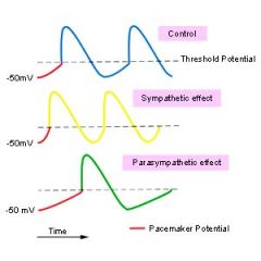
-Heart's natural pacemaker located in the upper right atrium (near opening of sup. vena cava)
-initiates intrinsic, regular, rhythmic emission of contractile signals at 120 impulses/min -receives nerve supply from both sympathetic and parasympathetic (vagus nerve) |
|
|
If the heart's SA node initiates 120 impulses/min, why do we have a slower heart rate?
|
Because the vagus nerve from the parasympathetic nervous system innervates the SA node to produce a resting heart rate of 50-75 impulses/min.
|
|
|
Pathway of impulse signal
|
SA node (upper right atrium) → AV node (interatrial septa, up near intersection of 4 heart chambers) → bundle of His (myocardial fibers along septum btwn ventricles)→ Purkinje fibers spread through bottom of ventricles (provide force for ventricular contraction, "systole")
|
|
|
Which have thinner walls, atria or ventricles?
|
Atria
|
|
|
Which have thick, muscular walls, atria or ventricles?
|
Ventricles
|
|
|
What is different about the pulmonary arteries compared to others?
|
transport DEOXYGENATED blood away from the heart, to the lungs
|
|
|
How do large veins in legs prevent back flow caused by gravity?
|
They have valves
|
|
|
Since there is low blood pressure in veins, what are mechanisms that allow return of blood to heart?
|
-Muscle contraction.
-Decreased venous compliance. Sympathetic activation of veins decreases venous compliance, increases central venous pressure and promotes venous return indirectly by augmenting cardiac output through the Frank-Starling mechanism, which increases the total blood flow through the circulatory system. -Respiratory activity. During respiratory inspiration, the venous return increases because of a decrease in right atrial pressure. -Gravity. |
|
|
blood pressure is lowest in the ____ and highest in the ____ when the ventricles contract.
|
blood pressure is lowest in the VEINS, and highest in the ARTERIES when the ventricles contract.
|
|
|
what is normal blood pressure?
what is 120 in normal blood pressure? what is 80 in normal blood pressure? |
-120/80
-systolic number = pressure when the ventricles contract -diastolic number = pressure when heart relaxes (and atrial contraction) |
|
|
What does the vagus nerve do?
|
-Innervates the heart and digestive system.
-Slows the rate of heart contractions and increases digestive activity in the intestines |
|
|
How do the impulses travel from the SA node?
|
Through electrical synapses made from gap junctions
|
|
|
Structure of arteries
|
-Elastic and stretch when filled with blood
-Contain most smooth muscle. |
|
|
Medium size arteries contain more smooth muscle than larger arteries.
|
True. More efficient in rerouting blood.
|
|
|
Name 4 methods for materials to cross capillary vessels:
|
1) pinocytosis
2) diffusion or transport through capillary cell membranes 3) movement through pores in the cells called fenestrations 4) movement through the space between the cells |
|
|
List blood vessels in descending order based on: cross-sectional area, velocity, and blood pressure.
|
Cross-sectional area: Capillaries>veins>arteries
Velocity: arteries>veins>capillaries Blood Pressure: Systemic arteries (some capillaries)> pulmonary arteries> systemic capillaries> pulmonary capillaries and veins> systemic veins |
|
|
Blood pressure
|
(arterial blood pressure), pressure exerted by circulating blood upon the walls of blood vessels
-increases near the heart and decreases to its lowest in the capillaries |
|
|
Velocity and cross-sectional area follow which equation
|
continuity equation, Q=Av (blood flow)
|
|
|
How do pressures create fluid exchange in capillaries?
|
As blood flows into capillary (arterial end):
-hydrostatic pressure>osmotic pressure →net fluid flow is out of capillary & into interstitium As blood flows out of capillary (venule end): -osmotic pressure>hydrostatic pressure →net fluid flow is out of interstitium & into capillary |
|
|
What is the net result of fluid exchange by the capillaries from aterial to venule end?
|
10% loss of fluid to interstitium
|
|
|
Contrasts between arteries and veins.
|
VEINS ARTERIES
Blood Volume (64%) Blood Volume (15%) Valves No Valves Larger diameter Smaller diameter Thinner walls Thicker walls |