![]()
![]()
![]()
Use LEFT and RIGHT arrow keys to navigate between flashcards;
Use UP and DOWN arrow keys to flip the card;
H to show hint;
A reads text to speech;
65 Cards in this Set
- Front
- Back
|
Cicrulatory is what in origin... |
Mesoderm |
|
|
Made up of... |
2 branches systems + pump |
|
|
Circulatory system is a .... system? |
Closed & Blood vascular system & Lymphatic System |
|
|
Blood vascular system is made up of... |
Areteries, arteioles (conducts blood away from the heart) Capillaries Veins, Venules (Conducts blood towards the heart) |
|
|
Lymphatic system consists of |
Lymph capillaries, vessels, sinuses, nodes and variety of lymphoid organs |
|
|
Heart |
Basic 2 stroke pump with valves to ensure unidirectional flow |
|
|
Arteries Walls of arteries, veins and large lymph vessels are |
similar |
|
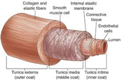
Artery Layers |
Tunica intima, Tunica media and Tunica externa |
|
|
Layer of Artery: Tunica Intima |
Made up of Endothelium Supported by Internal elastic membrane |
|
|
Layer of Artery: Tunica Media |
Made up of Thick circular smooth muscle and supported by External elastic membrane |
|
|
Layer of Artery: Tunica Externa (Adventitia) |
Made up of Loose Connective Tissue Supported by Longitudinal collagenous and elastic fibers |
|
|
Arteries in general |
Thickness varies with vessel Generally arteries are thick and muscular (made up of simple squamous epithelium lining) Blood forced into arterial system by contraction of heart Lumen diameter regulates amount of blood to area of body ( pressure) |
|
|
Steps of... Blood forced into arterial system by contraction of heart |
Distends vessels (swells) Backflow into heart prevented valves Tension in vessels forces blood along in distention - rebound push due to elastic nature of walls. |
|
|
Lumen diameter regulates amount of blood to area of body ( < diameter, >pressure) ,>
|
Vasoconstriction and Vasodilation |
|
|
Veins |
Little muscle in walls Elastic fibers in wall Clear connective tissue layers Paired semilunar valves prevent backflow (gravity) Lumen acts as "reservoir" for body |
|
|
Capillaries -Fenestrated -Continuous -Dis-continuous |
Lining 1 cell layer -Endothelium -Basal laminar layer Sphincter at beginning and end to help regulate flow. Not all open at once |
|
|
Fenestrated capillary |
have pores in the endothelial cells and allow small molecules and limited amounts of protein to diffuse
|
|
|
Continuous capillary |
endothelial cells provide an uninterrupted lining only allow smaller molecules, such as water and ions to pass through their intercellular clefts
|
|
|
Dis-continuous |
have larger openings in the endothelium. These types of blood vessels allow red and white blood cells and various serum proteins to pass, aided by a discontinuous basal lamina.
|
|
|
Origin of circulatory system Vessels (in Chick for Eg) Part 1 |
Clusters of mesodermal cells form blood islands in splanchnic mesoderm of yolk sac Fluid filled spaces gradually appear in islands. -Seperate peripheral layers of cells appear to enclose central cluster -Space enlarges -> endothelium lining --Blood cells = plasma |
|
|
Origin of circulatory system
Vessels (in Chick for Eg) Part 2 |
Vessels expand and collapse Network expands to meet spreading yolk sac -Paired vitelline veins now meet Develop dorsally and forward to form aortic arches Dorsal aorta forms Vitelline arteries join plexus to complete arc |
|
|
Heart 3 layers |
Endocardium Myocardium Pericardium |
|
|
Heart formation part 1 |
Pulsating tube derived from fusion of paired primordial epimyocardium and endocardium alongside developing gut. After foregut forms, a portion of coelom pinches of around developing heart primordia. --amniocardiac vessel or pericardium. Endocardial tubes + external layer of epimyocardium meet, fuse -> internal cardiac jelly. |
|
|
Heart formation part 2 |
Endocardium joins vitellines, aortae and 1rst aortic arches Epimyocardium becomes myocardium or visceral peritoneum Cardiac jelly transmits initial waves of contraction from myocardium. -endocardium closes off lumen between 2 endocardial cavities. --Allows contraction to have something to work against. |
|
|
Heart formation part 3 |
Eventually 4 chambers of the heart are seen Sinus venosus Atrium Ventricle Conus (truncus) Arteriosus |
|
|
Union of tubes segmental |
Ventricle - truncus (conus) -Begins to contract due to autorhythmicity of cardiac muscle As Atrium formed, contracts faster -Ventricle speeds up Sinus joins in -Eventually becoming the pacemaker and speeds up other areas Enlargement of atria = bulbus arteriosus -conus is the actual joining of the truncus and ventricle -Truncus = embryonic 4th region of the heart. --is a functional part of the heart in some lower vertebrates |
|
|
Comparative anatomy Amphixous |
No discrete heart Single median contractile vessel ventral to the pharynx Blood pushed by peristaltic contraction About 1 min per circuit enters afferent aortic arches -Contractile enlargement at entry helps push to paired dorsal aorta (anterior = internal carotid) --Fused posteriorly |
|
|
Cyclostomes |
2 chambered heart Simplest heart Only deoxygenated blood passing through |
|
|
Cyclostomes pathway |
Common cardinal veins (anterior cardinals etc, sinuses, hepatic veins -> sinus venosus -> SA valves -> large thin walled atrium -> AV valve -> small thick walled ventricle -> conus (truncus) arteriosus ( 2 semilunar valves stop backflow) -> steady stream of blood to gills via ventral aorta)
|
|
|
Fishes |
Similar to cyclostomes Greater development of semilunar truncus vlaves Some Dipnoans show tendency to a 3 chambered heart with partial division of atrium Single circulatory system -Hepatic sinuses (veins) leading to heart my become postcavas Lung breathing heart more posterior |
|
|
Amphibians part 1 |
1st double circulatory system (11, not series) SV -> R atrium Both +- oxygenated blood in heart + oxygen from lungs (pulmonary circulation) -> L side single chambered ventricle Partially oxygenated blood from body -> SV + blood from buccal region and skin (+) oxygen -> RA -> R side of ventricle |
|
|
Amphibians continued |
Partially oxygenated blood from body -> SV + blood from buccal region and skin (+) oxygen -> RA -> R side of ventricle Minimal mixing Seperate in time with oxygenated blood through first Separation aided by ventricular trabeculae (ridges) Spiral valves in some species |
|
|
Reptiles part 1 |
3- chambered heart with 2 systemic arches -Similar to amphibians but have incomplete partition in ventricle -Also ventricular trabeculae to seperate oxygenated from deoxygenated blood - + oxygen directed to 2 systemic branches --Directed to cavum venosum - (-) oxygen directed to pulmonary truck |
|
|
Reptiles part 2 |
Crocodiles and Alligators -Partiton complete -> double circulation -4 chambered heart, although can still shunt blood from RA to RV to LA and LV and bypass lungs during a dive |
|
|
Birds |
Complete double circulation No sinus venosuus -3 vessels ( 2 pre and 1 post cava) -> RA Pulmonary veins bring oxygenated blood to LA 2 bicuspid valves at AV septum No left aorta -Only R branch (unlike crocodiles) and pulomary trunk leaving heart. Well developed coronary system -Supports high MR, flight, thermoregulation |
|
|
Mammals |
See cat 4 chambered Major difference from birds = loss of R aorta and retention of L systemic arch |
|
|
Arterial Systems Common to all vertebrates, at least in embryo |
Usually 6 pairs of aortic arches -Join ventral to dorsal aortae (R and L) --Posteriorly, 2 dorsal aortae fust to single aorta As aortic arches change during evolution and development, primary changes are in arterial system. -Progressive reduction in # of aortic arches left in adult |
|
|
Cyclostomes arterial system |
Along with many other fish >st number of arches Each arch has afferent + effernt portion with capillary bed in gill lamellae for respiration |
|
|
Most teleosts arterial system |
Only last 4 pair of arches remain |
|
|
Most other vertebrates arterial system |
Lose 1, 2 and 5 + radix (L-R connection) on 3 and 4 R and L #3 + remnants of ventral aorta + anterior radices -> carotid complex |
|
|
Amphibians arterial system |
#4 stays but L4 splits off ventral aorta to form new connection with right ventricle |
|
|
Birds arterial system |
L4 loses connection with dorsal aorta and degenerates R4 remains |
|
|
Mammals arterial system |
L4 stays R4 lost |
|
|
In reptiles birds and mammals arterial system |
Pulmonary arteries come from a separate pulmonary aorta via splitting of the truncus arteriosus when true right ventricle develops. Rest of truncus forms systemic aorta base out of left ventricle |
|
|
Portal systems |
Vein between 2 capillary networks ([pre-heart_ |
|
|
3 types of portal systems |
Hepatic portal (all vertebrates) -Liver sinusoids Renal portal (Adults of lower vertebrates and embryos of all others) Pituitary or Hypophyseal portal (To some degree in all veretbrates) |
|
|
Hepatic portal system |
From sub intestinal and vitelline veins As liver grows, it interrupts the path of these to the heart Carries all digestive products for assimilation |
|
|
Renal Portal system |
In fish and amphibians Reduced in birds and reptiles Absent in mammales Originates in caudal region -Terminates in capillaries in opisthonephric or mesoephric kidney on the way to the heart Primary function probably water conservation, especially in early marine forms |
|
|
Hypophyseal portal system |
Part of endocrine system Blood from hypothalamus of brain to adenohyposphysis -Local hormones |
|
|
Basic venous channels In all vertebrate embryos (May be modified in adults) |
Primary streams -Cardinals -Renal portal -Lateral abdominal -Hepatic portal -Coronary stream -Pulmonary and Postcaudal (lungfish and tetrapods) |
|
|
Anterior Cardinals (interior jugulars) |
Drain head to common cardinals (postcava) |
|
|
Postcardinals |
Not in anurans, reptiles or birds In mammals = azygous or hemiazygous |
|
|
Abdominal Vein |
Drain pectoral and pelvic fin in cartilaginous fishes Lose connection with forelimb in tetrapods and with hindlimb in birds and mammals In mammals, remain as umbilical cord |
|
|
Renal Portal |
Drains fish tail Connects to hind limbs in amphibians Crocodiles and birds -Partially bypasses kidney and goes postcava Lost in mammals above monotremes |
|
|
Postcava |
Increased prominence in higher vertebrates Began as alternate route to heart from kidneys (dipnoans and amphibians) Finally drains hindlimbs, most of trunk and tail |
|
|
Lymphatic System part 1 |
From fluid filled spaces in mesenchyme long after vein develops Highly branched and anastomosing system spread through body 1st lymphatics arose close to larger veins Smaller vessels may lie near companion veins. Lymph sinuses -Result from enlargement of some lymphatic networks in certain regions |
|
|
Lymphatic System part 2 |
Vessles have valves to stop back flow Lymphatic nodules and nodes formed when connective tissue elements condense about lymphatic plexes associated with mesenchymal cells |
|
|
Lymphatic system part 3 |
3 major functions Returns interstitial fluids leaked from capillaries due to pressure -> blood stream Source of lymphocytes Route for absorption of fats from digestive tract |
|
|
Flow of lymph is sluggish |
Moved by muscular activity of body Arterial pulsing next to vessels Pressure buildup in small vessels by osmosis ad absorption of tissue fluid. Action of pulsating lymph hearts (enlargements of lymphatic vessels which have contractile walls) -Valves in "hearts" direct flow - In bony fishes, amphibians, reptiles and bird embryos - Amphibians with much fluid have many lymph hearts |
|
|
Other parts of the lymph system |
Thymus (source of lyphocyes-. antibodies (possibly not in cyclostomes) Tonsils Adenoids Peyer's patches (mucosa of small intestine of aminiots -. lymphocytes) Bursa Fabricius (young birds -> lymphocytes) Lymph nodes in mammals (Groin, axilla, neck, intestinal mesentery -> lymphocytes and phagocytes (filter out bacteria as lymph moves through nodes) Spleen |
|
|
Spleen |
Largest organ From mesenchyme near stomach Colonized with lymphocytes from thymus Site for erthrocyte production in embryo, storage in adutlt Via phagocytosis, breaks down old rbc's |
|
|
Circulation in mammalian fetus (part 1) |
Blood from caudal end of the dorsal aorta -> umbilical arteries via umbilical cord -> placenta From placenta oxygenated blood returns to fetus via umbilical vein -Limb vein via falciform ligament to liver -Some blood to liver capillaries -> ductus venosus -> heart -Most continues -> postcava ->RA |
|
|
Circulation in mammalian fetus
|
From RA most through foramen ovale (intraatrial foramen) -> LA Rest of aertated blood + blood from heart to RA -> RV -> pulmonary trunk -> ductus arteriosus (degenerats at birth) -> DA DA-> umbilical arteries ->placenta Some blood which does get to lungs returns to heart via pulmonary vein -> LA with some blood from RA via foramen ovale to LV -> ascending aorta |
|
|
At birth part 2 |
Ductus arteriosus closes
-Reflex when lungs filled since blood via pulmonary trunk to lungs. -Becomes arterial ligament Inter-arterial valve closes against foramen by increase in pressure in LA from blood returning to lungs . -Prevents mixing -Sealed permanently in few days |
|
|
At birth part 2 |
Umbilical arteries and veins severed From bladder to navel -Becomes lateral umbilical ligaments (ventral messentary of bladder) -Umbilical vein becomes round ligament of vein Vessels from liver -> postcava = ductus venosus-> ligamentum venosum |