Reading...
![]()
Play button
![]()
Play button
![]()
Use LEFT and RIGHT arrow keys to navigate between flashcards;
Use UP and DOWN arrow keys to flip the card;
H to show hint;
A reads text to speech;
28 Cards in this Set
- Front
- Back
- 3rd side (hint)
|
3 ligaments at AC joint
|
Coracoacromial ligament
Coracoclavicular ligament Acromioclavicular ligament |
|
|
|
Coracoacromial ligament function:
|
Controls anterior & posterior translation of lateral clavicle
|
|
|
|
Coracoclavicular ligament function:
|
Controls vertical stability; restrains superior & anterior displacement
|
|
|
|
Acromioclavicular ligament function:
|
Provides stability across the joint;
Restrains posterior translation and displacement of the clavicle. |
|
|
|
2 ligaments at GH joint
|
Capsular ligaments
Coracohumeral ligament |
|
|
|
Capsular ligaments function:
|
Joins the GH joint capsule anteriorly, inferiorly & posteriorly
|
|
|
|
Coracohumeral ligament function:
|
Provides stability superiorly, preventing superior translation
|
|
|
|
Facts of the MM of the rotator cuff:
|
The four major muscles of the rotator cuff rotate the humerus and properly orient the humoral head in the glenoid fossa (socket). The tendons of these four muscles merge, forming a cuff around the glenohumeral joint.
|
|
|
|
Supraspinatus:
|
abducts the humeral head and acts as a humeral head depressor
|
|
|
|
Infraspinatus:
|
externally rotates and horizontally extends the humerus
|
|
|
|
Teres minor:
|
externally rotates and extends the humerus
|
|
|
|
Subscapularis:
|
internally rotates the humerus
|
|
|
|
Teres minor:
|
externally rotates and extends the humerus
|
|
|
|
Subscapularis:
|
internally rotates the humerus
|
|
|
|
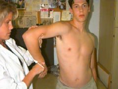
ER ROM test
|
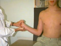
The patient is positioned sitting and the elbow is flexed 90 degrees. While the elbow is held against the patient's side, the examiner externally rotates the arm as permitted.
|
|
|
|
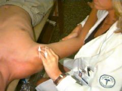
IR ROM test
|

The patient should be positioned sitting. Again with the elbows at the patient's side, the examiner should raise the thumb up the spine, and record the position in relation to the spine (reaching T7 is normal, unless bilateral symmetry is observed).
|
look up what spine level is associated with what anatomical level
|
|
|
IR ROM at 90
|
The patient is positioned sitting with the elbow and shoulder supported to prevent muscle contraction. The arm is at 90 degrees with the fingers pointing downward and palm facing posteriorly. The examiner attempts to rotate the forearm posteriorly as far as possible.
|
|
|
|
Forward Flexion ROM
|
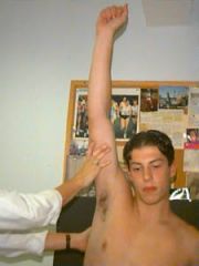
The arm is kept straightened and brought upward through the frontal plane, and moved as far as the patient can go above his head. Note: for recording purposes, 0 degrees is defined as straight down at the patient's side, and 180 degrees is straight up.
|
|
|
|
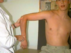
Shoulder ABd ROM test
|

|
The arm is again kept straightened, while raised and abducted. Observe the twisting of hand -- facing outward, not forward, as in forward flexion. The ROM is measured in degrees as decribed for forward flexion. As pictured, this test is being done actively by the patient, but may be performed by the examiner as well.
|
|
|
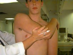
Cross Arm Horizontal Add
|
|
The patient places his hand on the opposite shoulder, while the examiner exerts force horizontally. Again, the presence of pain indicates possible pathology.
|
|
|
Apprehension Test
|
|
Have the patient in the supine position, with the arm abducted 90 degrees. Rotate the shoulder externally by pushing the forearm posteriorly. If patient feels instability, they typically will balk when the test is performed.
|
|
|
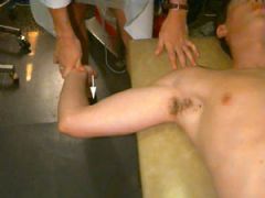
Laxity test
|
|
Have the patient in the supine position. Stabilize the scapula, and slide the humeral head anteriorly and posteriorly within the glenoid fossa to evaluate the stability of the joint. Note the axial load being applied to the elbow.
|
|
|
Hawkin's test
|
|
Position the patient standing with the shoulder abducted 90 degrees, and internally rotate the forearm. The presence of pain with movement is indicative of possible pathology. |
|
|
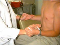
IR RC strength
|
|
Same as above, but the patient is attempting to rotate internally (and examiner resisting externally).
|
|
|
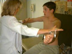
Supraspinatus strength
|
|
The patient is positioned sitting with arms straight out, elbows locked, thumbs down, and arm at 30 degrees (in scapular plane). The patient should attempt to abduct his arms against the examiner's resistance.
|
|
|
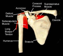
Anterior view shoulder- muscle & cony prominences
|
|
|
|
|
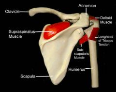
Shoulder posterior view-muscle & bony prominences
|
|
|
|
|
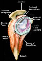
Sagittal view of shoulder with muscle attachments
|
|
|