![]()
![]()
![]()
Use LEFT and RIGHT arrow keys to navigate between flashcards;
Use UP and DOWN arrow keys to flip the card;
H to show hint;
A reads text to speech;
25 Cards in this Set
- Front
- Back
|
Skeletal System: |
Bones, cartilage and ligaments. |
|
|
Bone "Organ": |
Bone tissue, cartilage, fibrous, connective tissue, blood and nerve |
|
|
1. Functions |
A. Supports soft tissues. B. Protects organs. C. Provides shape for the body. D. Provides attachment for muscles/movement. E. Storage of materials. F. Hematopoiesis/Hemopoiesis (form blood cells) |
|
|
2. Classification |
Classified 5 ways according to shape. 1. Long bones - long axis, expanded ends. 2. Short bones - cube-like. 3. Flat bones - Plate-like, ribs, scapula. 4. Irregular bones - variety, connects to other bones. 5. Round (sesamoid) bones - small with tendons, compressions. |
|
|
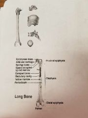
3. Four major parts of long bones |
1. Epiphysis- expanded portion on ends of long bones, covered with articular cartilage, composed of spongy bone. 2. Diaphysis - shaft of bone. 3. Periosteum- tough, vascular covering of fibrous tissue that covers bone, functions in bone formation and repair. 4. Medullary cavity- hollow cavity in diaphysis, filled with marrow, continuous with spongy bone in epiphysis.
Processes: bony projections provide sites for ligaments and tendons to attach. |
|
|
4. Type of bone tissues: |
A. Spongy/Cancellous •many large spaces with red marrow (produced rbc) •plates between spaces called trabeculae. •found in epiphysis (keep end of long bones light) B. Compact •tightly packed, solid and strong, resists bending. •diaphysis (shaft of long bones) •contains osteocytes. •lined with endosteum (thin squamous layer) •yellow marrow: fat storage.
Review: Osteocytes - bone cells. Lacunae - cavities. Central/Haverisan Canal Canaliculi - allow osteocytes transport nutrients and wastes to and from nearby cells. Collagen and inorganic salts. Collagen gives bone it's strength and resilience and inorganic salts make it hard and resistance to crushing. |
|
|
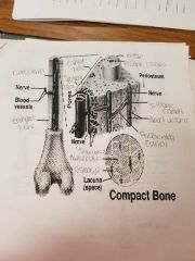
5. Microscopic Structure Compact Bone |
•Osteons/Haversian System. •Cylinder - shape unit. •Haversian canals •Caniliculi. •Volkman's canals. Transverse channels with large blood vessels, connect surface to medullary cavity. •Lamellae. •Osteocytes (Bone cells) |
|
|
6. Microscopic Structure spongy bone |
•Osteocytes. •Trabeculae - branching bony plates. •Matrix. •Canaliculi. •NO Haverisan Canals. |
|
|
7. Bone development and growth |
Ossification: process of bone formation.
Parts of the skeletal system begin to form during the first few weeks of prenatal development and bony structures continue to grow and develop into adulthood. Bones grow by replacing connective tissue.
2 Types of ossification: A. Intramembranous ossification. B. Endochondral ossification.
|
|
|
A. Intramembranous ossification |
Intramembranous ossification: process of forming intramembranous bones by replacement of connective tissue.
1. Location: broad, flat bone of skull. 2. Steps in development: A. Primitive connective tissue (unspecialized) appears at site of future bones. B. They become arranged around blood vessels & differentiate into osteoblasts which form spongy bone. C. Osteoblasts become osteocytes when surrounded by bony matrix. D. Connective tissue then forms periosteum over surface of developing bone. E. Osteoblasts on inside of periosteum form combat bone over spongy. |
|
|
B. Endochondral ossification |
Endochondral ossification: process of forming endochondral bones by the replacement of hyaline cartilage. 1. Location: most bones of the skeleton. 2. Stages of development: A. Hyaline cartilage forms a model for future bones. B. Chondrocytes enlarge and destroy cartilage; periosteum forms. C. Osteoblasts form periosteum invade tissue and form spongy bone in space left by cartilage. D. Combat bone is deposited around spongy bone by osteoblasts. |
|
|
8. Growth of Endochondral Bones |
A. Primary ossification center •Occurs in the diaphysis. •Bone develops in middle, grows outward. •Osteoblasts in periosteum deposit thin layer of compact bone around POC. (Primary ossification center) •Epiphysis remains cartilagenous & grows. B. Secondary ossification center •Occurs in the epiphysis •Spongy bone is formed. •Between diaphysis and epiphysis a cartilage bond is left: epiphyseal disk/plate. |
|
|
4 Types of epiphyseal disk |
Function of epiphyseal disk: band of cartilage, growth occurs here. 4 layers: Layer 1- closet to epiphysis, anchors epiphyseal plate to bone tissue; resting cells. Layer 2- undergoes mitosis, thickens disc. Layer 3- thickens disc, bone lengthens, salts deposited. Layer 4- dead cells, calcified.
•osteoclasts remove calcified matrix & osteoblasts deposit bone. •ends about 25. (Length but can grow in width) •epiphyseal plate - band of cartilage; growth occurs here Growth in thickness: •Underneath periosteum. •Osteoblasts form compact bone. •Actually a type of intramembranous ossification. Growth in thickness: •Underneath periosteum. •Osteoblasts form compact bone. •Actually a type of intramembranous ossification. •Note!!!- osteoclasts secrete acids to break down home and form the medullary cavity. •Note!!!- osteoclasts secrete acids to break down home and form the medullary cavity. |
|
|
9. Two Types of fractures |
Compound- exposed to outside by break in skim microbial. Simple- break with uninjured skin.
A. Causes Traumatic - due to injury. Pathologic (spontaneous) - resulting from disease.
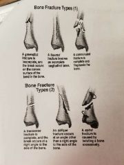
B. Six types of compound fractures. 1. Green stick - incomplete, one side breaks, one side bends; most common. 2. Fissured - incomplete longitundial break. 3. Comminuted - complete, several bony fragments. 4. Transverse - complete, right angles for axis. 5. Oblique - complete, angle other than right. 6. Spiral - complete, excessive twisting.
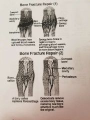
C. Fracture repair four steps 1. Blood escapes from ruptured vessels and forms hematoma (blood outside blood vessels; internal bleeding.) 2. Spongy bone forms near blood vessels, fibrocartilage in distant regions. 3. Fibrocartilage is replaced by bone tissue. 4. Osteoclasts remove excess bony tissue and new is like original. |
|
|
10. Four factors affecting bone development, growth & repair. |
A. Nutrition. B. Exposure to sunlight. C. Hormones. D. Physical exercise. |
|
|
A. Nutrition |
Vitamin D. •needed for proper absorption of calcium. •riskets (children) caused by too little vitamin D. •osteomalacia (adults) caused by too little vitamin D; suffering of the bones. What does vitamin D do for you? •help prevent a growing list of chronic diseases, including type 2 diabetes, heart disease, hypertension, osteoporosis, breast cancer, colon cancer and ovarian cancer. Foods with vitamin D Vitamin A: •needed for bone development resorption (osteoblasts and osteoclasts) •insufficient amount delays bone development Vitamin C: •important for collagen synthesis. •deficiency results in slender, fragile bones. |
|
|
B. Sunlight |
•Stimulates the subcutaneous layer of the skim to produce vitamin D (needed to absorb calcium) |
|
|
C. Hormones |
1. Pituitary Growth Hormone •stimulates epiphyseal plate. •Abnormalities: Gigantism: too much HGH through life (exceed 8') Dwarfism: absence of HGH. Acromegaly: adults; begin to secrete too much HGH; enlarged hands, feet and jaws. 2. Thyroid Hormone: Decreased levels: delays bone growth because it falls to stimulates release of growth hormone. Overactive: premature ossification of epiphyseal disk. 3. Sex hormones •Androgen •Estrogen •Most active during puberty. •Promotes bone formation and increases in bone growth. •Estrogen more powerful hormone in bone development. |
|
|
D. Physical Exercise |
If your bones are not called upon to work, they do not receive any messages that they need to be strong. A lack of exercise, may contribute to lower bone mass. Two Types of exercise are important for building and maintaining bone mass and density: 1. Weight-bearing. 2. Resistance exercises. 1. Weight-bearing exercises are those in which your bones and muscles work against gravity. Any exercise in which your feet and legs are bearing your weight. Jogging, walking, stair climbing, dancing and soccer are examples of weight bearing exercise. 2. Resistance exercises are activities that are muscular strength to improve muscle mass and strengthen bone. These activities include weight lifting, such as using free weights and weight machines found as gyms. |
|
|
11. Bone marrow |
Hematopoiesis: blood cell formation in the bone marrow. 2 types of bone marrow: 1. Red- located in cavities of infant bones (replaced by yellow as we age.) •Adults: spongy bone of skull, ribs, sterum, clavicle, vertebrae, and pelvis. 2. Yellow- •inactive in blood cell formation •can revert back to red bone marrow if blood supply •stores fat |
|
|
12. This and that |
A. Osteoporosis •Excessive demineralization of bone •associated with aging, menopause in women. C. Disorders of the vertebral column •Changes in intervertebral •Ruptured or herniated disk. •Curvature problems •hyphosis hunchback, exaggerated thoracic curvature) •scoliosis (abnormal lateral curvature) •lordosis (accentuated lumbar curve), swayback. •spina bifida (open spine, congenital) D. Sexual differences •skulls female is lighter and smaller. •less obvious muscular attachment sites. •jaw line is smaller. Pelvis •cavity is wider in females (assist in childbirth) •sacrum is wider. •Coccyx is more moveable. E. Life span changes 1. Decrease in height beginning at 30. 2. Skeleton loses strength. 3. Bones become brittle. 4. Boneless exceeds bone replacement. Trabecular/spongy - shows bone loss first around 30. (Occurs in women before men) Compact - bone loss beings around 40. F. Five actions steps to preserve skeletal health 1. Exercise 2. Get enough vitamin D. 3. Avoid falls. 4. Take calcium suoplements 5. Avoid carbonated beverages. |
|
|
Compact Bone |
|
|
|
Long bone |
|
|
|
Bone fracture types |
|
|
|
Bone fracture repair |
|