![]()
![]()
![]()
Use LEFT and RIGHT arrow keys to navigate between flashcards;
Use UP and DOWN arrow keys to flip the card;
H to show hint;
A reads text to speech;
88 Cards in this Set
- Front
- Back
|
Cutaneous Membrane |
*****is the skin***** *maintains homeostasis - stable internal environment *protection *regulates body temp *contains sensory receptors *excretes waste products-sweat glands
|
|
|
Epidermis |
*contains stratified squamous epithelium ***NO BLOOD VESSELS |
|
|
Stratum Basale |
*deepest layer *attached to basement membrane *forms epidermal ridges - projects down into ridges *where new cells are generated Melanocytes: pigments that provide color |
|
|
Stratum Spinosum |
*Several layers thick |
|
|
Stratum Granulosum |
->By the time the epithelium cells reach this layer, they have stopped dividing. |
|
|
Stratum Lucidum |
->Located in only thick skin (ex.) palms of hands and soles of feet |
|
|
Movement of cells from Basale to Corneum |
->It takes 2-4 weeks for cells to move from Stratum Basale to the Stratum Corneum. During this time, the cells loses its oxygen supply and becomes packed with keratin and finally dies. The dead cells remain in the Stratum Corneum for an additional 2 weeks before they are shed or washed away. (most superficial-dead skin) |
|
|
Keratin |
tough water protein |
|
|
Dermis |
->contains sensory receptors and blood vessels (cold to touch, hot to touch) ->2 layers ->Reticular Layer *Superficial (towards the surface) *Dense, irregular connective tissue *Dermal papille: projection of the dermis into the epidermis. |
|
|
Subcutaneous Layer |
->Loose connective tissue with many fat cells (adipose) *Insulation keeps us warm |
|
|
Skin color |
->Pigment composition (2) *CAROTENE: orange color pigment *MELANIN: brown-yellow or black pigment ->produced by melanocytes which inject pigment into the epidermis ->increased activity with sunlight exposure ->melanin absorbs UV light to prevent damage to the DNA. |
|
|
What is the skin color when blood vessels are dilated and blood is highly oxygenated? |
Bright red, or pink - Erythrema |
|
|
What is the skin color when blood vessels are constricted and blood flow is decreased? |
pale/white or pallor |
|
|
What is the skin color when there is sustained reduction in blood supply? |
bluish---cyanosis |
|
|
Hair Follicles |
->where hair originates **projects into the: dermis and contains the hair root. ->Hair is formed at the deepest portion where cells divide. ->Keratization occurs and a shaft is made which projects above the skin and is composed of dead, keratized cells. ***ARRECTOR PILI: muscle attaches and pulls on the hair follicle. *****smooth muscle --involuntary****(Goosebumps - happens when you are cold, or scared.) |
|
|
4 Functions of Hair |
1.) protects scalp - UV light 2.) provides insulation head 3.) guards insulation ears, eyes, and nose 4.) helps to feel something and nerve fibers |
|
|
Sebaceous glands |
->also called holocrine glands ->discharge a waxy, oily secretion into the hair follicles ->secretion is called: Sebum (greasy feel) |
|
|
Sweat glands |
->also called exocrine glands ->Function: Perspiration (cools the body) ***2 types ->Apocrine ****Location: Axillary-Armpit, groin ->Merocrin ****Location: Forehead, palm, back, soles |
|
|
Regulation of Body Temperature |
->normal body temp - 98.6* ->Increased Body Temp: sweat glands activated, blood vessels dilated, bring temp back 98.6. ->Decreased Body Temp: Start shiver, generate heat, start to constrict to get temp to 98.6 |
|
|
Redness (inflammation) |
vasodilation, more blood in area. |
|
|
Heat (inflammation) |
large amount of blood accumulating in area and as a by-product of increased metabolic activity in tissue. |
|
|
Swelling (inflammation) |
increased premeability of blood vessels, fluids leaving blood go into tissue spaces (edema) |
|
|
Pain (inflammation) |
injury to neurons and increased pressure from edema. |
|
|
Pressure Ulcers |
->if a person is a in a position for too long, blood flow can be blocked by the body weight ->without oxygen, cells die and tissue breaks down and a pressure ulcer can develop ->Prevention: changed position every 2 hours |
|
|
Functions of Skeletal System |
->Support ->Protection (lungs, heart, brain) ->Movement (muscles) ->Storage of Calcium and (homeopoesis) *When calcium increases: move from blood to bone to be stored. *When calcium decreases: move from bone to blood. |
|
|
Homeopoesis |
blood cell formation. where the RBC's are made in the bone marrow which is in the epiphyses |
|
|
Long Bone |
Length is greater than width ****Femur, tibia, humerus, ulna, radius, and Fibula. |
|
|
Short Bone |
Cube like, short ****Carpals, tarsals |
|
|
Flat |
Platelike structures ****Frontal, ilium, scapula |
|
|
irregular |
variety shapes and connected to others ****vertebrae |
|
|
Diaphysis |
->Shaft of the bone *made of: compact bone |
|
|
Medullary Cavity |
->Hollow area inside the bone *contains: yellow, bone marrow - stores fat |
|
|
Epiphyses |
->End of long bones *made of spongy bone/cancellous *contains red bone marrow in small spaces of spongy bone (red bone cells produced) *blood cells, RBC's, WBC's, and Platelets. ****Produces****RED BLOOD CELLS |
|
|
Articular <-(joining or connecting) Cartilage |
(two bones join with one) ->Thin layer of hyaline cartilage that covers the epiphysis ->Function: absorb shock and protection; helps to protect joint underneath (rubber cushion) |
|
|
Periosteum (outside) |
->Strong fibrous membrane that covers the long bone. *not at ends of long bone - (that's where muscles are attached too.) |
|
|
Endosteum (within) |
->Fibrous membrane that lines the medullary canal |
|
|
Spongy |
'porous' bone - holes contains many spaces that are filled up with: RED BONE MARROW Epiphysis 'Cancellous home'
|
|
|
Compact Bone |
outer layer of bone appears to be solid ****Diaphysis |
|
|
Osteon |
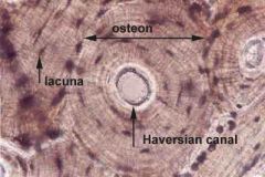
Cylindrical unit calcified matrix arranged in layers - forms rings |
|
|
Central Canal |
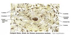
located in the center of the osteon Contains: blood vessels and nerves |
|
|
Osteocytes |

bone cells Lacunae: Space that they occupy; very small bony chambers central canal |
|
|
Communication within the compact bone |
-> central canals communicate with each other through perforating canals ->osteocytes communicate with each other through canaliculi - (process that allows communication) |
|
|
Bone formation and growth |
->skeletal system appears in fetus during the 1st few weeks of prenatal development ->skeletal model is initially cartilage which is then replaced by bone **Osteoblast: bone building cells **Osteoclast: breakdown cells ->combines action of osteoblasts and oseoclasts sculpt bones into correct shape=remodeling. |
|
|
Endochondral Ossification |
->'formed cartilage' ->most bones go through this process ->start with cartilaginous model ->1*ossification center - Primary Ossification *Diaphysis: first place where bone replaces cartilage ->2*Ossification Center *Epiphysis: second place where bone replaces cartilage *Epiphyseal plate: band of cartilage that remains between the 2 ossification centers Also called: Growth plate Purpose: make them grow longer |
|
|
Fibrous Joint |
Between bones that are very close together Movement? No movement Example? Skull |
|
|
Cartilaginous Joint |
Bones connected by hyaline or fibrocartilage Movement? Some/very little (child birth) Example? pubic symphysis (child birth), between bones of disc, fibrocartilage, intervertebrae |
|
|
synovial Joint |
Most of the joints in the body Movement? free movement More complex End of long bones are covered with hyaline cartilage and bones are held together by a joint capsule. ***decreased stability after time; ex...hip, shoulder, knee |
|
|
Associated with Synovial Contents |
Bursa: Fluid filled sacs, - help protect reduce friction. ex...knee and shoulder
Menisci: fibro cartilage disc between two bones (to absorb shock) ex...knee |
|
|
Ball and Socket (Types of Synovial Joints) (there are 6, but we cover only 2) |
One bone fits into a concave socket of another bone movement: alot of movement sagittal, frontal,transverse all 3 ex. convex, concave |
|
|
Hinge |
resembles a door hinge movement: not as much; flexion and extension ex. elbow, knee |
|
|
Flexion |
bending of joint |
|
|
Extension |
increasing angel |
|
|
Dorsiflexion |
bring foot up |
|
|
Plantarflexion |
bring foot down |
|
|
hyperextension |
excessive extension of joint |
|
|
abduction |
moving a part away from midline |
|
|
adduction |
moving a part toward midline |
|
|
rotation |
around verticle access |
|
|
circumduction |
moving through a circular path |
|
|
pronation |
turning palm down |
|
|
supination |
turning palm up |
|
|
eversion |
turning foot out (laterlly) |
|
|
inversion |
turning foot in (medially) |
|
|
protraction |
moving part forward |
|
|
retraction |
moving part backward |
|
|
elevation |
moving part up (raising) |
|
|
depression |
moving part down (lowering) |
|
|
Structure of Skeletal Muscle |
Attached to bone via a tendon Muscle-Fascicle-Muscle Fiber-Myofibril-Filaments |
|
|
Epimysium (outer) |
Connective tissue that holds a muscle together and separates it from another muscle = fascia |
|
|
Perimysium (middle) |
Surrounds each fascicle |
|
|
Endomysium (within) |
Surrounds each muscle fiber (kind of like a cell membrane) |
|
|
Protein filaments ( in a muscle fiber) |
Actin: thin myofilaments Myosin: Thick myofilaments
Actin and Myosin give skeletal muscle its stiated appearance. |
|
|
Sarcomere |
Basic contractile unit of skeletal muscle I bands (light bands) -> made of:Actin ->attached to: Z lines A bands (dark bands) -> made of: Actin and Myosin -> contains: H Zone H Zone: just has Myosin Z Lines: ends of the Sarcomere |
|
|
SR and T-Tubules |
Sarcoplasmic Reticulum: membranous channels that surround each muscle cell - parallel Transverse Tubules: membranous channels that extend inward into each cell/perpendicular Function of both: carry calcium and they initiate muscle contraction |
|
|
Neuromuscular Junction |
-connection between a muscle fiber and motor neuron - (goes to skeletal) -also called the motor end plate
|
|
|
neurotransmitters |
communicate with the cells they control by releasing chemicals -they cross the synaptic cleft -they send messages to the SR and T-Tubules which release calcium resulting in a muscle contraction |
|
|
synaptic cleft |
gap between neuron and motor end plate of muscle fiber |
|
|
Actin |
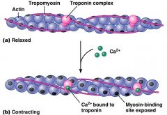
thin myofilaments contains 2 specific proteins **Troponin **Tropomysin |
|
|
Myosin |
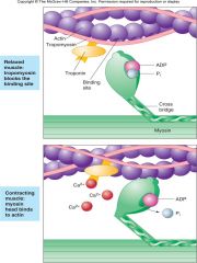
Thick myfilament Contains cross bridges that extends toward the actin |
|
|
Sliding Filament Model |

*Calcium from SR binds to troponin on the Actin filament *As calcium and troponin combine, a binding site on actin is expected *cross bridges of myosin bind with actin *cross bridges pulls actin towards the center of the sarcomere and the sarcomere shortens ******this process needs energy (ATP) |
|
|
Origin (proximal) |
immovable end of a muscle - When a muscle contracts, the insertion is pulled towards the origin |
|
|
insertion (distal) |
movable end of a muscle - When a muscle contracts, the insertion is pulled towards the origin. |
|
|
defining Agonist and Antagonist |
Agonist=Prime mover Antagonist=Opposite of the prime mover and resists the prime mover's action
(ex.) elbow ext - triceps=agonists biceps=antagonist knee flex - hamstring=agonist quadricep=anagonist knee ext - quadricep=agonist hamstring=antagonist |
|
|
hypertrophy |
enlargement of muscle |
|
|
atrophy |
decreased size of muscles |
|
|
slow fibers |
fatigue resistant used during endurance activities neck
|
|
|
fast fibers |
fatigable used from sprinting short duration activites |
|
|
Epidermis |
Stratum Basale - basement membrane Stratum Spinosum - several layers thick Stratum Granulosum - stopped dividing Stratum Lucidum - thick skin (palms hands/feet) Stratum Corneum - top of surface/dead cells |