![]()
![]()
![]()
Use LEFT and RIGHT arrow keys to navigate between flashcards;
Use UP and DOWN arrow keys to flip the card;
H to show hint;
A reads text to speech;
97 Cards in this Set
- Front
- Back
|
Skeletal Cartilage |
Made of some variety of cartilage tissue molded to fit its body location and function Consists primarily of water, which accounts for its resilience, its ability to spring back to its original shape |
|
|
What is cartilage surrounded by? What does it not contain? |
It is surrounded by a layer of dense irregular connective tissue It has no nerves or blood vessels |
|
|
Perichondrium |
around the cartilage contains blood vessels which nutrients diffuse through the matrix to reach the cartilage cells interally |
|
|
What are the 3 types of cartilage tissue? |
Hyaline, Elastic, Fibrocartilage |
|
|
What are the basic components of the 3 types of cartilage tissue? |
cells called Chondrocytes encased in small cavities within an extracellular matrix containing a jelly-like substance and fibers |
|
|
Hyaline Cartilage |
look like frosted glass when freshly exposed provide support with flexibility and resilience most abundant skeletal cartilages their chondrocytes are spherical the only fiber type in their matrix is fine collagen fibers |
|
|
What are the four types of hyaline cartilage? |
articular cartilage (moveable joints), costal cartilage (ribs), respiratory cartilage (form the skeleton of the larynx), nasal cartilage (support external nose) |
|
|
Elastic Cartilage |
resemble hyaline cartilage, contain more stretchy elastic fibers, this makes them better able to stand up to repeated bending, found in 2 skeletal locations-the external ear & epiglottis |
|
|
Fibrocartilage |
Highly compressible with great tensile strength, consist of roughly parallel rows of chondrocytes, occur in the sites that are subjected to both pressure and stretch, such as the pad like cartilages in the knee and the disks between the vertebrae |
|
|
Cartilage |
has a flexible matrix that can accommodate mitosis, is the ideal tissue to use to rapidly lay down the embryonic skeleton and to provide for new skeletal growth |
|
|
What two ways do cartilage grow? |
Appositional growth: cartilage forming cells in the surrounding perichondrium secrete new matrix against the external face of the existing cartilage tissue Interstitial growth: the lacunae found chondrocytes divide and secrete new matrix expanding the cartilage from within |
|
|
When does cartilage growth end? |
During adolescence when the skeleton stops growing |
|
|
What happens when a bone becomes calcified? |
It is hardened due to the deposit of calcium salts... it does NOT become a bone!! |
|
|
What are the 7 important functions our bones produce? |
1. Support 2. Protection 3. Anchorage 4. mineral and growth factor storage 5. bone cell formation 6. triglyceride storage 7. hormone production |
|
|
Hematopoiesis |
blood cell formation, occurs in the red marrow cavities of certain bones |
|
|
What does the osteocalcin hormone produced by bone help regulate and protect against? |
It helps regulate insulin secretion, glucose homeostasis, and energy expenditure. It protects against obesity, glucose intolerance, and diabetes. |
|
|
What are the two most common minerals produced by bone? |
calcium and phospate |
|
|
How many bones are there in the human body? |
206 |
|
|
What are the 2 groups bones are divided into? |
Axial and Appendicular |
|
|
The Axial Skeleton |
forms the long axis of the body includes the bones of the skull, vertebral column, and the rib cage these bones protect, support, or carry other body parts |
|
|
The Appendicular Skeleton |
consists of the bones of the upper and lower limbs and the gurdles that attach the limbs to the axial skeleton |
|
|
Long Bones |
considerably longer than they are wide, has a shaft and two ends |
|
|
What bones are NOT long bones? |
patella, wrist, and ankle bones ALL LIMB BONES ARE LONG BONES |
|
|
Short Bones |
roughly cube shaped, the bones of the wrist and ankle are examples, DO NOT have a shaft or an epiphyses |
|
|
What are sesamoid bones? |
A special type of short bone that form in the tendon, for example the patella. |
|
|
Flat bones |
Bend, Flattened, and usually a bit curved. The sternum, scapulau, ribs and most skull bones are flat bones. They consist of a layer of spongy bone sandwiched between two thin layers of compact bone. |
|
|
Irregular Bones |
They have complicated shapes that fit in none of the preceding classes. Examples include the vertebrae and the hip bones. |
|
|
Why are bones considered organs? |
Because they contain different types of tissue. |
|
|
What do bones contain? |
Osseous tissue, nervous tissue in their nerves, cartilage in their articular cartilages, dense connective tissue covering their external surface, and muscle and epithelial tissues in their blood vessels. |
|
|
What are the three levels of bone structure? |
1. Gross 2. Microscopic 3. Chemical |
|
|
What does every bone have in common? |
Every bone has a dense outer layer that looks smooth and solid to the naked eye. This external layer is compact bone. |
|
|
Compact bone |
external layer that looks smooth and solid to naked eye in every bone |
|
|
Spongy bone |
Internal to the compact bone, also called the trabecular bone, and it is a honeycomb of small needle-like flat pieces called trabeculae. |
|
|
In living bones, what are the open spaces between the tribeculae filled with? |
Red or yellow bone marrow. |
|
|
What do short irregular and flat bones share? |
They all consist of thin plates of spongy bone covered by compact bone. |
|
|
Diaphysis |
tubular shaft, forms long access of bone,constructed of a relatively thick collar of compact bone that surrounds a central medullary cavity or marrow cavity. In long bones. |
|
|
long bones |
All have general structure: a shaft, bone ends, and membranes. There is one diaphysis and 2 epiphyses. |
|
|
In adults, the medullary cavity contains what? |
It contains fat and is called the yellow marrow cavity. |
|
|
Epiphyses |
The bone ends. In many cases they are broader than the diaphyses. outer shell of compact bone forms the exterior and the interior contains spongy bone. A thin layer of articular hyaline cartilage covers each joint surface. |
|
|
Where is the growth plate in long bones located? |
The epiphyseal plate. |
|
|
Epiphyseal line |
Located between the diaphyses and each epiphyses of an adult long bone. it is a disk of hyaline cartilage that grows during childhood to lengthen the bone. |
|
|
Periosteum |
-glistening white double layered membrane -covers the external surface of the entire bone except for joint surfaces -richly supplied with nerve fibers and blood vessels that pass through the shaft to enter the marrow cavity via a nutrient foramen -Provides anchoring points for tendons and ligaments |
|
|
The outer fibrous layer of the periosteum |
Dense irregular connective tissue |
|
|
The inner ostiogenic layer |
located next to the bone surface and consists primarly of primitive stem cells, osteogenic cells, that give rise to all bone cells except bone destroying cells. |
|
|
Perferating (sharpeys) fibers |
tufts of collagen fibers that extend from its fibrous layer into its bone matrix. they secure the periosteum to the underlying bone. |
|
|
Bones broken in the __________ heal easier |
Diaphyses |
|
|
Endosteum |
A delicate connective tissue membrane that covers internal bone surfaces. It covers the tribiculae of spongy bone and lines the canals that pass through spongy bones. |
|
|
What do the periosteum and the endosteum have in common? |
They both cover bones and they both have osteogenic cells that can differentiate between other bone cells. |
|
|
Red marrow |
Also called hematopoietic tissue. It is typically found in the tribicular cavity of spongy bone of long bones and in the diploe of flat bones. For this reason, both these cavities are often called red marrow cavities. Red marrow makes red blood cells. It is in the epiphyses |
|
|
In new born infants, the medullary cavity of the diaphyses and all areas of the spongy bone contain what? |
Red bone marrow |
|
|
In adult bones, how much red marrow is present in spongy bone cavities? |
very little. For this reason, blood cell production in adult long bones routinely occurs only in the heads of the femur and humerus. |
|
|
The red marrow found in the diploe of flat bones and some irregular bones are ___________ in Hematopoiesis? |
much more active |
|
|
If a person becomes anemic and needs more red blood cells, what can happen? |
yellow marrow in the medullary cavity can revert to red marrow |
|
|
What are projections? |
bone markings that bulge outward from the surface-include heads, trochanters, spines, and others |
|
|
What are the five major cell types that populate bone tissue? |
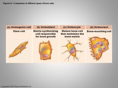
Osteogenic cells Osteoblasts Osteocytes Bone lining cells Osteoclasts |
|
|
Osteogenic cells |
aka osteoprogenic cells mitotically active stem cells found in the membranous periosteum and endosteum |
|
|
Osteoblasts |
bone-forming cells that secrete the bone matrix; actively mitotic |
|
|
Osteocytes |
-mature bone cells that occupy spaces (lacunae) that conform to their shape -monitor and maintain the bone matrix -act as stress or strain "sensors" and respond to mechanical stimuli -communicate this info to osteoblasts and osteoclasts so that bone matrix can be made or degraded as necessary to preserve calcium homeostasis |
|
|
Bone lining cells |
-flat cells found on bone surfaces where bone remodeling is not going on -also thought to help maintain the matrix -called periosteal cells if they are located on the external bone surface -called endosteal cells if they are located on the internal surface |
|
|
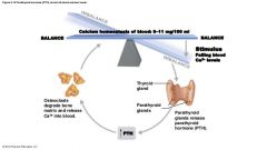
Osteoclasts |
giant multinucleate cells located at sites of bone resorption
when active they display a ruffled border which increases surface area for enzyme degradation of bone and seals off area surrounding matrix |
|
|
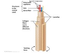
Osteon (Haversian System) |
-the structural unit of compact bone -each osteon is an elongated cylinder oriented parallel to the long axis of the bone -tiny weight bearing pillars -designed as a "twister resister" |
|
|
Osteon |
|
|
|
Central canal (Haversian Canal) |
runs through the core of each osteon contains small blood vessels and nerve fibers that serve the osteon's cells |
|
|
Perforating canals (Volkmann's canals) |
-lie at right angles to the long axis of the bone and connect the blood and nerve supply of the medullary cavity to the central canals -lined with endosteum -larger ones that connect everything together |
|
|
Interstitial lamellae |
incomplete lamellae that lie between intact osteons in compact bone |
|
|
Circumferential lamellae |
-located just deep to the periosteum and just supericial to the endosteum -extend around the entire circumference of the diaphysis and effectively resist twisting of the long bone |
|
|
Hydroxyapatites |
mineral salts |
|
|
Organic components |
include bone cells and osteoid |
|
|
inorganic components |
mineral salts |
|
|
How strong is healthy bone? |
-1/2 as strong as steel in resisting compression -fully as strong as steel in resisting tension |
|
|
Ossification |
-synonym: osteogenesis -the process of bone formation -in embryos, this leads to the formation of the bony skeleton -ossification in adults serves mainly for bone remodeling and repair |
|
|
endochondral ossification |
-a bone develops by replacing hyaline cartilage -the resulting bone is called a cartilage, or endochondral, bone -forms essentially all bones below the base of the skull except for the clavicles |
|
|
intramembranous ossification |
a bone develops from a fibrous membrane and the bone is called a membrane bone |
|
|
primary ossification center |
a region in the center of the hyaline cartilage where long bone formation begins |
|
|
5 steps of endochondral ossification |
1. A bone collar forms around the diaphysis of the hyaline cartilage model. 2. Cartilage in the center of the diaphysis calcifies and then develops cavities. 3. The periosteal bud invades the internal cavities and spongy bone forms. 4. The diaphysis elongates and a medullary cavity forms. 5. The epiphyses ossify. |
|
|
Intramembranous ossification |
forms the cranial bones of the skull (frontal, parietal, occipital, and temporal bones) and the clavicles most bones formed by this are flat bones |
|
|
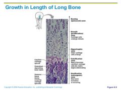
Figure 6.10 Growth in length of a long bone occurs at the epiphyseal plate. |
|
|
|
During infancy and childhood, what is the single most important stimulus of epiphyseal plate activity? |
growth hormone released by the anterior pituitary gland |
|
|
What two processes constitute bone remodeling? |
bone deposit and bone resorption |
|
|
osteoid seam |
an unmineralized band of gauzy-looking bone matrix 10-12 micrometers wide marks areas of new matrix deposits by osteoblasts |
|
|
calcification front |
an abrupt transition between the osteoid seam and the older mineralized bone |
|
|
remodeling units |
"packets" of adjacent osteoblasts and osteoclasts that coordinate bone remodeling |
|
|
Parathyroid hormone (PTH) |
produced by the parathyroid glands primarily involved in the hormonal controls |
|
|
Hormonal Controls |
|
|
|
Wolff's Law |
holds that a bone grows or remodels in response to the demands placed on it |
|
|
Nondisplaced vs Displaced Fractures |
Nondisplaced: the bone ends retain their normal position Displaced: the bone ends are out of normal alignment |
|
|
Complete vs Incomplete Fractures |
Complete: if the bone is broken through Incomplete: if it is NOT broken through |
|
|
Open vs Closed fractures |
Open (compound): when the bone ends penetrate the skin Closed (simple): when the bone ends do NOT penetrate the skin |
|
|
Reduction |
the realignment of the broken bone ends |
|
|
Closed vs open reduction |
Closed (external): the physician's hands coax the bone ends into position Open (internal): the bone ends are secured together surgically with pins or wires |
|
|
4 stages of repair in a simple fracture |
1. A hematoma forms. 2. Fibrocartilaginous callus forms 3. Bony callus forms 4. Bone remodeling occurs |
|
|
Common types of fractures |
Comminuted: bone fragments into three or more pieces Compression: bone is crushed Spiral: ragged break from excessive twisting Epiphyseal: epiphysis separates from the diaphysis along the epiphyseal plate Depressed: broken bone portion is pressed inward Greenstick: bone breaks incompletely |
|
|
Osteomalacia |
includes a number of disorders in which the bones are poorly mineralized |
|
|
Rickets |
the analogous disease in children |
|
|
Osteoporosis |
refers to a group of diseases in which bone resorption outpaces bone deposit |
|
|
Paget's Disease |
characterized by excessive and haphazard bone deposit and resorption |
|
|
Pagetic bone |
is hastily made and has n abnormally high ratio of spongy bone to compact bone |