![]()
![]()
![]()
Use LEFT and RIGHT arrow keys to navigate between flashcards;
Use UP and DOWN arrow keys to flip the card;
H to show hint;
A reads text to speech;
59 Cards in this Set
- Front
- Back
|
Where is bone deposited and resorbed according to wolff's law? |
-deposited in sites subjected to stress -resorbed in sites deprived of stress |
|
|
ABCs stands for? |
-Alignment -bone density -cartilage spaces -soft tissue |
|
|
What is the definition of assessing alignment & appearance? |
-general outline, size, contour, and postition in relation to other bones |
|
|
What is the deffinition of assessing bone? |
-bone density, texture abnormalities, bone thickness changes |
|
|
What is the definition of assessing cartilage, cortex, consistency? |
-joint space width, subchondral bone, epiphyseal plates, disks, breaks or inconistencies |
|
|
What is the definition of assessing soft tissue? |
-muscle (wasting, swelling). fat pads, periosteum, and joint capsules (none, should not be seen) |
|
|
What are the advantages of radiology? |
-quick -easy -portable -relatively inexpensive |
|
|
What are the disadvantages of radiology? |
-ionizing radiation -poor at visualizing soft tissues and small fractures |
|
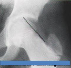
What is this showing? |
Normal Klein's line alignment (line of mensuration) |
|
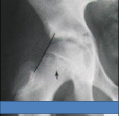
What is this showing? |
-abnormal Klein's Line Alignment |
|
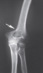
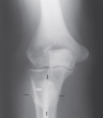
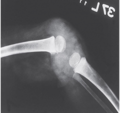
What is seen here? |
impact fracture on the supracondylar area of the humerus |
|
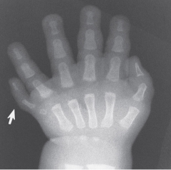
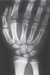
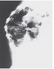
What is being shown? |
-plydactyly in a 10 mnt old child -note: there is only a trace carpal bone on the 6th digit |
|
What can be seen in this picture? |
Normal radiograph -regualar spaces formed by the growth plates (epiphyseal plates, or physis) in 8yr old |
|
|
What are the 6 major categories of bone pathology? |
1. congenital 2. inflammatory 3. metabolic 4. neoplastic 5. traumatic 6. vascular (there is a 7th category, miscellaneous, that encompasses conditions that do not fall strictly into one category ex. osteoarthritis) |
|
|
What does radio-graphic diagnosis of skeletal pathology begin with? |
-defining the distribution of the lesion -applying predictor variables to the lesion |
|
|
What are bone tumors categorized by? |
-weather the tumor is benign or malignant -by the tissue of orgin |
|
|
What things help radiologists defferentiate types of tumors from a radiograph? |
-site of the lesion -margin of the lesion -weather matrix is osteoid, chondroid, or mixed -the type of destruction (geographic, moth-eaten, or permeative) -an interrupted or uninterrupted periosteal response -the presence of a soft tissue extension of the lesion |
|
|
DO NOT HAVE TO KNOW THE TYPES OF THE TUMORS! |
|
|
|
What is a predictor variable? |
-factors that further limit diagnostic choices |
|
|
What are Daffner's 11 predictor variables? |
-behavior of the lesion -the bone or joint involved -the locus within a bone -the age, gender, or race of the patient -the margin of the lesion -the shape of the lesion -involvement of the joint space -bony reaction -matrix production by the lesion -soft tissue changes -history or trauma of surgery |
|
|
If you see a sharp, clearly defined border what type of tumor does this typically indicate? |
-slow growing benign lesion |
|
|
If you see a wide, poorly defined lesion border what type of tumor does this typically indicate? |
-a fast growing malignant lesion |
|
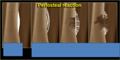
If you see these what does it mean and what are they called?? |
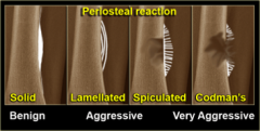
lamellated aka: onion skin spiculated aka: sunburst |
|
|
What do infections do that tumors dont? |
-cross joint spaces -Tumors do not cross joint spaces, infections do! |
|
|
What is a lamellated periosteal reaction also known as? |
-onionskin |
|
|
What is a spiculated periosteal reaction also known as? |
-sunburst |
|
What is this showing? |
osteosarcoma -note: tumor extends to joint space but does not cross it |
|
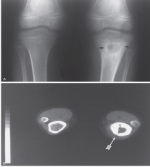
What is this showing? |
-chondroblastoma |
|
What is this showing? |
-diffuce lytic metastasis to bone from a rhabdomyosarcoma note: poorly defined borders of the lesions which is seen in an aggressive (malignant) lesion |
|
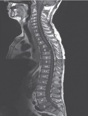
What is this showing? |
-lung cancer metastasized to the spine -lesions seen at T1, 3, 5, 6, 9, 10, 12, and L1, L4, and S1 ***largest at T6 |
|
|
What are the radiologic characteristics of adult rheumatoid arthritis? |
-periarticular soft tissue swelling -articular eriosions -minimal or absent reparative processes -concentric joint space narrowing -rarefaction of periarticular regions in early stages -generalized osteoporosis in later stages -joint deformities |
|

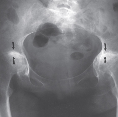
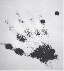
What is this showing? |
-advanced rheumatoid arthritis of hands with joint subluxations |
|
|
Bone scans are what?? |
very sensitive-next to 0 false positives but not very specific--if you can define what they have |
|
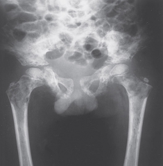
What is this showing? |
-advanced RA of the hip joints -RA destroys both sides of the joint space and appears bilaterally **not osteoarthritis because not this type of symmetry |
|
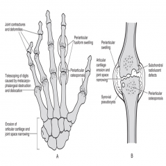
What are these showing ... A, B, ? |
A- Hallmarks of RA in small joints B- hallmarks of RA in large joints |
|
What is this showing? |
-bone scan **radiographs were normal, but the bone scan shows the inflammatory phase of RA |
|
|
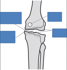
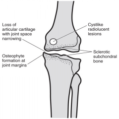
What are the osteoarthritis radiologic characteristics? |
-joint space narrowing -sclerosis of subchondral bone -osteophyte formation at joint margins |
|
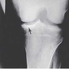
What is being shown? |
-Degenerative joint disease (DJD) (osteoarthritis) of the knee Note: sclerotic subchondral bone of the medial tibial plateau in response to the thinning of the articular cartilage |
|
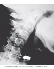
What is this showing? |
Degenerative disc disease (DDD) of cervical spine -demonstrated by thinning disc space and osteophyte formation |
|
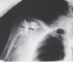
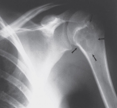
What is this showing? |
-osteoarthritis of the shoulder joint |
|
What are the lines representing? |
|
|
|
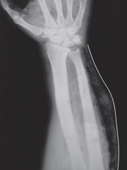
What is FBI sign? |
-Fat -Blood -Interface |
|
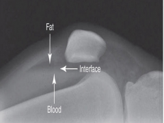
When you see this what does it suggest? |
-FBI sign -sugggestive of a fracture even when one is not readily visible |
|
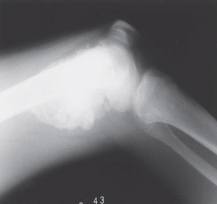
What is this showing? |
-myositis ossificans -AP view of elbow |
|
|
What are the radiologic characteristics of osteoporosis? |
-loss of cortical thickness -generalized osteopenia -associated fracture |
|
|
What are the most common sites for osteoporosis? |
-vertebrae (compression fracture) -proximal humerus (FOOSH) -Distal radius (FOOSH) -proximal femur ("I fell and broke my hip". Usually the femur breaks, and the patient falls) |
|
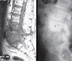
What is this showing? |
-osteoporosis of the spine with multiple compression fractures -severe kyphosis (gibbus deformity) |
|
|
What is the difference between gibbus deformity vs. dowager's hump? |
-Gibbus deformity: often used to define a sharp angle in the back with 1 or 2 vertebrae involved
-Dowager's hump: often used for 3 or more vertebrae |
|
|
When do you see buffalo hump? |
-it involves fat -seen in Cushing's disease |
|
|
When there is a decreased number of trabeculae, and the remaining trabeculae are thin what is it? |
-osteoporosis |
|
|
What is osteomyelitis? |
-infeciton of bone |
|
|
What is septic or infectious arthritis? |
-infections of joints |
|
|
What is cellulitis, myositis? |
-infections of soft tissues |
|
|
What is the earliest sign of an infection? |
-swelling in soft-tissue |
|
What is being shown? |
osteomyeliis in left tibia note: how the body tried to isolate lesion |
|
What is this showing? |
-osteomyelitis and diskitis of the spine due to IV drug use |
|
What is this showing? |
-infectious arthritis note: distension of the knee joint capsule with pus. note: no patella because 3yr. old.. patella forms at age 4 |
|
What is this showing? |
-gas gangrene and cellulitis due to clostridium organism -medical emergency |
|
|
What are the parts of a radiological report? |
-heading -clinical information -findings -conclusions -recommendations -signature |