Reading...
![]()
Play button
![]()
Play button
![]()
Use LEFT and RIGHT arrow keys to navigate between flashcards;
Use UP and DOWN arrow keys to flip the card;
H to show hint;
A reads text to speech;
47 Cards in this Set
- Front
- Back
|
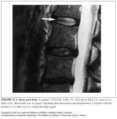
What does a degenerated disk look like on MR?
|
Loss of signal on T2
|
|
|
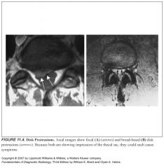
A broad-based disk bulge=
|
A bulging annulus fibrosus
|
|
|
A focal disk bulge=
|
Herniated nucleus pulposus
|
|
|
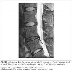
What does disruption of the annular fibers look like on MR?
|
High signal on T2— “high intensity zone”
|
|
|
Most common cause of failed back surgery?
|
Missed free fragment disk protrusion
|
|
|
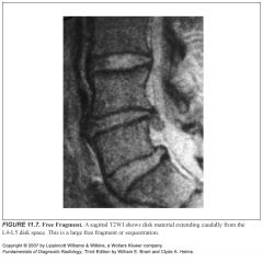
What is a free fragment?
|
Disk protrusion that has broken off from the parent disk
|
|
|
What does a free fragment look like?
|
-On CT: soft tissue density with a higher attenuation than that of the thecal sac, located away from the disk space
-On MR: disk material that has moved away from the disk space -No documented preference for cephalad vs. caudal location. |
|
|
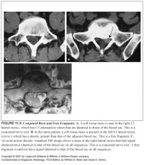
What entities can look similar to a free fragment?
|
-Conjoined root
-Tarlov cyst |
|
|
What is a conjoined root?
|
-Normal variant
-Two nerve roots exiting the thecal sac together -Has attenuation similar to the thecal sac |
|
|
What is a Tarlov cyst?
|
-Normal variant
-Dilated nerve root sleeve -Has attenuation similar to the thecal sac |
|
|
Why is a free fragment important to surgeons?
|
It means they have to explore more cephalad or caudally during surgery to remove the free fragment
|
|
|
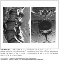
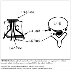
Which spinal nerve gets stretched by a lateral disk protrusion?
|
-The nerve that has already exited the central canal
-Symptoms mimic disk protrusion one level cephalad |
|
|
Define spinal stenosis:
|
Encroachment of the bony or soft tissue structures in the spine on one or more of the neural elements
|
|
|
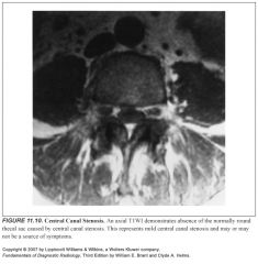
Define central canal stenosis:
|
-Compression of the thecal sac
-Described as mild, moderate, or severe |
|
|
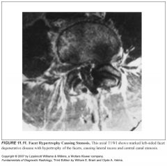
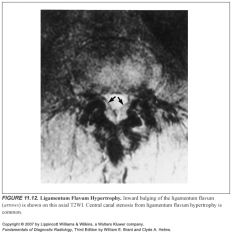
Most common cause of central canal stenosis?
|
-Degenerative disease of the facets with associated bony hypertrophy that encroaches on the central canal
-When facets are degenerative, they often have some slippage, which results in buckling of the ligamentum flavum, termed ligamentum flavum hypertrophy |
|
|
Less common causes of canal stenosis?
|
Paget disease, achondroplasia, posttraumatic changes, severe spondylolisthesis
|
|
|
Causes of neuroforaminal stenosis:
|
-Facet hypertrophy (most common)
-Free disk fragments -Post-operative scar -Lateral disk protrusion |
|
|
Is the exiting nerve above or below the disk space?
|
It’s above
|
|
|
Where are the lateral recesses?
|
They are the bony canals in which the nerve roots lie after they leave the thecal sac and before they enter the neuroforamen
|
|
|
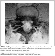
Define spondylolysis:
|
Defects in the bony pars interarticularis
|
|
|
What does it look like?
|
Seen better on CT
Identified on axial images through the midvertebral body as a break in the normally intact bony ring of the laminae |
|
|
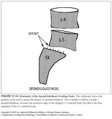
Causes of spondylolisthesis:
|
-Slippage of vertebral bodies due to bilateral spondylolysis (can be severe)
-DJD of the facets with slippage (usually minimal) |
|
|
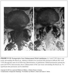
How can you tell post-op scar tissue from disk material?
|
Scar tissue enhances, disk material only has peripheral enhancement (presumably due to inflammation)
|
|
|
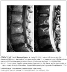
Describe type 1 bony changes in DJD:
|
T1: low signal bands that parallel the endplates
T2: these bands get brighter |
|
|
Causes of type 1 bony changes?
|
-They represent inflammatory or granulomatous response to degenerative disk disease
-Fibrovascular marrow replacement |
|
|
Describe type 2 bony changes in DJD:
|
T1: high signal bands that parallel the endplates
T2: these bands remain high signal |
|
|
Causes of type 2 bony changes?
|
They represent fatty marrow conversion
|
|
|
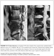
How do you distinguish type 2 changes from disk space infection?
|
In disk space infection, the disk should be bright on T2
A degenerative disk is not bright on T2 |
|
|
Describe type 3 bony changes in DJD:
|
Parallel bands of low signal adjacent to the endplates on both T1 and T2
|
|
|
Causes of type 3 bony changes in DJD?
|
They represent bony sclerosis with little residual marrow
|
|
|
1
|
|
|
2
|
|
|
3
|
|
|
4
|
|
|
5
|
|
|
6
|
|
|
7
|
|
|
8
|
|
|
9
|
|
|
10
|
|
|
11
|
|
|
12
|
|
|
13
|
|
|
14
|
|
|
15
|
|
|
16
|
|
|
17
|