Reading...
![]()
Play button
![]()
Play button
![]()
Use LEFT and RIGHT arrow keys to navigate between flashcards;
Use UP and DOWN arrow keys to flip the card;
H to show hint;
A reads text to speech;
178 Cards in this Set
- Front
- Back
|
Nervous system
|
The master controlling and communicating system of the body
|
|
|
Sensory input
|
Monitoring stimuli occurring inside and outside the body
|
|
|
Integration
|
interpretation of sensory input
|
|
|
Motor Output
|
Response to stimuli by activating effector organs
|
|
|
Central Nervous System (CNS)
Where is it located? |
-Integration and command center
-Brain and spinal cord |
|
|
Peripheral Nervous System (PNS)
Where is it located? |
-Carries messages to and from the spinal cord and brain
-Paired spinal and cranial nerves |
|
|
(PNS): Sensory Afferent Division
-Sensory Afferent Fibers -Visceral Afferent Fibers |
-Carry impulses from skin, skeletal muscles, and joints to the brain
-Transmit impulses from visceral organs to the brain |
|
|
(PNS) Motor Efferent Division
|
-Transmits impulses from the CNS to effector organs
|
|
|
Somatic Nervous System
|
-Conscious control of skeletal muscles
|
|
|
Autonomic Nervous System (ANS)
|
-Regulates smooth muscles, cardiac muscles, and glands
|
|

|
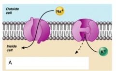
A) Sensory input
B) Integration C) Motor Output |
|

|
A) Capillary
B) Neuron C) Astrocyte |
|
|
Microglia
|
Small, ovoid cells with spiny process
|
|
|
Function of Microglia
|
Phagocytes that monitor the health of neurons
|
|
|
Ependymal Cells
|
Range in shape from squamous to columnar
|
|
|
Function of Enendymal Cells
|
They line the central cavities of the brain and spinal column
|
|

|
A) Neuron
B) Microglial cell C) Enendymal Cell D) Brain or spinal cord tissue |
|
|
Oligodendrocytes
|
Branches cells that wrap CNS nerve fibers
|
|
|
Schwann Cells (neurolemmocytes)
|
Surround fibers of the PNS
|
|
|
Satellite Cells
|
Surround neuron cell bodies with ganglia
|
|

|
A) Schwann Cells
B) Satellite Cells C) Sensory neuron with schwann cells and satellite cells D) Oligodendrocytes |
|

|
A) Schwann Cells
B) Satelite Cells C) Cell body (soma) of sensory neuron |
|
|
Neurons
|
Structural units of the nervous system
|
|
|
What are neurons composed of
|
Body, axon and dendrites
|
|
|
What nerve cell is long lived, amitotic, and have a high metabolic rate
|
Neurons
|
|
|
Name the two functions of the plasma membrane in neurons
|
-Electrical Signaling
-Cell to cell signaling during development |
|
|
Perikaryon or Soma ( Nerve Cell Body)
|
Is the major biosynthetic Center
|
|
|
What does the Perikaryon contain
|
-The nucleus and a nucleolus
-Has no centrioles though |
|
|
What is an important feature of the Perikaryon cell
|
Has well developed Nissl Bodies (Rough ER)
|
|
|
Processes
|
Armlike extensions from the soma
|
|
|
What are the two types of processes
|
Axons and dendrites
|
|
|
Dendrites of Motor Neurons
|
-Are the receptive, or input regions of the neurons
-Elecrical signals are conveyed as graded potentials |
|
|
What are the features of Dendrites
|
-Short, tapering, and diffusely branched processes
|
|
|
Axons: Structure
|
-Slender processes of uniform diameter arising from the hillock
-Usually only one unbranced axon per neuron |
|
|
Axon Terminal
|
Branched terminus of an axon
|
|
|
Axon Function
|
-Generate and transmit action potentials
-Secrete neurotransmitters from the axonal terminals |
|
|
Anterograde
|
Toward axonal terminal
|
|
|
Retrograde
|
Away from axonal terminal
|
|
|
Myelin Sheath
|
Increase the speed of nerve impulse transmission
|
|
|
Mylein Sheath physical description
|
Whitish, fatte (protien-lipoid), segmented sheath around most long axons
|
|
|
The mylein sheath is formed by?
|
-Schwann Cells in the PNS
|
|
|
What are two things a schwann cell does for an axon cell
|
-Envelopes an axon in a trough
-Encloses the axon with its plasma membrane |
|
|
Neurilemma
|
Remaning nucleus and cytoplasm of a schwann cell
|
|

|
A) Schwann Cell Cytoplasm
B) Axon C) Schwann cell plasma membrane D) Schwann cell nucleus E) Neurilemma F) Mylein Sheath |
|
|
Nodes of Ranvier
|
Gaps in the myelin sheath between adjacent schwann cells
|
|

|
A) Oligodendroglial Cells
B) Axon C) Myelin Sheath D) Node of Ranvier |
|
|
Axons of the CNS
|
-Both myelinated and unmyelinated fibers are present
-Nodes of Ranvier are widely spaced |
|
|
White Matter
|
Dense collections of myelinated fibers
|
|
|
Dark Matter
|
Mostly soma and unmyelinated fibers
|
|
|
Multipolar
|
Three or more processes
|
|
|
Bipolar
|
Two processes (axon and dendrite)
|
|
|
Unipolar
|
Single, short process
|
|
|
Sensory (Afferent)
|
Transmit impulses toward CNS
|
|
|
Motor (Efferent)
|
Carry impulses away from the CNS
|
|
|
Interneurons (Associated neurons)
|
Shuttle signals through CNS pathways
|
|
|
Action potentials, or nerve impulses are
|
always the same regardless of stimulus
|
|
|
Voltage
|
Meausre of potential energy generated by seperated charge
|
|
|
Potential difference
|
Voltage measured between two points
|
|
|
Current
|
The flow of electrical charge between two points
|
|
|
Resistance
|
Hindrance to change flow
|
|
|
Insulator
|
Substance with high electrical resistance
|
|
|
Conductor
|
Substance with low electrical resistance
|
|
|
Electrical current and the body
|
reflects the flow of ions rather than electrons
|
|
|
What are the two potentials on either side of the membrane
|
-The number of ions is different across the membrane
-The membrane provides a resistance to ion flow |
|
|
Passive or leakage channels
|
Always open
|
|
|
Chemically gated channels
|
open with binding of a specific neurotransmitter
|
|
|
Voltage-gated channels
|
open and close in response to membrane potential
|
|
|
Mechanically gated channels
|
open and close in response to physical deformation of receptors
|
|
|
A gated channel is open when
|
a neurotransmiter is attached to the receptor
|
|
|
A gated channel is closed when
|
a neurotransmitter is not bound to the extracellular receptor
|
|

|
A) Receptor
B) Neurotransmitter chemical attached to receptor C) Chemically gated ion channel closed D) Chemically gated ion channel opens |
|
|
When is a voltage gated channel close
|
When the intracellular enviroment is negative
-Na cannot enter the cell |
|
|
When is a voltage gated channel open
|
When the intracellular enviroment is positive
-Na can enter the cell |
|

|
A) Voltage-gated ion channel (Closed)
B) Membrane voltage changes C) Voltage-gated ion channel opens |
|
|
When gated channels are open
|
movement is along their electrochemical gradients
|
|
|
When gated channels are open where does voltage change
|
across the membrane
|
|
|
When do ions move across their chemical gradient
|
When they move from an area of high concentration to an area of low concentration
|
|
|
When does ions move along their electrical gradient
|
When they move toward an area of opposite charge
|
|
|
Electrochemical Gradient
|
The electrical and chemical gradients taken together
|
|
|
What causes a change in resting membrane potential
|
-different concentrations of Na, K, Cl, and protien anions (A-)
|
|
|
Ionic differences are the consequence of
|
Operation of the sodium-potassium pump
|
|

|
Know
|
|
|
Membrane Potentials: Signals
|
Used to integrate, send, and recieve information
|
|
|
Membrane potential changes are produced by (2)
|
-Changes in membrane permeability to ions
-Alterations of ion concentration across the membrane |
|
|
Two types of membrane potential signals
|
-Graded potentials
-Action potential |
|

|
A) Myelinated axons
B) Soma of oligodendrocyte C) Microtubule D) Node of Ranvier E) Mitochondrion in axoplasm F) Node of Ranvier |
|
|
Depolarization
|
the inside of the membrane becomes less negative
|
|
|
Repolarization
|
the membrane returns to its resting membrane potential
|
|
|
Hyperpolarization
|
the inside of the membrane becomes more negative than the resting potential
|
|

|
A) Depolarizing Stimulus
B) Hyperpolarizing Stimulus C) Depolarization D) Resting Potential E) Resting Poteltial F) Hyperpolarization |
|
|
Graded potentials
|
Short-lives, local changes in membrane potential
|
|
|
Sufficiently strong graded potentials can initiate
|
Action Potentials
|
|

|
A) Depolarized region
B) Stimulus C) Depolarization D) Spread of Depolarization |
|
|
Graded potentials can only travel over
|
short distances
|
|
|
Action potentials are only generated by
|
muscles cells and neurons
|
|
|
These do not decrease in strength over distance
|
Action Potentials
|
|
|
Nerve Impulse
|
An action potential in the axon of a neuron
|
|
|
Action Potential: Resting State
Activation gates: |
Closes in the resting state
|
|
|
Action Potential: Resting State
Inactivation gates: |
Open in the resting state
|
|

|
A) Resting State: All gated Na and K channels closed (Na+ activation gates closes; inactivation gates open)
|
|
|
What happens to Na+ in the depolarization phase
|
Permeability increases; membrane potential reverses
|
|
|
What happens to Na+ and K+ gates in the depolarization phase
|
Na+ gates are opened
K+ gates are closes |
|
|
Threshold
|
a critical level of depolarization
(-55 to -50 mV) |
|
|
At threshold depolarization
|
becomes self-generating
|
|
|
A) Depolarizing phase: Na+ channels open
|
|
|
Action Potential: Repolarization
Sodium inactivation gates |
Close
|
|
|
In repolarization (AP) as sodium gates _______, voltage-sensitive K+ gates ________
|
Close, Open
|
|

|
A) Repolarization phase: Na+ channels closing and K+ channels open
|
|
|
Action Potential: Hyperpolarization
What happens to potassium gates |
They remain open, causing an excessive efflux of K+
|
|

|
A) Hyperpolarization: K+ channels remain open; Na+ channels closed
|
|
|
Role of the sodium-potassium pump in repolarization
|
Restores the resting electrical conditions of the neuron.
Does not restore the resting ionic conditions |
|
|
What is restored by the sodium potassium pump in repolarization
|
Ionic resdribution back to resting conditions is restored
|
|
|
What are the 4 phases of action potentials
|
1) Resting State
2) Depolarization Phase 3) Repolarization Phase 4) Hyperpolarization |
|
|
Propagation of an Action Potential
(Time=0ms) |
-Na influx causes a patch of the axonal membrane to depolarize
-Positive ions in the axoplasm move toward the polarized (negative) portion of the membrane |
|
|
Propagation of an Action Potential (Time=1ms)
|
-Ions of the extracellular fluid (Na+) move toward the area of greatest negative charge
|
|
|
Propagation of an Action Potential
(Time=2ms) |
-The action potential moves away from the stimulus
-Where sodium gates are closing, potassium gates are open and create a current flow |
|
|
Threshold
|
-Membrane is depolarized by 15 to 20 mV
|
|
|
Weak (Subthreshold) stimuli
|
are not relayed into action potentials
|
|
|
Strong (Threshold) stimuli
|
are relayed into action potentials
|
|
|
All-or-none phenomenon
|
Action potentials either happen completely, or not at all (off-on)
|
|
|
How does the central nervous system determine stimulus intensity
|
By the frequency of impulse transmission (FM)
|
|
|
Coding for Stimulus Intensity
-Upward Arrow -Downward Arrow |
-Stimulus applied
-Stimulus stopped |
|
|
AM vs. FM
|
-Amplitude Modulation
-Frequency Modulation |
|
|
Coding for Stimulus Intensity
Length of Arrows: Action Potentials: |
-Strength of stimulus
-Vertical Lines |
|
|
Absolute Refractory Period
|
Time from the opening of the Na+ activation gates until the closing of inactivation gates
|
|
|
What are the characteristics of the absolute refractory period
|
-Prevents the neuron from generating an action potential
-Ensures that each action potential is seperate -Enforces one-way transmission of nerve impulses |
|
|
o
|
A) Absolute refractory period
B) Relative refractory period C) Threshold D) Resting membrane potential |
|

|
A) Absolute refractory period
B) Relative refractory period C) Threshold D) Resting membrane potential |
|
|
Relative Refractory Period
|
-Sodium gates are closed
-Potassium gates are open -Repolarization is occuring |
|
|
Rate of impulse propagation is determined by:
-Axon diameter -Presence of a myelin sheath |
-The larger the diameter, the faster the impulses
-Myelination dramatically increases impulse speed |
|
|
Saltatory Conduction
|
Current passes through a myelinated axon only at the nodes of ranvier
|
|
|
Saltatory conduction is much faster than conduction along
|
unmyelinated axons
|
|
|
Multiple Sclerosis (MS)
|
-A autoimmune disease that mainly affects young adults
-Nerve fibers are severed and myelin sheaths in the CNS become nonfunctional scleroses |
|
|
Nerve fibers are classified according to
|
-Diameter
-Degree of myelination -Speed of Conduction |
|
|
Synapses
|
A Junction that mediates information transfer from one neuron
-To another neuron -To an effector cell |
|
|
Presynaptic Neuron
|
Conducts inmpulses toward the synapse
|
|
|
Postsynaptic Neuron
|
Transmits impulses away from the synapse
|
|

|
A) Axosomatic Synapses
B) Axodendritic Aynapses C) Axoaxonic Synapses D) Axosomatic Synapses E) Soma of postsynaptic neuron |
|
|
Electrical Synapses
|
-Correspond to gap junctions found in other cell types
|
|
|
Electrical synapses are important in the CNS in
|
-Arousal from sleep
-Mental attention -Emotions and memory -Ion and water homeostasis |
|
|
Chemical Synapses
|
Specialized for the release and reception of neurotransmitters
|
|
|
Chemical Synapses are typically composed of two parts
|
-Axonal terminal of the presynaptic neuron, which contains synaptic vesicles
-Receptor region on the dendrites or soma of the postsynaptic neuron |
|
|
Synaptic Cleft
|
Fluid-filled space seperating the presynaptic and postsynaptic neurons
|
|
|
Transmission across the synaptic cleft
|
-Is a chemical event
-Ensures unidirectional communication between neurons |
|
|
In synaptic cleft nerve impulses reach the axonal terminal of the
|
presynaptic neuron and open Ca channels
|
|
|
______ is released into the synaptic cleft via exocytosis in response to ______
|
-Neurotransmitter
-Synaptotagmin |
|

|
Know
|
|
|
Neurotransmitter bound to a postsynaptic neuron (2)
(TQ) |
-Produces a continous postsynaptic effect
-Must be removed from its receptor |
|
|
Name the three ways that neurotransmitters are removed
|
-When they are degraded by enzymes
-Are reabsorbed by astrocytes -Diffuse from the sunaptic cleft |
|
|
Synaptic Delay
|
The rate-limiting step of the neural transmission
|
|
|
Name the two ways that neurotransmitter receptors mediate changes in membrane potential
|
-The amount of neurotransmitter released
-The amount of time the neurotransmitter is bound to receptors |
|
|
What are the two types of postsynaptic potentials
|
-EPSP and IPSP
|
|
|
Excitatory Postsynaptic Potentials
|
ESPS are graded potentials that can initate and action potential in an axon
|
|
|
EPSP only use _____ _______ channels
|
-Chemically Gated
|
|
|
______ ______ do not generate action potentials
|
Postsynaptic membranes
|
|
|
Nerotransmitter binding to a receptor at inhibitory synapses reduces the
|
postsynaptic neurons ability to produce an action potential
|
|
|
ESPS must summate ________ or ______ to induce an action potential
|
-temporally or spatially
|
|
|
Temporal summation
|
presynaptic neurons transmit impulses in rapid-fire order
|
|
|
Spatial Summation
|
postsynaptic neuron is stimulated by a large number of terminals at the same time
|
|
|
IPSP can also summate with _______, canceling each other out
|
EPSP
|
|
|
How many different neurotransmitters have been identified
|
50
|
|
|
What are three chemical neurotransmitters
|
-Acetylcholine
-Biogenic Amines -Amino Acids |
|
|
Acetylcholine
What is it degraded by |
-Released at the neuromuscular junction
-Degraded by the enzyme acetylcholinesterase |
|
|
Acetylcholine is released by (2) (TQ)
|
-All neurons that stimulate skeletal muscle
-Some neurons in the autonomic nervous system |
|
|
Biogenic Amines include
|
-Catecholamines, indolamines
|
|
|
Catecholamines
|
-dopamine, norepinephrine, and epinephrine
|
|
|
Indolamines
|
-serotonin and histamine
|
|

|
A) presynaptic axon terminal
B) Synapse C) Postsynaptic dendrite D) Synaptic cleft E) Synaptic vesicles |
|
|
Neurotransmitters: Amino Acids include
|
-GABA
-Found only in the central nervous system |
|
|
Which neurotransmitter acts as natural opiates, reducing our preception of pain
|
Peptides
|
|
|
Peptides bind to the same receptors as _______ and ______
|
-Opiates and morphine
|
|
|
What are the two classifications of neurotransmitters
|
-Excitatory and inhibitory
|
|
|
Excitatory neurotransmitters cause
|
depolarization
|
|
|
which neurotransmitters cause hyperpolarization
|
inhibitory
|
|
|
Excitatory neurotrasmitters
|
neuromuscular junctions with skeletal muscle
|
|
|
Inhibitory neurotransmitters
|
neuromuscualr junction with cardiac muscle
|
|
|
Direct receptor mechanisms
|
-neurotransmitters that open ion channels
-Promote rapid responses |
|
|
Indirect receptor mechanisms
|
-neurotransmitters that act through second messengers
-Promote long lasting effects |