![]()
![]()
![]()
Use LEFT and RIGHT arrow keys to navigate between flashcards;
Use UP and DOWN arrow keys to flip the card;
H to show hint;
A reads text to speech;
82 Cards in this Set
- Front
- Back
|
Anatomy |
The study of structure of the body parts and their relationships to one another, it is concrete. |
|
|
Physiology |
Concerning the function of the body , how the body parts work and carry out their life-sustaining activities. |
|
|
Gross or Macroscopic anatomy |
the study of large body structures visible to the naked eye. ie. Heart Lungs. |
|
|
Anatomy (greek) |
to cut apart |
|
|
Regional anatomy |
all the structures bone, muscles, blood vessels nerves in a particular region of the body are examined at the same time. |
|
|
Systemic Anatomy |
Body structures are studied system by system, cardiovascular or skeletal. |
|
|
Surface Anatomy |
A subdivision of gross anatomy, that is the study of internal structure as it relates to the underlying structures. These are useful for clinicians to located appropriate blood vessels and things. |
|
|
Microscopic Anatomy |
Structures too small to see with the naked eye. |
|
|
Cytology |
the study of the cells of the body |
|
|
Histology |
The study of the tissues of the body |
|
|
Developmental Anatomy |
Traces structural changes that occure in the body throughout the life span. |
|
|
Embryology |
A subdivision of developmental anatomy that is, concerns developmental changes that occur before birth. |
|
|
Pathological anatomy |
Studies structural changes caused by disease. |
|
|
Radiographic anatomy |
Studies internal structures as visualized by X-ray images or specialized scanning procedures |
|
|
Molecular biology |
the structures of biological molecules are investigated. |
|
|
Palpation |
feeling organs with your hands |
|
|
Auscultation |
Listening with a stethoscope. |
|
|
Tissues |
The 4 basic tissues of the body are epithelial, connective, muscular, and nervous. |
|
|
Organ |
is a discrete structure composed of at least two tissue types, four is more common. |
|
|
Organ, organ system and organism |
The organ allows for extremely complex functions. Organ systems are organs that work together. and organism is the sum total of all structural levels working together to keep us alive. |
|
|
Maintaining Boundaries |
The internal environment remains isolated and distinct from the external environment. |
|
|
Movement |
The activities promoted by the muscular system. of blood movement of food movement. The cells ability to shorten is called contractility. |
|
|
Responsiveness |
or excitability is the ability to sense changes in the environment and respond to them. like cutting your hand and the withdrawal reflex. |
|
|
Digestion |
the breaking down of ingested foodstuffs to simple molecules that can be absorbed into the blood. |
|
|
Metabolism |
is the sum of all the chemical reactions that occur withing the body cells. |
|
|
Excretion |
the removal of waste |
|
|
Reproduction |
Occurs at both the cellular an organismal level. producing offspring |
|
|
Growth |
increase in size of a body part of the organism as a whole. |
|
|
Nutrients |
Chemical substances taken in via the diet, used for cell building and energy. |
|
|
Oxygen |
oxidative reactions that release energy require O2 |
|
|
Water |
Accounts for 60-80% of our body weight and is the single most abundant chemical substance in the body. It provides the watery environment necessary for chemical reactions and the fluid base for body secretions and excretions. |
|
|
Normal Body Temperature |
for chemical reactions to be maintained at a life sustaining rate, normal body temperature must be maintained. metabolic reactions stop at low body temperatures. At high temperatures proteins denature and lose their activity. the activities of the muscle systems generate most body heat. |
|
|
Appropriate Atmospheric Pressure |
the force the air exerts on the surface of the body. Breathing and gas exchange in the lungs depend on appropriate atmospheric pressure. At high altitudes gas exchanges is inadequate |
|
|
Homeostasis |
Coined by Walter Cannon, an american physiologist. the ability to maintain relatively stable internal conditions even though the outside world changes continuously. Literally translates into unchanging. Although it is a dynamic state of equlibrium. internal states vary within normal limits. |
|
|
three components of homeostasis |
The receptor, that senses or monitors the environment and responds to stimuli The control center, determines the set point and analyzes the impute from the receptor. The effector provides the means for the control centers response. |
|
|
Negative feedback |
The response is negative to the initiating stimulus. shutting of the original effect of the stimulus or reducing it. |
|
|
Positive feedback |
The result or response enhances the original stimulus. Child birth, clotting, Action potentials, ovulation. |
|
|
Homeostatic imbalance |
most diseases can be regarded as a disturbance in homeostasis. |
|
|
Superior (cranial) |
Toward the head end or upper part of a structure or the body above. |
|
|
Inferior |
Away from the head or toward the lower part of a structure of the body, below |
|
|
Ventral (anterior) |
Toward of at the front of the body, in front of |
|
|
Dorsal (posterior) |
toward or at the back of the body, behind. |
|
|
Medial |
toward or at ht midline of the body on the outer side of |
|
|
Lateral |
away from the midline of the body, on the inner side of |
|
|
intermediate |
between a more medial and a more lateral structure. |
|
|
Proximal |
closer to the origin of the body part of the point of attachment of a limb to the body trunk. |
|
|
Distal |
Farther from the origin of the body part of the point of attachment of a limb to the body trunk. |
|
|
Superficial (external) |
toward or at the body surface |
|
|
Deep (internal) |
Away from the body surface; More internal |
|
|
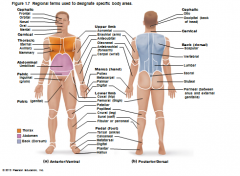
Regional terms used to designate specific body areas. |
|
|
|
Axial Part |
Makes up the main axis of our body, including head neck and trunk |
|
|
Appendicular part |
consists of the appendages, or limbs |
|
|
Sagittal Plane |
a vertical plane that divides the body into right and left parts. All sagittal planes that offset from the midline are parasagittal planes. |
|
|
Median Plane or Midsagittal plane |
A sagittal plane that lies exactly in the midline. |
|
|
Frontal Plane |
divide the body into anterior and posterior parts. also called a coronal plane. |
|
|
Transverse or horizontal planes |
Run horizontal from right to left dividing the body into superior and inferiror parts, and is also called a cross section. |
|
|
oblique section |
diagonal cuts made between the horizontal and verticle planes. |
|
|
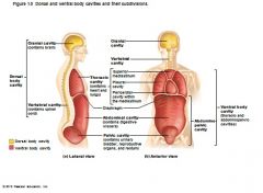
Dorsal Body Cavity |
Contains the skull (crainal cavity) and the spinal cavity |
|
|
Ventral body Cavity |
The more anterior and larger of the closed body cavities. Composed of the Thoracic and abdominopelvic cavity. |
|
|
The Thoracic Cavity |
the superior subdivision of the dorsal body cavity. is surrounded by the ribs and muscles of the chest. The thoracic cavity is divided into lateral pleural cavities each enveloping a lung and the medial mediastinum, that contains the pericardial cavity. which encloses the heart and it also surrounds the remaining thoracic organs. (esophagus, trachea, and others) |
|
|
Abdominopelvic cavity |
A subsection of the ventral body cavity, seperated from the thoracic cavity by the diaphram. a dome-shaped muscle. The supperior portion is the abdominal cavity. containing the stomach, intestines, spleen, liver and other organs. The inferior part, the pelvic cavity, lies in the bony pelvis and contains the urinary bladder, some reproductive organs and the rectum. these two cavities are not aligned with eachother. |
|
|
Dorsal and ventral body cavities and their subdivisions. |
|
|
|
X-Rays |
Basically a negative image, X rays are absorbed by hard dense materials. The film behinds these gets less exposure producing light sections, fat and air provides little resistance thus these sections show up as black from over exposure. Dense structures like tumors and tuberculosis are imaged most easily with xrays. |
|
|
Computed tomography (CT, formally a CAT scan) |
An xray machine in a tunnel that can spin all around the patient. this produces slices through the body about the width of a dime. The computer compiles the data. Used for evaluating most problems in the brain and abdomen. and with their clarity there is almost no need for exploritory surgery. |
|
|
Xenon CT |
A CT with xenon gas that allows the quick tracing of blood flow, inhaling the gas delivers it to the blood and blood flow can be checked, especially in the brain, if there is no sign of xenon, this usually indicates pour blood profussion and may indicate a stroke. |
|
|
Dynamic Spatial Reconstruction (DSR) |
Uses ultrafast CT scanners to provide 3D images of body organs from any angle. It also allows for monitoring of changes in size of the organ in slow motion, normal speed or at a specific moment. Great for visuallizing the heart beating and blood flow through blood vessels. this allows for the evaluation of blockages or the results of artery bypass grafts. |
|
|
Digital Subtraction Angiography (DSA) |
uses xrays to provide a picture of small arteries. Images are taken before and after a contrast dye is added and the computer subracts the similarities so only the artery is seen. These are used to visualized the heart and brain. |
|
|
Positron emission Tomography (PET) |
the PET excells in visuallizing metabolic activity. The patient is giving radioactive glucose and positioned in the scanner. As the radioisotopes are absorbed by the most active brain cells High energy gamma rays are produced. These gama rays are detected and a picture of the brain in live action color pictures. This has been used to diagnose people affeced by mental illness, stroke alzheimers disease and epliepsy. or to show what parts of the brain are used during specific tasks. |
|
|
Sonography, or ultrasound. |
this is cheaper and safer than most ionizing forms of imaging. Sound waves are shot into tissues and their echos are recovered. the handpeice emits and detects the echos, and thus different planes can be easily examined. |
|
|
Magnetic resonance imaging (MRI) |
produces high contrasting images of soft tissue, something that xrays can not do. the body is surrounded by a magnetic field that is 60,000 times greater than the on produced by the earth. These magnetic fiels cause the hydrogen atoms in the body to act like magnets themselves. As radio waves are directed into the mix the energy released is transmitted into an image. Body tissues are distinguished based on water content so fatty white matter and watery gray matter of the brain can be distinguished. Delicate nerve fibers can also be seen. good at detecting tumors and degernerative diseases like MS. Magnetic gas in the lungs can be used to diagnose asthma. |
|
|
Magnetic resonance spectroscopy |
Map other elements besides hydrogen, allowing the observer to track how disease alters body chemistry. |
|
|
Functional MRI |
Tracks blood flow in the brain in real time. MRIs do not require dies and can pinpoint much smaller brain areas then PET scans. |
|
|
Serosa or serous membranes |
The walls of the ventral cavity contain a layer of thin double layered membranes. The membrane lining the cavity wall is called the parietal serosa, and then folds in on itself covering organs in the viceral serosa. The membranes are seperated from eachother by a serous fluid which is secreated by both membranes which lubricates the two. |
|
|
Serous membranes of the body |
Pariatal pericardium lines the pericardial cavity and folds back on the visceral pericardium, which covers the heart. Likewise the parital plerurae line the walls of the thoracic cavity. the viceral pleurae cover the lungs. Parietal peritoneum is associated with the walls of the abdominaopelvic cavity while the viceral peritoneum covers most of the organs within that cavity. |
|
|
Pleurisy |
inflamation of the pleurae, or peritonitis, inflamation of the peritoneum. |
|
|
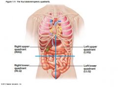
Abdominopelvic quadrants |
|
|
|
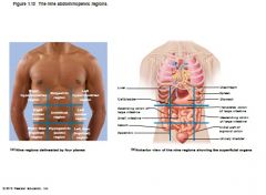
Abdominopelvic regions |
|
|
|
Oral and digestive cavities |
mouth contains the teeth and tongue and is continuous with the cavity of the digestive organs an open to the body exterior at the anus. |
|
|
Nasal Cavity |
located within and posterior to the nose, the nasal cavity is part of the respirtory system passageways. |
|
|
orbital cavities |
in the skull and house the eyes and present them in an anterior position |
|
|
Middle ear cavities |
medial to the eardrum, contain the tine bones that transmit sound vibrations to the hearing receptors |
|
|
synovial cavities. |
Joint cavities, enclosed with fibrous capsules that surround freely moving joints of the body. these secreate synovial fluid to lubricate the joints. and reduce friction. |