![]()
![]()
![]()
Use LEFT and RIGHT arrow keys to navigate between flashcards;
Use UP and DOWN arrow keys to flip the card;
H to show hint;
A reads text to speech;
113 Cards in this Set
- Front
- Back
- 3rd side (hint)
|
anterior
|
in front or in the front part
|
|
|
|
anteroinferior
|
in front and below
|
|
|
|
anterolateral
|
in front and to the outside
|
|
|
|
anteromedial
|
in front and toward the inner side or midline.
|
|
|
|
anteroposterior
|
Relating to both front and rear
|
|
|
|
Anteriosuperior
|
in front and above.
|
|
|
|
bilateral
|
Relating to the right and left sides of the body or of a body structure such as the right and left extremities
|
|
|
|
caudal
|
below in relation to another structure, inferior.
|
|
|
|
cephalic
|
above in relation to another structure; higher, superior.
|
|
|
|
Contralateral
|
Pertaining or relating to the opposite side.
|
|
|
|
Deep
|
beneath or below the surface; used to describe relative depth or location of muscles or tissue.
|
|
|
|
Dexter
|
Relating to, or situated to the right or on the right side of, something.
|
|
|
|
Distal
|
Situated away from the center or midline of the body, or away from the point of origin.
|
|
|
|
Dordal
|
Relating to the back, being or located near, on, or toward the back, posterior part, or upper surface of; also relating to the top of the foot.
|
|
|
|
Fibular
|
Relating to the fibular (lateral) side of the lower extremity
|
|
|
|
inferior (infra)
|
below in relation to another structure; caudal
|
|
|
|
inferolateral
|
below and to the outside
|
|
|
|
Inferomedial
|
Below and toward the midline or inside.
|
|
|
|
Ipsilateral
|
on the same side
|
|
|
|
lateral
|
On or to the side, outside, farther from the median or midsagittal plane
|
|
|
|
Medial
|
Relating to the middle or center; near to the median or midsagittal plane
|
|
|
|
Median
|
Relating to, located in, or extending toward the middle, situated in the middle, medial.
|
|
|
|
Palmar
|
Relating to the palm or volar aspect of the hand.
|
|
|
|
plantar
|
Relating to the sole or under surface of the foot
|
|
|
|
posterior
|
behind, in back,or in the rear
|
|
|
|
posteroinferior
|
behind or in back and below.
|
|
|
|
posterolateral
|
Behind and to one side, specifically to the outside.
|
|
|
|
Posteromedial
|
Behind and to the inner side
|
|
|
|
postetosuperior
|
behind or in back and above.
|
|
|
|
Prone
|
Face downward position of the body; laying on the stomach
|
|
|
|
Proximal
|
Nearest the trunk or the point of origin
|
|
|
|
Radial
|
Relating to the radial (lateral) side of the forearm or hand
|
|
|
|
Scapular plane
|
In line with the normal resting position of the scapula as it lies on the posterior rib cage; movements in the scapular plane are in line with the scapular, which is at an angle of 30 to 45 degrees from the frontal plane
|
|
|
|
Sinister
|
Relating to, or situated to the left or on the left side of, something
|
|
|
|
Superficial
|
Near the surface used to describe relative depth or location of muscles or tissue
|
|
|
|
Superior (supra)
|
Above in relation to another structure; higher, cephalic
|
|
|
|
Superolateral
|
Above and to the outside
|
|
|
|
Superomedial
|
Above and toward the midline or inside
|
|
|
|
Supine
|
Face upward position of the body; lying on the back
|
|
|
|
Tibial
|
Relating to the tibial (medial) side of the lower extremity.
|
|
|
|
Ulnar
|
Relating to the ulnar (medial) side of the forearm or hand.
|
|
|
|
Ventral
|
Relating to the belly or abdomen, on or toward the front,anterior part of
|
|
|
|
Volar
|
relating to palm of the hand or sole of the foot.
|
|
|
|
Anteversion
|
Abnormal or excessive rotation forward of a structure such as femoral anteversion
|
|
|
|
Kyphosis
|
Increased curving of the spine outward or backward in the sagittal plane
|
|
|
|
Lordosis
|
Increased curving of the spine inward or forward in the sagittal plane
|
|
|
|
Recurvatum
|
Bending backward, as in knee hyperextension
|
|
|
|
Retroversion
|
Abnormal or excessive rotation backward of a structure, such as femoral retroversion
|
|
|
|
Scoliosis
|
lateral curving of the spine
|
|
|
|
valgus
|
Outward angulation of the distal segment of a bone or joint as in knock_knees
|
|
|
|
Varus
|
Inward angulation of the distal segment of a bone or joint, as in Bowlegs.
|
|
|
|
bone properties
|
Calcium carbonate, calcium phosphate, collagen, and water are the basis of bone composition.
|
|
|
|
Cortical bone
|
Is harder and more compact, with only about 5 percent to 30 percent of its volume being porous, with non mineralized tissue.
|
|
|
|
Cancellous bone
|
Is spongy, with around 30% to 90% of its volume being porous.
|
|
|
|
Arthrosis
|
Joint
|
|
|
|
3 classifications by joint structure
|
Fibrous, cartilaginous, or synovial.
|
|
|
|
3 Joint functional classification
|
Synarthrosis, amphiarthrosis, and diarthrosis.
|
|
|
|
synarthrodial joints
|
Innumerable, structurally, these articulations are divided into two types: suture, gomphosis.
|
|
|
|
Suture joints
|
Found in the sutures of the cranial bones. The sutures of the skull are truly immovable beyond infancy.
|
|
|
|
Gomphosis joint
|
Found in the sockets of the teeth. The socket of a tooth is often referred to as gomphosis. Normally, there should be essentially no movement of the teeth in the mandible or maxilla.
|
|
|
|
amphiarthrodial joints
|
Slightly movable joint. Structurally, oceans are divided into three types: syndesmosis, symphysis, synchondrosis.
|
|
|
|
Syndesmosis joint
|
type of joint held together by strong ligamentous structures that allow minimal movement between the bones. Examples are the coracoclavicular joint and the inferior tibiofibular joint.
|
|
|
|
symphysis
|
Type of joint separated by a fibrocartilage pad that allows very slight movement between the bones. Examples are the symphysis pubis and the intervertebral disks.
|
|
|
|
synchondrosis
|
type of joint separated by hyaline cartilage that allows very slight movement between the bones. Examples are the costrochondral joints of the ribs with the sternum.
|
|
|
|
diathrodial joints
|
Freely movable joints. Also known as synovial joints.
|
|
|
|
Joint capsule
|
surround the bony ends forming the joints.
|
|
|
|
Joint cavity
|
the ligamentous capsule is wine with us in vascular synovial capsule that secretes synovial fluid to lubricate the area inside the joint capsule known as the joint cavity.
|
|
|
|
enarthrodial
|

Ball and socket joint
|
|
|
|
ginglymus
|

Hinge joint
|
|
|
|
sellar
|

Saddle joint
|
|
|
|
trochoidal
|

Pivot joint
|
|
|
|
arthrodial
|

Gliding joint
|
|
|
|
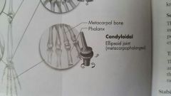
Condyloidal
|

Ellipsoid joint
|
|
|
|
Abduction
|
Lateral movement away from the midline of the trunk in the frontal plane.
|
|
|
|
Adduction
|
Movement medially toward the midline of the trunk in the frontal plane.
|
|
|
|
Flexion
|
Bending movement that results in a decrease of the angle in a joint by bringing bones together, usually in the sagittal plane.
|
|
|
|
Extension
|
Straightening movement that results in an increase of the angle in a joint by moving bones apart, usually in the sagittal plane.
|
|
|
|
Circumduction
|
Circular movement of a limb that delineates an arc or describes a cone. It is a combination of flexion, extension, abduction, adduction. Sometimes referred to as circomflexion.
|
|
|
|
diagonal abduction
|
Movement by a limb through a diagonal plane away from the midline of the body, such as in the hip or glenohumeral joint.
|
|
|
|
Diagonal adduction
|
movement by a limb through a diagonal plane toward and across the midline of the body, such as in the hip or glenohumeral joint.
|
|
|
|
External rotation
|
Rotary movement around the longitudinal axis of a bone away from the midline of the body. Occurs in the transverse plane and is also known as rotation laterally, outward rotation, and lateral rotation.
|
|
|
|
Internal rotation
|
In rotary movement around the longitudinal axis of a bone toward the midline of the body. Occurs in the transverse plane and is also known as rotation medially , inward rotation, and medial rotation.
|
|
|
|
Eversion
|
Turning the sole of the foot outward or laterally in the frontal plane; Abduction.
|
|
|
|
Inversion
|
Turning the sole of the foot inward or medially in the frontal plane; adduction.
|
|
|
|
Dorsiflexion or dorsal flexion
|
Flexion movement of the ankle that results in the top of the foot moving toward the anterior tibia in the sagittal plane.
|
|
|
|
Plantar flexion
|
Extension movement of the ankle that results in the foot and / or toes moving away from the body in the sagittal plane.
|
|
|
|
Pronation
|
the position of a foot and ankle resulting from a combination of ankle dorsiflexion, subtalar eversion , and forefoot abduction (toe-in).
|
|
|
|
Supination
|
A position of the foot and ankle resulting from a combination of ankle plantar flexion, subtalar inversion, and forefoot adduction (toe-in).
|
|
|
|
Depression
|
Inferior movement of the shoulder girdle in the frontal plane. An example is returning to the normal position from a shoulder shrug.
|
|
|
|
Elevation
|
Superior movement of the shoulder girdle in the frontal plane. an example is shrugging my shoulders.
|
|
|
|
Protraction ( abduction)
|
Forward movement of the shoulder girdle in a horizontal plane away from the spine.
|
|
|
|
Retraction (adduction)
|
Backward movement of the shoulder girl in a horizontal plane toward the spine.
|
|
|
|
Rotation downward
|
Rotary movement of the scapula in the frontal plane with inferior angle of the scapula moving medially and downward.
|
|
|
|
Rotation upward
|
Rotary movement of the scapula in the frontal plane with the interior angle of the scapula moving laterally and upward.
|
|
|
|
Horizontal adduction
|
Movement of the humerus or femur in the horizontal plane toward the midline of the body. Also known as horizontal flexion or transverse adducation
|
|
|
|
Horizontal abduction
|
movement of the humerus or femur in the horizontal plane away from the midline of the body. Also known as a horizontal extension or transverse abduction.
|
|
|
|
scaption
|
Movement of the humerus away from the body in the scapular plane. Glenohumeral abduction in a plane 35 degrees to 45 degrees between the sagittal and frontal plane.
|
|
|
|
Lateral flexion (side bending)
|
Movement of the head and / or trunk in the frontal plane laterally away from the midline. Abduction of the spine.
|
|
|
|
Reduction
|
return of the spinal column in the frontal plane to the anatomic position from lateral flexion. Adduction of the spine.
|
|
|
|
Dorsiflexion also known as dorsal flexion
|
Extension movement of the wrist in the sagittal plane with the dorsal or posterior side of the hand moving towards the posterior side of the forearm.
|
|
|
|
Palmar flexion
|
Flexion movement of the wrist in the sagittal plane with the volar or anterior side of the hand moving toward the anterior side of the forearm.
|
|
|
|
Radial flexion or radial deviation
|
Abduction movement of the wrist in the frontal plane of the thumb side of the hand towards the lateral forearm.
|
|
|
|
Ulnar flexion or owner deviation
|
adduction movement at the wrist in the frontal plane of the little finger side of the hand toward the medial forearm.
|
|
|
|
Opposition of the thumb
|
Diagonal movement of the thumb across the palmar surface of the hand to make contact with the fingers.
|
|
|
|
Reposition of the thumb
|
Diagonal movement of the thumb as it returns to the anatomical position from opposition with the hand and / or fingers.
|
|
|
|
osteokinematic motion
|

The motion of the bones relative to the 3 Cardinal planes resulting from these physiological movements.
|
|
|
|
Accessory motions of arthrokinematics
|

Spin, roll, and glide
|
|
|
|
synarthrodial
|
Immovable joints. Suture such as skull sutures.
|
|
|
|
amphiarthrodial
|
Slightly movable joints. Allow a slight amount of motion to occur. Examples would be syndesmosis, synchondrosis, and symphysis.
|
|
|
|
Syndesmosis
|
Two bones join together by strong ligament or an interosseous membrane that allows minimal movement between the bones.
|
|
|
|
synchondrosis
|
Type of joint separated by hyaline cartilage that allows very slight movement between the bones
|
Example joints of the ribs with the sternum, costochondral.
|
|
|
Symphysis
|
When separated by fibrocartilage pad that allows very slight movement between the bones. Example would be symphysis pubis and intervertebral discs.
|
|
|
|
Diarthrodial joints
|
Known as synovial joints. Freely movable. Composed of sleeve like joint capsule. Secretes synovial fluid to lubricate joint activity.
|
|

