![]()
![]()
![]()
Use LEFT and RIGHT arrow keys to navigate between flashcards;
Use UP and DOWN arrow keys to flip the card;
H to show hint;
A reads text to speech;
62 Cards in this Set
- Front
- Back
|
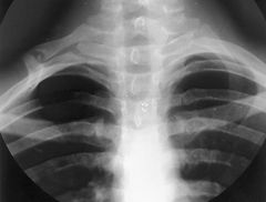
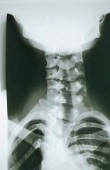
What does an A-P open mouth radiograph show? |
- atlantoaxial joint (C1-C2) - dens of C2 - lateral masses of C1 |
|
|
What does an A-P lower cervical radiograph show? |
lower 5 cervical vertebrae (C3-C7) |
|
|
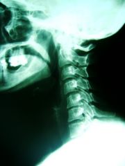
What does a lateral cervical radiograph show? |
alignment of all 7 vertebrae |
|
|
What do right and left oblique cervical radiographs show? |
single side IVF (intervertebral foramen) |
|
|
What does an anterior right/left oblique cervical radiograph show? |
It shows the IVF on the same side |
|
|
What does a posterior right/left oblique cervical radiograph show? |
It shows the IVF on the opposite side |
|
|
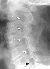
Where are the 3 nearly parallel lines drawn on a lateral view of the vertebrae? |
- anterior borders of vertebral bodies - posterior borders of vertebral bodies - junction of lamina to the spinous process (spinolaminar line) |
|
|
If the 3 nearly parallel lines do not line up, what could be suspected? |
- fracture - dislocation |
|
|
What does a marker on a radiograph represent? |
The view/side that is closest to the bucky, and the initials of the radiograph technician |
|
|
What are BBs called, and why are they used? |
- also known as Mitchell markers - can tell you if the radiographs were taken upright or supine - sometimes BBs are used to mark where pain is |
|
|
What is the likelihood of neurological damage with the fracture of a cervical vertebrae? |
- anterior column fracture = little chance of damage - middle or posterior column fracture = may be neurological damage - middle AND posterior column fracture = neurological damage is very likely |
|
|
What is contained in the prevertebral space? |
- prevertebral muscles (longus colli and capitis) - vertebral vessels - scalene muscles - phrenic nerve - proximal part of brachial plexus |
|
|
What are considered normal dimensions of prevertebral space in an adult? |
- Rule of 2's and 6's - Max = 6 mm at C2 and 22 mm at C6, anything greater is abnormal |
|
|
What are conditions that may cause the prevertebral space to become enlarged? |
- trauma of cervical spine - vertebral osteomyelitis - spondylodiscitis - vertebral metastasis |
|
|
What is radiculopathy? |
When a nerve group is being affected in the cervical spine, by nerve root near intervertebral foramen |
|
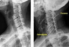
What view are these radiographs taken in? |
Oblique They also show growths in the intervertebral foramina |
|
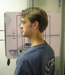
What view is this radiograph being taken in? |
Anterior oblique: facing the bucky, back to x-ray tube, and shows the same side IVF |
|
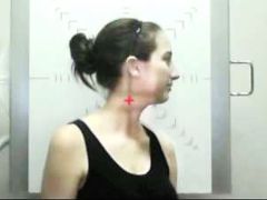
What view is this radiograph being taken in? |
Posterior oblique: back to the bucky, facing the x-ray tube, and shows opposite side IVF |
|
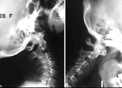
What type of views are these? What are we looking for in these views? |
- These views are lateral flexion and extension stress views - Looking for: preservation of 3 parallel lines and constant width of atlantodental interface (ADI space) |
|
|
What are the ABCs of analysis? |
A: alignment - following parallel lines on lateral x-ray B: bone - following the outline of each vertebra - check for steps and breaks C: cartilage - look for intervertebral discs and facet joints being displaced - disc space could also be widened of annulus fibrosus ruptures in DDD s: soft tissue - check for widening of soft tissues anterior to spine - the prevertebral space. Also look for widening of bony interspaces. |
|
|
When do we use CT scans? |
When there is any doubt about the integrity of the cervical spine on plain radiographs |
|
|
How are CT scans different than radiographs? |
- they provide greater detail of bony structures - show extent of encroachment on spinal canal by displacement or bone fragments - useful in assessing cervicothoracic junction, upper cervical spine, and any suspected fracture/misalignment |
|
|
What do MRI's provide that radiographs do not? |
- info about spinal cord and soft tissues - reveal cause of cord compression - show extent of cord damage and edema |
|
|
What are signs of trauma? |
- abnormal soft tissue - abnormal vertebral alignment - abnormal joint relationships |
|
|
What are some examples of stable injuries? |
- compression fractures - disc herniations - unilateral facet dislocations |
|
|
What is an unstable injury? |
- an injury that has immediate or potential risk to the spinal cord or nerve root - example: fracture dislocations and bilateral facet dislocation |
|
|
Which areas of the body are radiographs good at demonstrating fractures? |
They are good at representing fractures in the long bones - femur - tibia - fibula - humerus - radius - ulna |
|
|
Where in the body can fractures be missed by radiographs? |
They are missed in complex skeletal areas like: - carpals and tarsals - upper cervical spine - pars interarticularis (spondylolysis!) |
|
|
What is the Canadian C-Spine Rule? |
Following a head or neck trauma 1 or more of the following (who is at risk the most): - 65 years or older - dangerous mechanism of injury (MVA, fall from 3ft+, or axial load to head) - paresthesias in extremeties - <45 degrees of cervical rotation |
|
|
What are the minimum standard radiographic images taken to evaluate cervical spine trauma? |
- horizontal beam lateral - AP view - open mouth odontoid view |
|
|
When is it considered a bilateral dislocation/fracture? |
When the spine is displaced more than 50% |
|
|
Which radiograph views are always taken in stabilized supine in the trauma series? |
- AP - AP open mouth - lateral |
|
|
What are the common fractures in the cervical spine? |
Avulsion and compression |
|
|
What are the 2 types of compression fractures? |
- Top: tear drop fracture flexion injury - Bottom: flexion or diving |
|
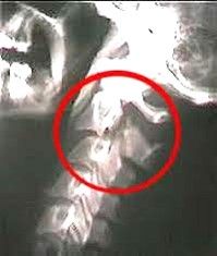
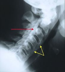
What type of fracture does this radiograph show? |
- Hangman's fracture - fracture of both pedicles of the axis |
|
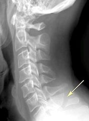
What type of fracture does this radiograph show? |
- Clay - shoveler's fracture - stable fracture through spinous process of vertebrae - occurs at any of lower cervical or upper thoracic (usually C6 or C7) |
|
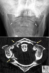
What type of fracture do these radiographs show? |
- Jefferson fracture - fracture of both anterior and posterior arches of C1 |
|
|
Why do isolated dislocations occur? |
They occur unilaterally at a facet joint because of a flexion rotation force |
|
|
When are unilateral dislocations stable? |
When there is no anterior translation |
|
|
Why do bilateral dislocations occur? |
They occur at a pair of facet joints due to hyperflexion force |
|
|
When are bilateral or unilateral dislocations unstable? |
when there is an anterior translation |
|
|
What indicates a facet dislocation in a radiograph? |
An anterior subluxation of one verterbrae on another. - < 50% of width of a vertebral body means unifacet dislocation - > 50% of width of a vertebral body means bilateral facet dislocation (accompanies by widening of interspinous and interlaminar spaces) |
|
|
Where do injuries caused by hyperflexion occur? |
It sprains the posterior ligaments and related soft tissues |
|
|
Where do injuries caused by hyperextension occur? |
It sprains the anterior ligaments and related soft tissues |
|
|
What is used to diagnose disc herniation? |
- MRIs (today) and myelography (past) - acutee disc herniation cannot be diagnosed by radiograph |
|
|
What marks a DDD (degenerative disc disease) on a radiograph? |
There is decreased disc space height on a lateral view radiograph |
|
|
What marks a DJD (degenerative joint disease) on a radiograph? |
In a lateral view there is decreased joint space, sclerosis, and osteophytes |
|
|
What marks cervical spine spondylosis on a radiograph? |
Osteophytes at the joint margins of the disc, and this is due to DDD |
|
|
What view is needed to see foraminal encroachment in the IVF area? |
an oblique view, this encroachment can cause impingement of spinal nerve |
|
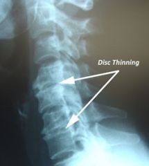
What pathology is found in this radiograph? |
Degenerative disc disease - shown with osteophytes |
|
What pathology is found in this radiograph? |
Degenerative joint disease of the facets (there is a decrease in the disc height of C5 - C6) |
|
What can you diagnose from this radiograph? |
There is foraminal encroachment at multiple levels which can: - result from DDD, DJD, spondylosis, dpinal stenosis, and disc herniation - result in spinal nerve compression - result in radiating arm pain |
|
|
What causes DDD? |
- > 60 years old - dehydration of disc - nuclear herniation - annular protrusion |
|
|
What can DDD cause? |
Decreased disk height, which causes: - endplate approximation - uncovertebral joint friction - spondylosis - schmorl's nodes - vacuum phenomenon |
|
|
What is spondylosis? |
spurring that forms at the vertebral end plates due to DDD |
|
|
What cervical degeneration diseases can show up with normal disc height on radiographs? |
- spondylosis deformans: extensive osteophytosis at anterior and lateral area of vertebral bodies - diffused idiopathic skeletal hyperostosis (DISH): ossification of at least 4 contiguous vertebrae with absence of DDD/DJD |
|
|
What can be found in the radiographs of 50% of patients with DISH? |
ossification of the posterior longitudinal ligament |
|
What pathology is present in this radiograph? |
- DISH: shown by large bridging of anterior osteophytes at level of C2 to upper thoracic - Ossification of posterior longitudinal ligament:shown by dense band of calcification along posterior area of vertebrae bodies **remember: disc heights are within normal limits** |
|
|
What type of anomalies can be found in radiographs? |
- failure of development - arrest in development - assymentrical development - development of accessory bones |
|
|
What could a small dens result in? |
instability of atlantoaxial joint and could be life threatening |
|
What pathology can you find in this radiograph? |
cervical rib |
|
What pathology can you find in this radiograph? |
Spina bifida |