Reading...
![]()
Play button
![]()
Play button
![]()
Use LEFT and RIGHT arrow keys to navigate between flashcards;
Use UP and DOWN arrow keys to flip the card;
H to show hint;
A reads text to speech;
33 Cards in this Set
- Front
- Back
|
Specialization
Muscle cells or fibers are highly differentiated to allow |
contraction
Elongated form allows shortening along axis, elongation give rise to the use of fiber Contain longitudinally oriented rod like contractile elements (myofibers) |
|
|
Classification of muscle fibers (cells)
|
Muscle fibers are divided into two categories based on the presence or absence of light microscopically visible alternating light and dark banks running transversely across the muscle fiber
|
|
|
Striated
|
Skeletal
Associated with the skeleton and fascial tissues of the body Is involved in gross body movement and tends to be under voluntary or conscious control Cardiac Confined to the heart Is involved in pumping blood though the body and is under involuntary control |
|
|
Smooth
|
No striations are visible under light microscope
Muscle usually occurs as an integral part of an organ where it makes up a major part of the organs wall Under involuntary control |
|
|
Size and shape of skeletal muscle
|
Long cylindrical cells with blunt/tapered ends
Fibers are multinucleated with nuclei located at the periphery of the cell just beneath the scacrolemma |
|
|
Nuclei of skeletal muscle
|
Nuclei are oval
Pale Basophilic Peripherally located chromatin 1 or 2 nuclei |
|
|
Sarcoplasm of skeletal muscle
|
Compromises organelles
Elongated sacrosomes Poorly developed Golgi Ribosomes Glycogen granules |
|
|
Myofibrils of skeletal muscle
|
Make up the bulk of the skeletal muscle fiber
Specific structure of these give rise to the striations visible Stain Eosinophilic |
|
|
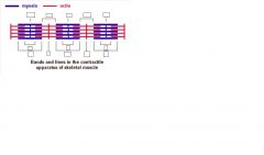
Striations of skeletal muscle
|
Alternating transversely orientated light and dark bands
Striations due to the arrangement of the myofibrils relative to each other as well as their own special structure |
|
|
the A BAND
H ZONE the I BAND Z LINE |
Dark band-
Pale region at middle Light band Dark line in middle |
|
|
Sacromere of skeletal muscle
|
The sacromere is the region of the muscle fiber between adjacent Z lines
It is the smallest regularly repeating unit and the basic contractile unit of the myofibril |
|
|
Myofibrils and myofilaments
|
Myofibrils are long rod like structures arranged parallel to each other and run longitudinally within the cell
They are made up of regularly arranged groups of myofilaments which are two types: Thick myosin myofilaments 15-16 nm diam Thin actin myofilaments 5-6 nm diam |
|
|
A BAND-
|
thick myosin filament span this region, resulting in dark staining
|
|
|
I BAND-
|
only thin actin filaments are present which results in pale staining
|
|
|
Actin filaments extending into the A BAND give an outer darker region and the paler
|
H ZONE (myosin only)
|
|
|
Z LINE-
|
this region is where actin filaments are anchored and kept in a particular array such that they lie between the myosin filaments within the A BAND
|
|
|
M LINE-
|
this is in the middle of the H ZONE where myosin filaments are anchored and kept in a particular spatial arrangement that accommodates the actin filaments
|
|
|

|
|
muscle type?
|
skeletal muscle
|
|
|
The sarcoplasmic reticulum
|
Extensive network of longitudinal tubules with transverse anastamoses and terminal dilations
Extends around and between the myofibrils forming interconnected sleeve like structures around these They are connected transversely but separated into discrete units at the A/I junctions These discrete units are either centered on the M line or Z line All myofibrils in a given region o f a muscle fiber are surrounded by an interconnecting network of sarcoplasmic reticulum which contains a high concentration of calcium ions. |
|
|
The transverse tubule system (T-system)
|
At the A/I junctions a system of slender transversely running tubules penetrate the muscle fibers and separate the terminal cisternae of adjacent sarcoplasmic reticulum units
These tubules also surround each myofibril in a given region The T-system is formed by invagination of sarcolemma and therefore contains extracellular fluid The region where the T-system tubules contact adjacent cisternae is called a TRIAD and is important in the initiation of muscle fiber contraction |
|
|
Contraction and innervations
Skeletal muscle fibers are innervated by |
motor nerves originating in the central nervous system
An action potential traveling down the nerve axon sets up an endplate potential which is propagated in an action potential in the sarcolemma This action potential then travels down the T-system tubules into the depth of the muscle fiber. At the triad this causes depolarization of the sacroplasmic reticulum with the result that Ca++ ins are released into the sarcoplasm surrounding the myofibrils On exposure to Ca++ ions the myosin and actin filaments interact such that the actin filaments are drawn between the myosin filaments toward the M line causing shortening of the sacromere and the muscle fiber. Relaxation occurs as Ca++ ions are withdrawn from the sacroplasm and returned to the sacrplasmic reticulum. |
|
|
Size and shape of cardiac muscle
|
Cylindrical in shape
Fibers branch and anastomse with each other |
|
|
Nuclei of cardiac muscle
|
Usually 1 but can be 2
Centrally located Oval with blunt ends Pale Basophilic |
|
|
Sarcoplasm and myofibrils of cardiac muscle
|
Eosinophilic
|
|
|
Striations for cardiac muscle
|
As for skeletal muscle but tissue possess intercalated discs
|
|
|
Intercalated discs
|
Distinct transverse dark bands scattered thought cardiac muscle tissue
Appear as straight or step like lines In addition to striation Regions of end to end contact between fibers and contain junctional complexes GAP JUNCTIONS on lateral part of discs transmits contractile impulses between fibers, allows sequential contraction DESMOSOMES AND FASCIA ADHERENS on transverse parts of discs anchor adjacent cells |
|
|
Control and innervations of cardiac muscle
|
Contraction is initiated and synchronized by specialized regions within the heart-the sinoatrial and atroventricular nodes
The transmission of contractile impulses via purkinje fibers and the gab junctions between adjacent muscle fibers Some modulation via autonomic nervous system |
|
|
Size and shape of smooth muscle
|
An elongated spindle shaped cell with finely tapered ends
|
|
|
Nuclei of smooth muscle
|
Single, located in middle widest part of the cell
Cylindrical in shape, but often wrinkled, twist or helical Basophilic with fine granular chromatin 1 or 2 |
|
|
Sarcoplasm and myofibrils of smooth muscle
|
Uniformly Eosinophilic
|
|
|
Multi unit innervations of smooth muscle
|
Each fiber has its own nerve ending which initiates contraction much like skeletal muscle
Allows small precise contraction |
|
|
Unitary innervations of smooth muscle
|
Single nerve will innervate a muscle bundle or syncytium of many fibers
Tends to have inherent spontaneous contractility Neural input tends to modify activity rather than initiate it Fibers tend to be linked by junctional complexes allowing for sequential coherent contractions as occurs in peristalsis |