![]()
![]()
![]()
Use LEFT and RIGHT arrow keys to navigate between flashcards;
Use UP and DOWN arrow keys to flip the card;
H to show hint;
A reads text to speech;
50 Cards in this Set
- Front
- Back
|
Mounting slides |
Dry mount - thin slice and then cover slip over Wet mount - suspended in liquid such as water or immersion oil Squash slide - wet mount then press down cover slip Smear slide - add small drop then use edge of cover slip to push along e.g. blood |
|
|
Light microscope |
Low resolution - 0.2um Low magnification - x1500 Advantages Easy to set up Can use live specimen Cheap Small and portable |
|
|
Magnification and resolution |
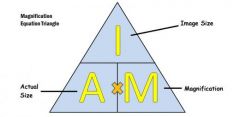
Magnification - how much bigger an image appears compared to the original object Resolution - the ability of the optical instrument to see or produce an image with fine detail. How well it can distinguish between 2 points close together without showing them together |
|
|
Magnification triangle |
|
|
|
Staining |
Some objects are completely transparent so need a stain to be able to view them Stains increase the contrast between different components as take up stains differently Methylene blue stains DNA as attracted to negative charge Differential staining - identify between 2 types of organism e.g. gram-positive/negative bacteria Counterstain - using 2 dyes to differentiate Need to chemically fix specimen before staining e.g. dehydrating |
|
|
Calibrating a light microscope |
To find the size of a specimen Use eyepiece graticule and stage micrometer Stage micrometer - 1mm and 100 divisions so 1mm = 0.1nm 0.1 (size of 1 stage division) / how many divisions on eyepiece Measure length of specimen on eyepiece x by answer of previous Need to recalibrate for different magnifications |
|
|
Units for size |
1nm - x1000 - 1um - x1000 - 1mm - x10 - 1cm - x100 - 1m 1m - /100 - 1cm - /10 - 1mm - /1000 - 1um - /1000 - 1nm |
|
|
Electron microscope |
Use beam of electrons as wavelength smaller than light Can see ultrastructure High magnification and resolution Expensive Lots of preparation as need specific conditions Need skill and training to use Need installing and are large Specimen must be dead |
|
|
TEM |
Transmission electron microscope (TEM) Beam of electrons is transmitted through the specimen Denser parts absorb more electrons so look darker Resolution - 0.0002um Magnification - x1,000,000 Specimen must be dead - dehydrated and stained so chemically fixed Specimen stained with metal salts Produces 2D black and white image False colour can be added |
|
|
SEM |
Scanning electron microscope (SEM) Beam of electrons across specimen and reflected electrons are collected Resolution - 0.002um Magnification - x500,000 Gives a 3D image in black and white but can add false colour Specimen must be dead as coated in a film of fine metal Done in a vacuum |
|
|
Magnifications and resolutions numbers |
Light 0.2um x1500 TEM 0.0002um x 1,000,000 SEM 0.002um x500,000 |
|
|
Laser scanning microscope |
Moves a single spot of focused light across causing fluorescence (gives off light) with components labelled with a dye Light emitted from specimen is filtered through a pinhole to a detector to produce an image than can be 3D Only light from close to focal plane is detected to give a clearer image Look at thick specimen in different depths High magnification and resolution Can use living specimen Used in biological research and diagnosis of diseases |
|
|
Plasma membrane structure |
On surface of animal cells Just inside cell wall in plants and prokaryotes Mainly made of lipids and proteins |
|
|
Plasma membrane function |
Regulates the movement of substances into and out of the cell Receptor molecules to respond to chemicals e.g. hormones |
|
|
Cell wall structure |
Rigid structure that surrounds plant cells Plants from carbohydrate cellulose Prokaryotes from peptidoglycan Absent from animal cell Fungi from chitin |
|
|
Cell wall function |
Prevents bursting when turgid Provides strength and support Maintain the cell’s shape Permeable to allow solutions to pass through |
|
|
Nucleus structure |
Double membrane called nuclear envelope Pores in the nuclear envelope Nucleolus doesn’t have a membrane and contains DNA Contains chromatin - genetic material of DNA wound on histone proteins When not dividing chromatin is spread out but coils and condenses into chromosomes when about to divide |
|
|
Nucleus function |
Separate contents from rest of the cell At some regions the outer and inner membranes fuse so dissolved substances and ribosomes can pass through Pores allow larger substances to pass through e.g. mRNA Ribosomes made in the nucleolus Contains genome and genetic information for protein synthesis Control centre of the cell |
|
|
Lysosomes structure |
Small bags surrounded by a single membrane Contain powerful hydrolytic (digestive) enzymes Abundant in phagocytes such as neutrophils and macrophages that ingest and digest invading pathogens |
|
|
Lysosomes function |
Keep powerful hydrolytic enzymes separate from the rest of the cell Engulf old cell organelles and foreign matter, digest them and use components for reuse |
|
|
Ribosomes structure |
Small, spherical organelles 20nm in diameterMade of ribosomal RNA Made in nucleolus as 2 separate sub-units and combine when pass through to cytoplasm Free in cytoplasm or on RER No membrane |
|
|
Ribosomes function |
Site where proteins are made RER ribosomes for synthesising proteins to export out of the cell Cytoplasm ribosomes assembly of proteins to be used in the cell |
|
|
Rough endoplasmic reticulum (RER) structure |
System of membranes containing fluid-filled cavities (cisternae) that are continuous with the nuclear membrane Coated with ribosomes |
|
|
Rough endoplasmic reticulum (RER) function |
RER is the intercellular transport system - the cisternae form channels for transporting substances from one area of the cell to another Provide large surface area for ribosomes and proteins pass through membrane into the cisterna to be transported to the Golgi apparatus |
|
|
Smooth endoplasmic reticulum (SER) structure |
System of membranes containing fluid-filled cavities (cisternae) that are continuous with the nuclear membrane No ribosomes |
|
|
Smooth endoplasmic reticulum (SER) function |
SER contains enzymes that catalyse reactions involved with lipid metabolism such as the synthesis of cholesterol, lipids/phospholipids and steroid hormones which are all needed by the cell Involved with absorption, synthesis and transport of lipids from the gut Synthesises and processes lipids |
|
|
Golgi apparatus structure |
Consist of a stack of membrane-bound, flattened sacs Vesicles bring material to and from the Golgi apparatus |
|
|
Golgi apparatus function |
Processes and modifies proteins Adding sugar molecules to form glycoproteins Adding lipid molecules to form lipoproteins Folding into 3D shape Proteins are then packaged into vesicles and pinched off |
|
|
Vesicles structure |
Small fluid-filled sacs in the cytoplasm Surrounded by a membrane |
|
|
Vesicle function |
Transport substances in and out of the cell via the plasma membrane Formed by Golgi, ER or plasma membrane |
|
|
Mitochondria structure |
Usually oval shaped 2-5um long Double membrane - inner membrane folded into cristae Inner part is fluid-filled matrix with enzymes for respiration |
|
|
Mitochondria function |
Site of ATP production during aerobic respiration Self-replicating so more is made if energy needs increase Abundant where much metabolic activity e.g. synapses |
|
|
Chloroplast structure |
Large organelle 4-10um longPlants and protocists Double membrane Inner membrane continuous with stacks of flattened membrane sacs called thylakoids which each contain chlorophyll. Each stack of thylakoids is a granum Fluid-filled matrix is stroma Chloroplasts contain loops of DNA and starch grains Grana linked by lamellae - thin pieces of thylakoid membrane |
|
|
Chloroplast function |
Site of photosynthesis Abundant in leaf cells in particular the palisade mesophyll layer |
|
|
Vacuole structure |
Plant cells only Membrane called the tonoplast Contains fluid - cell sap |
|
|
Vacuole function |
Maintain cell stability as when full it pushed against cell wall to make the cell turgid Turgid supports the whole plant |
|
|
Cilia structure |
Protrusions from the cell and are surrounded by the cell surface membrane Hair-like structure Formed from centrioles Contains microtubules in 9+2 formation with a ring of 9 pairs and 2 in the middle |
|
|
Cilia function |
Microtubules allow cilia to move so can move substances along the cell surface Epithelial cells in airways have cilia to move mucus |
|
|
Flagellum structure |
Like cilia but longer Stick out from the cell surface and surrounded by plasma membrane 9+2 formation |
|
|
Flagellum function |
Microtubules contract to make flagellum move Propel cell forward E.g. bacteria and sperm cells |
|
|
Centriole structure |
2 bundles of microtubules at right angles to each other Microtubules are made of tubulin protein subunits arranged to form a cylinder Small, hollow cylindersIn animal cell but only some plants |
|
|
Centriole function |
Before a cell divides , the spindle made of tubulin threads forms the centrioles Chromosomes attach to the middle part of the spindle and motor proteins walk along the tubulin threads, pulling chromosomes apart Involved in formation of cilia and flagella - centrioles multiply and line up beneath cell surface membrane and microtubules sprout outwards to form cilia |
|
|
Cytoskeleton structure |
Network of protein structures in cytoplasm Microtubules - straight cylindrical and made of protein tubulin Microfilaments - small, solid strands made of protein actin Cytoskeleton motor proteins |
|
|
Cytoskeleton function |
Support and strengthen the cell’s shape Move materials within the cell - track for motor proteins Form spindle before a cell divides to separate chromosomes Form cilia and flagella and cause them to move Support organelles and keep position |
|
|
Organelles working together - protein production |
Genes to code for the protein are in the nucleus and transcription to form mRNA mRNA moves out of nuclear pore Translation at the ribosome Pass into the cisternae of the RER Vesicle to Golgi apparatus where modified Vesicle to plasma membrane Fuse with plasma membrane to be released by exocytosis |
|
|
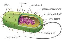
Prokaryotes |
Single-celled organisms No membrane-bound organelles Small cells 2um in diameter DNA is circular DNA is free in cytoplasm Cell wall peptidoglycan Flagella made of flagellin in a helix Small ribosomes |
|
|
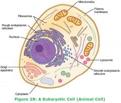
Eukaryotes |
Make up multi-cellular organisms Many organelles are membrane-bound Large cells 10-100umDNA is linear DNA in nucleus No cell wall in animals, plants cellulose, fungi chitin Flagella made of microtubules in 9+2 formation Large ribosomes |
|
|
Prokaryote cell diagram |
|
|
|
Eukaryotes cell diagram |
|
|
|
Bacteria |
Prokaryotic Protective waxy capsule surrounding cell wall Plasmids - small loops of DNA Flagella - enable movement Pili - hair-like structures to adhere to each other or host cells Nucleoid - area in the cytoplasm where the DNA is positioned Cell wall from peptidoglycan Prokaryotes divide by binary fission - DNA replicated in process No membrane-bound organelles |