![]()
![]()
![]()
Use LEFT and RIGHT arrow keys to navigate between flashcards;
Use UP and DOWN arrow keys to flip the card;
H to show hint;
A reads text to speech;
101 Cards in this Set
- Front
- Back
|
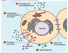
5 Functions of Membranes |
1) Boundary and permeability barrier. 2) Organization and localization of function. 3) Transport processes. 4) Signal detection. 5) Cell-to-Cell communication. |
|
|
5 Functions of Membranes Visual |
|
|
|
5 Roles of Membranes |
1) Define the boundaries of the cell and define its compartments – Permeability Barriers. 2) Serve as a loci (centers) of specific functions. 3) Possess transport proteins that facilitate and regulate the movement of substances [into and out of the cell and its compartment]. 4) Contains receptor [proteins] required for the detection of external signal. 5) Provide mechanisms for cell-to-cell communication. . |
|
|
Membranes: Defining Boundaries |
Physical separation. Desirable substances IN and undesirable substances OUT. Hydrophobic interior – permeability barrier for hydrophilic ions and molecules. Plasma Membrane - Permeability barrier of the cell. Intracellular membranes – Compartmentalize functions. |
|
|
Membranes: Sites of Specific Function |
Site of specific function - Special molecules and proteins embedded within membranes.
Membrane (Bio) Markers – protein or enzyme that associate with specific membranes e.g. Glucose phosphate – ER membrane protein. |
|
|
Membrane: Transport Regulation (Part 1) |
Concentration Gradients – molecule with no net charge.
Electrochemical potential – sum of its concentration gradient and the charge gradient across the membrane facilitate the movement of ions. |
|
|
Membrane: Transport Regulation (Part 2) |
Simple diffusion – water, oxygen, alcohol.
Facilitated diffusion – polar molecules e.g. sugars and amino acids moved across the membrane by transport proteins’.
Facilitated Water Movement – Aquaporins in Kidney cells. |
|
|
Membrane: Transport Regulation (Part 3) |
Active transport – against concentration gradient.
Direct active transport – requires ATP for ‘uphill’.
Indirect active transport – of the solute to ‘downhill’. |
|
|
Membrane: Transport Regulation (Part 4) |
Large Protein Transport
Endocytosis – into the cell – intracellular vesicles facilitate the movement of proteins.
Exocytosis – out of the cell – ER proteins or cytosolic proteins can be imported into membrane bound organelles such as lysosomes, peroxisomes, mitochondria. |
|
|
Membrane: Signal Detection (Part 1) |
Electrical or chemical signals on the outer surface of cell Signal Transduction – mechanisms that detect and direct specific signal in and out of the cell. |
|
|
Membrane: Signal Detection (Part 2) |
Chemical signal transduction – Hormone based
Estrogen – a polar steroid enter into target cell and interact – nonpolar molecule.
Some molecules need specific regulator protein.
Androgen – Androgen receptor.
Progesterone – Progesterone Receptor.
Hormones – ligands – binds to receptor and activate series of internal chemical signal – second messenger. |
|
|
Specific Transporters |
Glucose and Amino acid transporters. Nerve Cell – Na+ and K+ ion transporters (Channels). Muscle cell – Ca ++channels. Chloroplast – Phosphate Ions transporters. Mitochondria – Aerobic respiration intermediate transporters. |
|
|
Signal Detection |
Insulin receptor – on muscle and liver cells. Immune cell receptors. Light - sensing phytochromereceptors in plants. Membrane receptors. Hormone receptors. |
|
|
Cell Adhesion Mediators (Part 1) |
Intercellular communication by gap junctions in animal cells.
Intracellular communication by plasmodesmeta in plant cells. |
|
|
Cell Adhesion Mediators (Part 2) |
Intercellular communication by Cell to cell contact. Cadherins – membrane proteins extracellular domains – bind calcium ionsand stimulate adhesion between similar cells in a tissue. Pathogenic organisms – Listeria and Shigella use adhesive membrane proteinsto invade. |
|
|
Cell Adhesion Mediators (Part 3) |
Adhesive Junctions: holds cell together.
Tight Junctions: Form seals to block passage of fluids between cells.
Ankyrin – Membrane proteins – point of attachment to cell cytoskeleton. |
|
|
Important Functions of Biomembranes |
Compartmentalization Localization of function Transport Signal detection Intercellular communication |
|
|
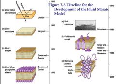
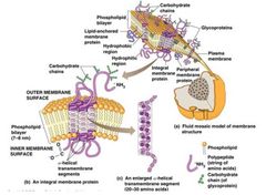
Fluid Mosaic Model of Membrane |
Different membranes. Composition of ‘material’ differ. Structure varies. Fluid mosaic model evolved. - Two fluid layers of lipids. - Proteins localized within and on the lipid bilayer. - Specific orientation. |
|
|
Fluid Mosaic Timeline Visual |
|
|
|
Lipid Monolayer (Part 1) (Experimental Perspective) |
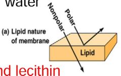
Overton: Lipids are important components – used cells of plant root hair (lipid soluble, water insoluble) - Lipophilic.
- lipids ‘coat’ on the cell surface.
- ‘coat’ a mixture of cholesterol and lecithin. |
|
|
Lipid Monolayer (Part 2) (Experimental Perspective) |
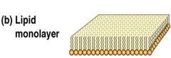
Langmuir: properties of purified phospholipids by dissolving in benzeneand layering on water. .
One molecule thick.
Phospholipids are amphipathic molecules. |
|
|
Lipid Bilayer (Experimental Perspective) |
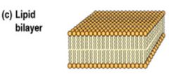
Gorter and Grendel: Lipids are important components – Extract lipids from known number of Erythrocytes - RBC.
Lipid film on water – lipid area twice the estimated total surface area of erythrocytes
Conclusion: Plasma membrane consists of two layers of lipids – LIPID BILAYER.
Wrong estimation: underestimated 1/3 of the surface and lipids. Also didn't account membrane proteins. |
|
|
Membrane Containing Protein (Experimental Perspective) |
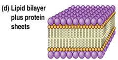
Davson and Danielli: studied Surface tension, Solute permeability, Electrical resistance of membranes.
Based on observations: movements of ions, hydrophilic solutes.
Importance of proteins in membrane structure.
Protein-lipid-protein Sandwich. |
|
|
Common Underlying Structures (Experimental Perspective) |
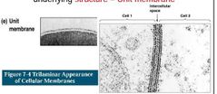
Robertson: Used electron microscope.
All cellular membranes share a common underlying structure – Unit membrane. |
|
|
Fluid Mosaic Model (Experimental Perspective) |
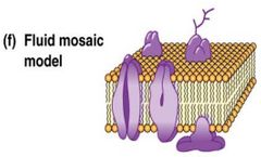
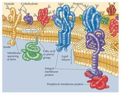
Singer and Nicolson: A membrane consists of a mosaic of proteins in a fluid lipid bilayer.
Mosaic of proteins.
Embedded in, attached to –fluid lipid bilayer. |
|
|
Membrane Proteins: Integral Membrane Proteins |
Integral Membrane Proteins – embedded within the lipid bilayerby affinity of hydrophobic segment of the proteins for the hydrophobic interior of the bilayer. |
|
|
Membrane Proteins: Peripheral Proteins |
Peripheral Proteins – hydrophilic and located on the surface – linked noncovalently to the polar head groups of phospholipids. |
|
|
Membrane Proteins: Lipid-Anchored Proteins |
Lipid-anchored Proteins – hydrophilic proteins – on membrane surface and covalently attached to embedded lipid molecules. |
|
|
Membrane Protein Visual |
|
|
|
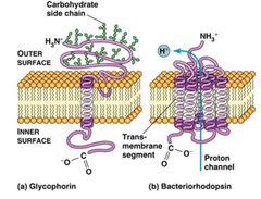
Transmembrane Segments (Experimental Perspective) |
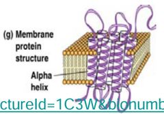
Unwin and Henderson: Most membrane contains transmembrane segments – bacteriorhodopsin – light absorbing molecule of Retinal (Halobacterium).
Seven transmembrane segments: anchor hydrophobic regions of proteins in lipid bilayer and align them. |
|
|
Recent Findings |
Membranes are NOT homogenous, freely mixing structures. Lipids and proteins are ordered within membranes. Outer membrane monolayer. Ordering occurs in dynamic microdomains call LIPID RAFTS. – Contains lot more cholesterol, sphingolipids (e.g. shingomylin) and less phosphatidylcholine. |
|
|
Recent Findings |
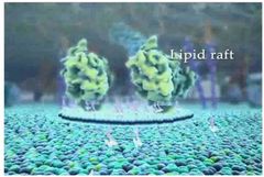
Serve as organizing centers for the assembly of signaling molecules. |
|
|
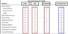
Protein, Lipid, Carbohydrate Ratios |
|
|
|
Singer and Nicolson: Fluid Mosaic Model Visual |
|
|
|
The Fluid Part of the Model: Phospholipid |
Phospholipid
- Glycerol based Phosphoglycerides.
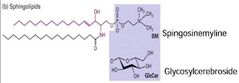
- Sphingosine based Sphingolipids. |
|
|
The Fluid Part of the Model: Glycolipids |
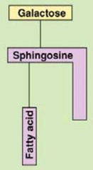
Glycolipids
- Carbohydrategroup attached to lipids.
- Glycerolas well as sphingosine based. |
|
|
The Fluid Part of the Model: Sterols |
Sterols
Animal Cell – cholesterol.
Plant Cell - phytosterol. |
|
|
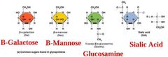
Glycolipids: Cerebrosidesand Gangliosides (Part 1) |
Glycolipids - Glycosphingolipids
Cerebrosides – Neutral glycolipids – single uncharged sugar attached as head group.
Gangliosides – One or more negatively charged sialic acid residues. |
|
|
Glycolipids:Cerebrosidesand Gangliosides(Part 2) |
Prominent in brain and nerve cell membranes.
Ganliosides on the surface – function as antigens.
Example – Cell surface markers (Glycosphingolipids) for ABO blood group. |
|
|
Tay-Sachs Disease |
Tay-Sachs Disease – absence of acetylhexosaminidasein lysosome.
– Accumulation of ganglioside in brain and nerve cells |
|
|
Sterols |
Mitochondria and chloroplast inner membrane - do not have sterols. Prokaryotes do not produce sterols. Prokaryotes produce - Hopanoids. |
|
|
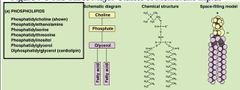
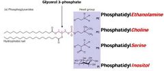
3 Major Lipid Membrane Classes: Phospholipid |
|
|
|
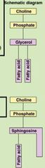
Schematic Diagram: (Part 1) Phospholipids (top) Sphingolipid (bottom) |
|
|
|
Schematic Diagram: Glycolipids |
|
|
|
Schematic Diagrams: Sterols |
|
|
|
Schematic Diagram: (Part 2) Phospholipids |
|
|
|
Schematic Diagram: (Part 3)Sphingolipids |
|
|
|
Cholesterol - The Steroid |
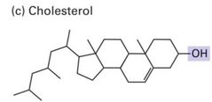
Third important membrane lipidAbundant in plasma membrane of mammalian cellsBasic structure –four membrane ringAmphipathic –due to the presence of a hydroxyl group. |
|
|
Sterol Substitutes in Bacteria and Cyanobacteria |
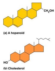
Rigid. Strong hydrophobic. Short hydrophilic side chain. Abundant in petroleum deposits – Membrane component of ancient prokaryotes. Sterols are not found in bacterium but found in MYCOPLASMA species. |
|
|
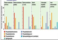
Different Membrane Compositions Visual |
|
|
|
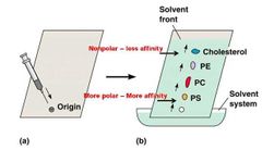
Lipid Analysis via Thin Layer Chromatography (TLC) |
Stationary Phase: Thin layer of silicic acid on glass or metal plate. Mobile Phase: Mixture of appropriate solvents. Chloroform, Methanol, Water |
|
|
Lipid Separation via Polarity |
Their Relative Affinities (based on polarity) for the stationary or mobile phase. Phospholipids (more polar) interact with silicic acid more strongly – slow their movement. Cholesterol (less polar) do not adhere strongly with silicic acid – move faster. |
|
|
Thin Layer Chromatography (TLC) Visual |
|
|
|
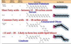
Fatty Acids and Membrane Function |
Fatty acids – components of all membrane lipids except the sterol. Long Hydrocarbon tails from hydrophobic barriers. Chain of fatty acids determine the thickness of bilayer. |
|
|
Common Fatty Acid Structures in Membranes Visual |
|
|
|
Membrane Asymmetry (Part 1) |
Membranes contains different kind of lipids.
Lipids are randomly distributed in TWO MONOLAYERS.
Variety of cell types – different lipids are unequally distributed between two monolayers – Membrane Asymmetry. |
|
|
Membrane Asymmetry (Part 2) |
Differences - both in the kinds of lipids present and in the degree of unsaturation of the fatty acids in the phopholipids molecules. Glycolipids – in animal membrane are mostly restricted to the OUTER of the two monolayer – facilitate signaling events. |
|
|
Membrane Asymmetry (Part 3) |
Phospholipids – prominent in the INNER monolayer – transmit signals from plasma membrane to interior of the cell. Phosphatidylethanolamine. Phosphotidylinositol. Phosphotidylserine. Phosphotidylcholine. |
|
|
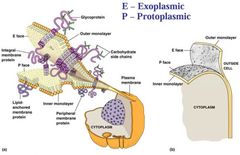
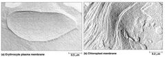
Freeze Fracture: Membrane Proteins |
Freeze the sample – Membranes. Slice through a diamond Knife. - Opens two layers. - Nonpolar lipid bilayer is the path of least resistance. - Split the lipid bilayer into its inner and other monolayers. |
|
|
Freeze Fracture Visual |
|
|
|
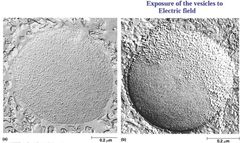
Phospholipid Movement within Membrane |
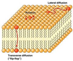
Phospholipid Molecules exhibit three kinds of movement in a membrane. Rotation along long axis. Lateral diffusion by exchanging place with neighboring molecule. Transverse diffusion OR Flip-Flop. Phospholipid translocators or Flippase. |
|
|
Recovery from Photobleaching Visual |
|
|
|
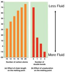
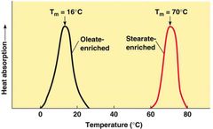
Fluid State: Proper Membrane Function |
Transition Temperature causes Phase Transition. Composition of Fatty acids affect membrane fluidity. Sterols also affect fluid state. Organisms can regulate membrane fluidity. |
|
|
MP of Fatty Acids vs. Chain Length and # of Double Bonds Visual |
|
|
|
Packing of Saturated Fatty Acids |
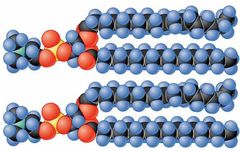
Saturated fatty acids pack well together. |
|
|
Packing of Saturated and Unsaturated Fatty Acids |
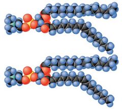
Lipids with a mixture of saturated and unsaturated fatty acids don't pack well together in the membrane. |
|
|
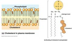
Cholesterol Orientation within Bilayer |
|
|
|
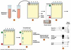
Separation of Proteins (Part 1) |
Isolation of Membrane proteins. SDS - Polyacrylamide Gel electrophoresis. SDS – sodium dodecyl sulfate (detergent). Polyacrylamide – matrix. |
|
|
Separation of Proteins (Part 2) |
Separation based on charge.
Molecular Weight markers to follow progress of electrophoresis.
Gel Stained with coomassie brilliant blue. |
|
|
SDS-PAGE Electrophoresis Gel |
|
|
|
Freeze Fracture of Membrane Protein Visual |
|
|
|
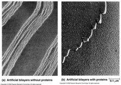
Freeze Fracture of Bilayer w/w-out Protein Visual |
|
|
|
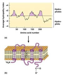
Hydropathy Analysis of Membrane Protein Visual |
|
|
|
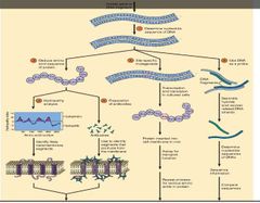
Application of Techniques to Study Membrane Proteins Visual |
|
|
|
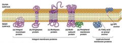
Main Classes of Membrane Proteins |
|
|
|
Membrane Proteins (Part 1) |
Integral Membrane proteins – embedded within the lipid bilayer by affinity of hydrophobic segment of the proteins for the hydrophobic interior of the bilayer. |
|
|
Membrane Proteins (Part 2) |
Peripheral Proteins – hydrophilic and located on the surface – linked noncovalently to the polar head groups of phospholipids.
Lipid-anchored Proteins – hydrophilic proteins – on membrane surface and covalently attached to embedded lipid molecules. |
|
|
Three Main Tyles of Membrane Proteins |
Based on the conditions required to extract membrane proteins – Nature of their association with the lipid bilayer. Integral Membrane Proteins. Peripheral Proteins. Lipid-Anchored Proteins. |
|
|
Integral Membrane Proteins (Part 1) |
Three parts - two hydrophilic and one hydrophobic.
Amphipathic.
Hydrophobic embedded in lipid bilayer. |
|
|
Integral Membrane Protein (Part 2) |
Integral Monotopic proteins. Transmembrane proteins. - Single pass. - Multipass. - Multisubunit multipass. |
|
|
Integral Membranes Protein (Part 3) |
20 –30 AA with hydrophobic R group. Helical conformation hydrophobic domains. Sheets or barrel. Examples Porins, glycoporeins, band 3 protein, anion exchange proteins. |
|
|
Structure of Two Membrane Proteins Visual |
|
|
|
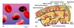
Peripheral Membrane Proteins |
Lack hydrophobic domains. Bound to membrane (polar heads) surface by (noncovalent) hydrogen bonding and electrostatic forces. Easily removed from membranes by changing pH and ionic strengths. Examples – Spectrin, ankyrin, band 4.1 in erythrocytes. |
|
|
Erythrocyte Plasma Membrane Visual |
|
|
|
Lipid-Anchored Membrane Proteins (Part 1) |
Fatty Acid Anchored Proteins. Synthesized in cytosol. Covalently attached to saturated fatty acid. Usually Myristic (14 carbon) acid or Palmitic (16 carbon) acid. |
|
|
Lipid-Anchored Membrane Proteins (Part 2) |
Prenylated Anchored Membrane Proteins •Synthesized as soluble cytosol proteins. •Covalently attached to saturated fatty acid. •attached to Isoprenyl (5 carbon) group. |
|
|
Lipid-Anchored Membrane Proteins(Part 3) |
Usually in the form of Farnesly(15 carbon) group or 20 carbon Geranylgeranylgroup.
GPI anchor – Glycosyl-phophatidyl-inositol. |
|
|
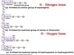
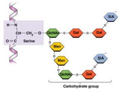
N-Linked and O-Linked Glycosylation Visual |
|
|
|
Carbs found in Glycoproteins Visual |
|
|
|
Carbohydrate Group Visual |
|
|
|
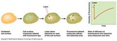
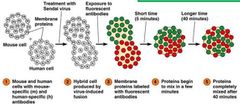
Cell Fusion Experiment |
David Frye and Michael Edidin.
Fluorescent tagged Antibodies – Anti-mouse Green (Fluorescein) and Anti-human Red (Rhodamine) colored.
Sendai Virus Infection which fuses eukaryotic cells.
Conclusion – Lateral diffusion of protein by fluid bilayer of plasma membrane. |
|
|
Cell Fusion Experiment Visual |
|
|
|
Mobility of Membrane Proteins Visual |
|
|
|
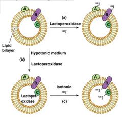
Labeling Membrane Proteins Inside and Out of Cell Visual |
|
|
|
Membrane Proteins: Variety of Functions (Part 1) |
Enzymes Electron Transport Proteins Transport Proteins – Sugar, Amino Acids Channel proteins –Na, K, Cl etc. |
|
|
Membrane Proteins:Variety of Functions(Part 2) |
Transport ATPases are Channel Proteins. Receptors. Autophagy – digest own organelles. Structural Roles – Stabilizing and Shaping. |
|
|
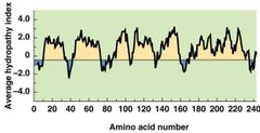
Integral Membrane Protein: Hydropathy Plot |
|
|
|
Heat Absorption of Membranes with Various Protein Concentrations Visual |
|
|
|
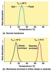
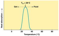
Heat Absorption of Normal Membrane |
|
|
|
Heat Absorption of Membrane Enriched with Oleate or Stearate Visual |
|
|
|
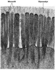
Glycocalyx of Intestinal Epithelial Cells Visual |
|