Reading...
![]()
Play button
![]()
Play button
![]()
Use LEFT and RIGHT arrow keys to navigate between flashcards;
Use UP and DOWN arrow keys to flip the card;
H to show hint;
A reads text to speech;
27 Cards in this Set
- Front
- Back
|
What are six causes of dilated CM?
|
1. idiopathic
2. infective 3. ischemic 4. toxic 5. peripartum 6. metabolic |
|
|
What are four categories of an infective cause?
|
viral
bacterial fungal parasitic |
|
|
What are four categories of a toxic cause?
|
alcohol
adriamycin lead cobalt |
|
|
What is a category of metabolic cause?
|
thiamin deficiency
|
|
|
What will be apparent in regard to LV walls and chamber?
|
Increased LV mass with thinned walls and
dilated chamber |
|
|
What function will be impacted and what happens to EF?
|
LV systolic function
is severely reduced, 15-25% |
|
|
What is a common infective cause of DCM?
|
Chagas disease
|
|
|
What will be apparent regarding LV walls with Chagas?
|
posterior and apical thinning with septum usually WNL
|
|
|
What is another infective cause of DCM?
|
AIDS
|
|
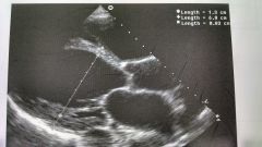
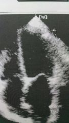
What does this show?
|
dilated cardiomyopathy
|
|
|
What are some physical signs?
|
symptoms of heart failure including dyspnea, fatigue, edema
|
|
|
What valves might be involved and how?
|
MR and TR
|
|
|
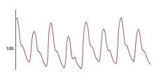
What two EKG findings might there be?
|
sinus tachycardia
pulsus alternans |
|
|
pic of pulsus alternans
|
|
|
What will be noted on echo in regard to chamber size?
|
multichamber enlargement
|
|
|
What will be noted in regard to LV contractility?
|
globally impaired
|
|
|
What is one M-Mode finding that may be noted and why?
|
B notch on MV due to increased LVEDP
|
|
|
pic of mv b notch
|
|
|
What is another M-mode finding?
|
reduced MV excursion shown as double diamond
|
|
|
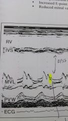
What is a another M-mode finding that may be pressent?
|
increased e-point to septal separation (EPSS) over >7mm
|
|

|
increased epss
|
|
|
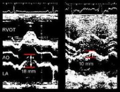
What may be seen in regard to the aortic root?
|
reduced aortic root excursion
|
|
|
pic of reduced aoric root excursion
|
|
|
What are two additional echo findings possible?
|
small pericardial effusion
thrombus |
|
|
What is a common solution for end-stage CM?
|
heart transplant
|
|
|
What two things will be significantly different on echo with a heart transplant patient?
|
double atria and 2 P waves on EKG
|
|
|
double atria following heart transplant
|