Reading...
![]()
Play button
![]()
Play button
![]()
Use LEFT and RIGHT arrow keys to navigate between flashcards;
Use UP and DOWN arrow keys to flip the card;
H to show hint;
A reads text to speech;
12 Cards in this Set
- Front
- Back
|
What is the Brugada Pattern?
|
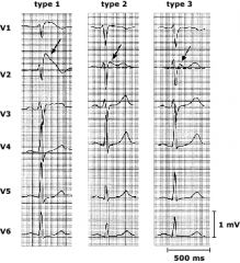
ECG findings in the precordial leads (especially V1, V2, and V3) consisting of a "pseudo-RBBB" (meaning rabbit ears are visible in V1 and V2) and ST-segment elevation.
Type 1 is the most obvious type, with down-sloping ST elevation in V1 or V2 descending to an inverted T-wave ("coved type" Brugada pattern). Type 2 and 3 have "saddleback" type ST segments, elevated at the beginning and end but drooping in the middle. The T-waves are usually upright or biphasic. |
|
|
What are the criteria for STEMI?
|
1) ST elevation of 1mm or more in 2 or more contiguous leads. The one exception is precordial leads V2 and V3 where 2mm is acceptable in men, and 1.5mm in women.
2) ST depression in the anterior leads and ST elevation in aVR, in a setting where true posterior MI is suspected. 3) New or presumed new LBBB with signs/symptoms suggestive of acute MI. |
|
|
What are the Sgarbossa Criteria?
|
Sgarbossa are criteria used to diagnose STEMI equivalents in patients with LBBB or pacemakers (in whom normal STEMI criteria cannot be applied).
1) ST segment elevation of 1mm or more that is CONCORDANT with the QRS complex, in any lead. 2) ST segment depression of 1mm or more in any of leads V1, V2, and V3. The above 2 are the most important criteria. There is also a third criterion which is less specific and which alone does not necessarily suggest a STEMI: 3) Excessively discordant ST segment elevation (5mm or more) in any lead. |
|
|
What are the hallmarks of the "pre-excitation" pattern on ECG?
|
The main feature is the DELTA WAVE.
The presence of the delta wave gives the appearance of: 1) Shortening of the PR internal 2) Widening of the QRS. |
|
|
What are the symptoms of hypokalemia?
What ECG changes are characteristic of hypokalemia? |
Symptoms of hypokalemia generally fall into 3 categories:
1) Muscle symptoms. Initially muscle weakness, then, as the K+ falls, muscle cramps and even muscle ischemia and rhabdo (since the blood supply to muscles is mediated in part by K+ concentrations). 2) GI symptoms, including decreased GI motility, leading to anorexia, nausea, vomiting, and constipation. 3) ECG changes/arrythmias. Features include: - Decreased T waves and presence of U waves (high K+ causes the T waves to rise and become peaked) - Prolonged QT with risk for torsades (high K+ shortens the QT) - ST depression (high K+ may cause ST elevation in a Brugada-type pattern) - Signs of myocardial irritibility such as PACs and PVCs, as well as basically any brady- or tachy-arrythmia. |
|
|
What ECG changes are characteristic of hyperkalemia?
What are the indications for giving Calcium in Hyper-K+? |
1) Peaked T-waves (low K+ shrinks the T wave)
2) QT shortening (low K+ prolongs the QT) 3) Widening of the QRS (can be any pattern, e.g. RBBB, LBBB, others) 4) ST elevation in a Brugada-type pattern (low K+ can cause ST depression) 5) The pre-terminal rhythm shows a loss of p-wave and the stretching out of the QRS into a sine-wave. Then you die. Calcium is indicated for widening of the QRS or loss of P wave on ECG. Most people would not consider peaked T waves an indication to give calcium. |
|
|
What are the symptoms of hypercalcemia?
What ECG changes are characteristic of hypercalcemia? |
Stones - kidney stones - also excessive urination.
Bones - bone pain Moans - neuropsychiatric symptoms such as depression, anxiety, altered mental status, confusion, coma. Groans - nausea, vomiting, anorexia, constipation. The main ECG change is shortened QT (hypocalcemia, on the other hand, causes prolonged QT). |
|
|
What's the DDx of ST elevation on ECG?
|
Mnemonic - ABC HELP
1) A - Acute STEMI 2) B - Brugada syndrome (leads V1-V3) 3) C - CNS catastrophe (e.g. SAH) 4) H - Left ventricular hypertrophy (generally seen in leads V1-V3) 5) E - Benign Early Repolarization 6) L - LBBB 7) P - Pericarditis |
|
|
What are the absolute contraindications to thrombolysis in acute MI?
|
1) Any prior intracranial hemorrhage - EVER!
2) Known intracranial pathology such as tumor or AVM 3) Stroke within the past 3 months 4) Active internal bleeding (e.g. GI bleed) - menstruation doesn't count 5) Suspected aortic dissection or pericarditis Relative contraindications include: Any other intracranial pathology not listed above (e.g. old stroke, cerebral aneurysm, etc). Severe hypertension Recent trauma (2 wks), recent surgery (3 wks), recent internal bleeding (4 wks) Known peptic ulcers (without bleeding) Pregnancy Known bleeding problems (include warfarin if INR is supratherapeutic) |
|
|
In patients with NSTEMI, what are indications for immediate coronary angiography?
|
1) Hemodynamic instability/cardiogenic shock
2) Severe left ventricular dysfunction / heart failure 3) Recurrent or persistent rest angina 4) Sustained ventricular arrythmias |
|
|
What features of a new cardiac murmur should prompt a referral and/or work-up?
|
1) Grade 3 or louder.
2) Holosystolic 3) Symptomatic 4) Evidence of cardiovascular disease. 5) Abnormal ECG or CXR. 6) Murmur intensity increases with Valsalva or standing (suggests HCM or mitral valve prolapse). |
|
|
What ECG changes distinguish the 4 stages of pericarditis?
|
1) Acute - ST elevation, especially in I, V5, V6, and PR depression.
2) ST segments normalize, T-wave flattening. 3) T-wave inversion. 4) Back to normal. |