Reading...
![]()
Play button
![]()
Play button
![]()
Use LEFT and RIGHT arrow keys to navigate between flashcards;
Use UP and DOWN arrow keys to flip the card;
H to show hint;
A reads text to speech;
123 Cards in this Set
- Front
- Back
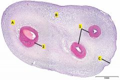
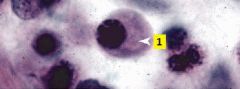
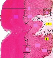
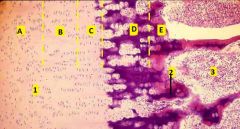
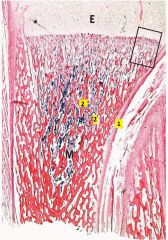
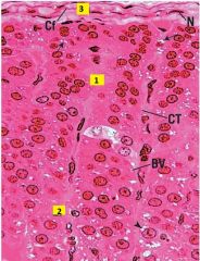
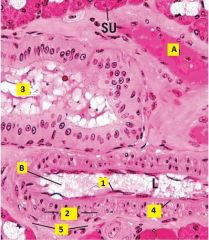
Where is this slide taken from?
Identify 1 and 2. Where is the best place to find mucous c.t., A, B, or C? |
Umbilical cord.
1 - umbilical arteries 2 - umbilical vein Mucous ct is best found is at the edge of the cord between the large vessel and one of the two small vessels. Mucous c.t. is a developmental c.t. that has far fewer collagen fibers than mature connective tissues. Hyaluronic acid comprises a large proportion of the ground substance in situ but is lost during tissue preparation. This results in the very loose appearance. |
|
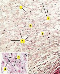
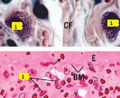
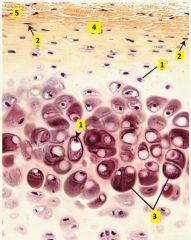
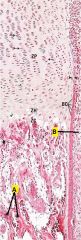
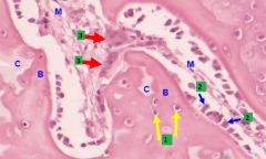
Image taken from umbilical cord. What type of tissue is shown?
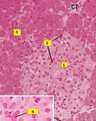
What type of cell is 1? Identify what kind of substance is labeled 2 and 3. What is 4 and 5? |
Mucous c.t.
1 - fibroblasts 2- ground substance (largely composed of hyaluronic acid) 3 - collagen fibers 4 - nucleus of fibroblast 5 - cytoplasm of fibroblast |
|
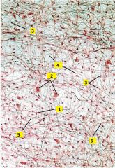
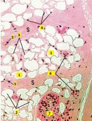
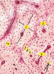
What type of connective tissue is this?
Identify the cells types of 1-3. Identify what type of substance is labeled 4-6. |
Areolar (loose) c.t. (whole mount of messentery)
1 - macrophage (small dense nucleus) 2 - mast cell (round nucleus with grainy cytoplasm) 3 - fibroblast (nucleus more pale than macrophage) 4 - ground substance 5 - elastic fibers (black, thin, straight, branching) 6 - collagen fibers (pink, thick, wavy, ribbon-like) |
|
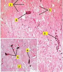
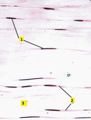
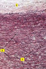
What type of connective tissue is this?
Identify 1-6. |
Dense irregular collagenous c.t. (from reticular layer of the dermis).
1 - collagen fiber bundles 2 - collagen fibril 3 - fibroblast nuclei. Inactive fibroblast, a fibrocyte, has a dense flat nucleus. Active fibroblastts have lighter, larger elongated nuclei. 4 - fibroblast cytoplasm 5 - blood vessel 6 - duct |
|
What type of cell is this?
|
Plasma cell
It has a round, eccentrically-located nucleus with the heterchromatin in a "clockface" pattern. Cytoplasm has light basophilia due to staining of the extensive RER. |
|
What type of cell is labeled 1?
|
Mast cell.
They have a round to oval nucleus and numerous small granules in the cytoplasm. |
|
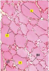
What type of connective tissue is this?
Identify 1-7. |
Adipose (unilocular) tissue (from the hypodermis).
1 - fat droplet 2 - adipocyte 3 - cytoplasm of adipocyte 4 - nuclei of adipocyte 5 - nuclei of fibroblast 6 - blood vessels 7 - sweat gland |
|
What type of connective tissue is this?
Identify 1-3. |
Dense regular collagenous c.t. (from muscle tendon)
1 - fibroblast (nucleus parallel with collagen) 2 - cytoplasm of fibroblast 3 - collagen fibers (thick pink, parallel bundles of collagen fibers) |
|
What type of connective tissue is this?
What type of fibers are 1 and 2? |
Dense regular elastic c.t. (from aorta, stained with orecin and H&E)
1 - collagen fibers 2 - elastic fibers Note: within tunica media collagen is secreted by smooth muscle cells instead of fibroblasts. Outermost layer (tunica adventitia) is loose areolar or dense irregular collagenous c.t. |
|
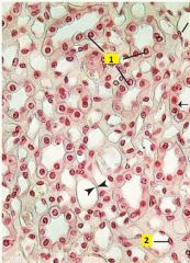
What type of cell is 1 and 2?
|
1 - Simple cuboidal epithelium.
2 - Simple squamous epithelium (appear flattened with nuclei bulging into lumen). Image of medulla of the kidney. |
|
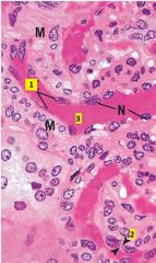
What type of cell is 1?
Identify 2 and 3. |
1 - simple squamous epithelium
2 - cytoplasm of simple squamous epithelium 3 - lumen of small arteriole (notice smooth muscle (M) lining of vessel) |
|
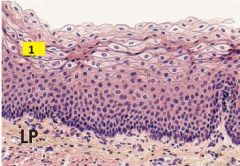
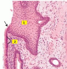
What of cells are labeled 1?
|
1 - stratified squamous epithelium.
Section from epithelium lining lumen of esophagus. |
|
What are the cell types of 1 and 2?
Identify 3 and 4. |
1 - stratified squamous epithelium
2 - simple squamous epithelium 3 - basal layer 4 - connective tissue |
|
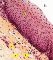
What type of cells are 1 and 2?
|
1 - stratified squamous epithelium
2 - simple columnar epithelium Image of esophagogastric junction (arrow). |
|
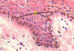
What of type of cells are labeled 1?
Identify 2-4. |
1 - transititonal epithelium (dome-shaped when bladder is relaxed, appear squamous when distended; occasionally binucleate)
2 - basement membrane 3 - veins 4 - arteriole (notice muscular layer) |
|
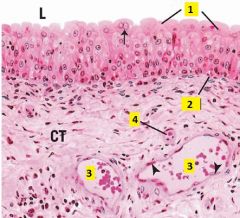
Identify the cell type of 1-3.
What are 4 and 5? |
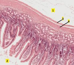
1 - simple columnar epithelium (nuclei are oval and at about the same level)
2 - goblet cell 3 - lymphocyte (small, deeply-basophilic round nucleus) 4 - brush border (microvili + glycocalyx) 5 - terminal web Image of villi of duodenum. |
|
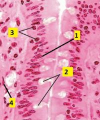
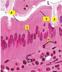
What type of cells are 1-4?
|
1 - simple columnar epithelium
2 - goblet cells (unicellular mucus-secreting glands) 3 - lymphocytes 4 - smooth muscle cell |
|
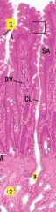
Identify 1-3.
|
1 - goblet cells
2 - multicellular gland (gland of Brunner) 3 - duct |
|
What type of cells are 1 and 2?
What is 3? |
1 - pseudostratified columnar epithelium (notice the several layers of nuclei on the basal side)
2 - simple squamous epithelium 3 - cilia |
|
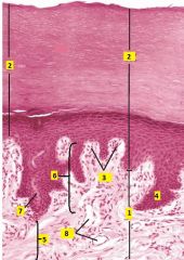
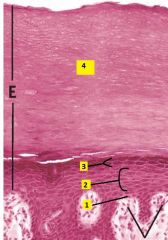
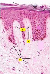
What type of tissue is this?
Identify 1-8. |
Thick skin
1 - dermis 2 - epidermis 3 - dermal ridges 4 - epidermal ridges 5 - reticular layer 6 - papillary layer 7 - duct 8 - blood vessels |
|
Identify the layers 1-4.
|
1 - stratum basale
2 - stratum spinosum 3 - stratum granulosum 4 - stratum corneum Stratum lucidum is not seen in this image |
|
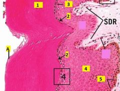
Identify the layers 1-5.
Identify A. |
1 - stratum corneum
2 - stratum lucidum 3 - stratum granulosum 4 - stratum spinosum 5 - stratum basale A - squames (very flattened, individual tile-like cells at apical surface which will be sloughed off). |
|
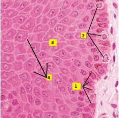
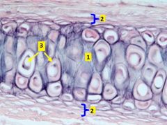
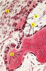
Image taken from dermal-epidermal junction. What type of cells are 1-3?
What is 4? |
1 - Basal keratinocytes (stem cells). Stratum basale is a single row of mitotic stems cells responsible for renewal of keratinocytes.
2 - Melanocytes. Pigment-synthesizing cells of skin, tend to be round and stain lighter; derived from neural crest, not keratinocytes. These are found in the stratum basale. 3 - Keratinocytes. Stratum spinosum is the thickest layer of keratinocytes; cells are cube-shaped and become flatter towards the stratum granulosum. 4 - Sites of desmosome attachments (structures have "spiny" appearance). |
|
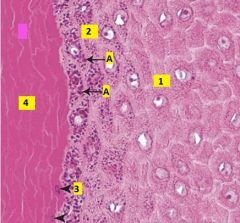
Image taken of epidermis of thick skin. What are the layers labeled 1-4?
Identify A. |
1 - Stratum spinosum
2 - Stratum granulosum. Usually 1-3 layers of flattened cells with numerous distinct keratohyalin granules. 3 - Stratum lucidum. Only present in thick skin, transitional cells in which nuclei and organelles are degenerated, and cytoplasm is becoming filled with keratin intermediate filaments. Stains lighter than corneum. 4 - Stratum corneum. Layer of dead, fully keratinized cells. A - keratohyalin granules (in keratinocytes of stratum granulosum). |
|
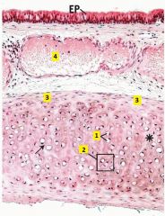
What type of skin is this?
|
Pigmented skin, A is highlighting a couple areas of melanin pigment. It appears brown and is mostly confined to thee basal keratinocytes.
|
|
Identify 1 and 2.
|
1 - capillary loop (persent in virtually every dermal papilla)
2 - dermal papillae (finger-like dermal projections) |
|
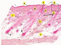
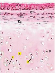
What type of tissue is this?
Identify the layers A,B, and C. Identify 1-6. |
Thin skin.
A - epidermis B - dermis C - hypodermis (loose c.t. with much adipose) 1 - sweat glands 2 - sebaceous glands 3 - duct 4 - hair follicle 5 - hair 6 - arrector pili muscle B - bulb of hair P - papilla of hair |
|
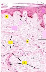
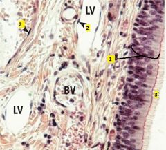
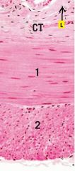
Identify 1 and 2.
What type of connective tissue is present at 3 and 4? |
1 - arrector pili muscles
2 - blood vessels 3 - papillary layer is composed of relatively loose irregular collagenous connective tissue (loose areolar c.t.). 4 - reticular layer is composed of dense irregular collagenous c.t. |
|
Identify 1-3.
|
1 - capillary loop
2 - dermal papillae 3 - collagen fibers |
|

This structure is found in the skin, what is it?
What portion of it is 1 and 2? Identify 3. |
Sweat gland. The base of a sweat gland (the secretory portion) is a highly coiled tube, hence a single section slices through the tube many times. This appears as multiple round profiles, each with a central lumen lined by cuboidal epithelium.
1 - secretory portion (simple cuboidal epithelium) 2 - duct portion (stratified cuboidal epithelium, stain darker) 3 - lumen |
|
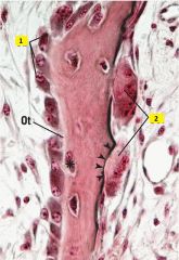
What type of structure is this that is found in the dermis?
Identify 1-3. |
Meissner's corpuscle - light touch sensory receptor present in those few dermal papillae that do not possess capillary loops. They lie just deep to the stratum basale.
1 - stratum basale 2 - capsule 3 - nerve fiber |
|
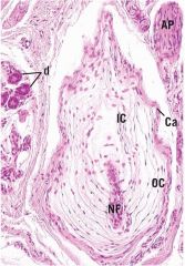
What type of structure is this which is found in the dermis?
|
Pacinian corpuscle - relatively large, light-staining structure that appears as a series of concentric rings. It is a mechanoreceptor.
NF - nerve fiber d - duct Ca - capsule AP - arrector pili OC,IC - outer, inner core |
|
What type of tissue is at the bottom of this image? Identify 1-4.
|
Hyaline cartilage (whole image is of trachea).
1 - chondrocytes 2 - isogenous group 3 - perichondrium 4 - vein |
|

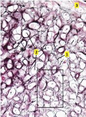
What type of tissue is this? What type of cells are 1 and 2?
Identify 3-5. |
Hyaline cartilage.
1 - chondrocytes 2 - chondrogenic cells (which differentiate into chondroblasts) 3 - isogenous groups 4 - chondrogenic perichondrium (inner cellular layer) 5 - fibrous perichondrium (outer fibrous layer) |
|
What type of cells are found in the area around the 5?
What type of cells are found in the area around the 4? |
Fibroblasts are found in the outer fibrous layer of perichondrium. They synthesize type I collagen.
Chondrogenic cells that divide and differentiate into chondroblasts are found in the inner cellular layer of the perichondrium. They are considered young chondrocytes. |
|
What is label 1?
What is responsible for the darker staining labeled 2? What is the predominant constituent of the area labeled 3? |
1 - lacuna.
2 - proteoglycan-rich matrix deposited by chondrocytes. 3 - predominant constituent of hyaline cartilage matrix is type II collagen. |
|
What type of tissue is in the center of this image? Identify 1-3.
|
Elastic cartilage (earlobe). Elastic cartilage has the same components of hyaline cartilage except that it has a high proportion of elastic fibers. Elastic fibers stain purple with orecin stain.
1 - matrix 2 - perichondrium 3 - chondrocytes in lacunae |
|
What type of tissue is this?
Identify 1-3. |
Elastic cartilage.
1 - elastic fibers 2 - chondrocytes 3 - perichondrium |
|
What type of tissue is this?
Identify 1-3. What type of fibers are located at 4? |
Elastic cartilage.
1 – inner cellular layer of perichondrium (chondrogenic) 2 – outer fibrous layer of perichondrium (fibrous) 3 – chondrocyte 4 – type II collagen and elastic fibers predominantly make up elastic cartilage matrix (elastic fibers are stained with orecin and appear purple). |
|
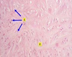
What type of tissue is this. What type of fibers are in this tissue that make it unique?
What type of cells are labeled 1? |
Fibrocartilage. Has characteristics of both hyaline cartilage and dense regular collagenous connective tissue. Relatively little cartilaginous matrix is present, but many parallel type I collagen fibers are packed together in bundles. Fibrocartilage does not have perichondrium.
1 - chondrocytes |
|

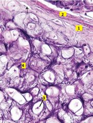
What type of tissue is this? Where is it normally found?
Identify 1 and 2. |
Fibrocartilage, primarily found in the pubic symphysis, intervertebral discs, and where tendons and ligaments insert into bone.
1 - chondrocytes 2 - fibrocartilage matrix (has type I collagen) |
|
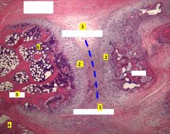
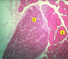
What is this image taken from?
Identify the tissue types 1-4. |
Pubic symphysis.
1 - fibrocartilage 2 - hyaline cartilage 3 - bone 4 - skeletal muscle 5 - bone marrow with adipose tissue Bone is deep red. Cartilage is variously gray with pink (collagenous) bands. The darker hyaline cartilage staining is due to glycosaminoglycans of its matrix. The spongy tissue (dark purple with white spots) is bone marrow with fat cells. Bottom left corner is skeletal muscle attached to the periosteum/perichondrium. |
|
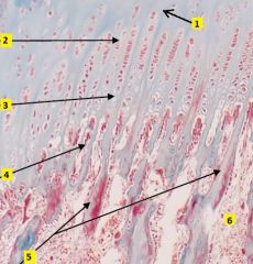
Where is this image taken from?
Identify 1-6. |
Epiphyseal plate.
1 - zone of reserve cartilage (chondrocytes randomly distributed) 2 - zone of proliferation (chondrocytes divide, stacked in columns) 3 - zone of hypertrophy (chondrocytes mature and hypertrophy) 4 - zone of calcification (chondrocytes die, matrix calcified) 5 - zone of ossification (bone matrix laid over calcified cartilage) 6 - marrow cavity of diaphysis |
|
Identify the zones A-E.
Identify tissue type for 1-3. |
A - reserve cartilage
B - proliferation C - hypertrophy D - calcification E - ossification 1 - hyaline cartilage 2 - bone formation (calcified cartilage is deep purple, bone is red) 3 - bone marrow |
|
Identify B.
What is occuring at A? |
Periosteum.
Bone matrix is being laid over calcified cartilage, resulting in bone spicules with pink bone matrix on the periphery and blue calcified cartilage in the center. |
|
What type of tissue is this?
What type of cells are 1 and 2? Identify 3 and 4. |
Developing bone.
1 - osteoblasts 2 - osteocytes in lacunae 3 - osteoid 4 - periosteum |
|
What type of cells are 1 and 2?
What is the depression that cell 2 makes? |
1 - osteoblasts
2 - osteoclasts (large, multinuclear) Osteoclasts form Howship's lacunae as they reabsorb bone. |
|
Identify 1 and 2.
|
1 - periosteum. Periosteum has two layers: outer fibrous layer made of dense irregular collagenous connective tissue; inner cellular layer and has osteoprogenitor cells.
2 - trabecula |
|
Identify the cell types for 1-3.
|
1 - osteocytes in lacunae
2 - osteoblasts lined along bone surface 3 - osteoclasts (mutilnucleate) |
|
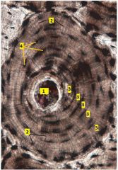
What type of tissue is this?
Identify 1-6? |
Compact bone.
1 - osteon (haversian canal with its surrounding lamellae of bone containing canliculi radiating to it from the osteocytes trapped in the lacunae) 2 - haversian canal (center of each osteon) 3 - lamella (ring layer of bone around haversian canal) 4 - interstitial lamella (portion of old osteon) 5 - Volkmann's canal 6 - lacuna (where osteocytes reside in situ, lost during preparation and filled with debris) |
|
What type of tissue is this?
Identify 1-4. |
Compact bone, stained with india ink.
1 - haversian canal 2 - canaliculum (in the living tissue were tiny "tunnels" possessing osteocyte cytoplasmic extensions) 3 - lamella 4 - lacuna |
|
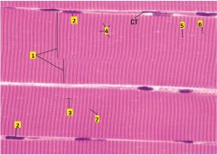
What type of tissue is this? Identify all the labels.
|
Skeletal muscle
1 - myofibrils (slightly out of register) 2 - myonuclei 3 - sarcomere 4 - Z disc 5 - A band 6 - I band 7 - H zone |
|

Which areas will get shorter when the muscle contracts?
|
6 and 7: Both the I band and H zone will get shorter when the muscle contracts. A band remains the same.
|
|
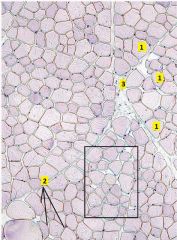
What type of tissue is this? Identify the labels.
|
Skeletal muscle, cross-section.
1 - muscle fibers 2 - myonuclei 3 - perimysium (surrounds fascicle) |
|
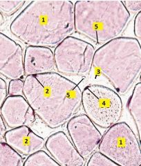
Cross-section of skeletal muscle. Identify the labels.
|
1 - muscle fiber
2 - myonuclei 3 - endomysium (surrounds muscle fibers) 4 - satellite cell (flatter and darker than myonuclei) 5 - capillary |
|

This is a TEM of what type of muscle?
|
Skeletal muscle. Cardiac muscle has a centrally located nucleus, and has many mitochondria and glycogen deposits in the sarcolemma.
|
|
Identify all the labels. What type of filaments are 7 and 8?
|
1 - Z disc
2 - I band 3 - A band 4 - H zone 5 - M disc (line) 6 - mitochondria 7 - thick myofilaments 8 - thin myofilaments 9 - triad (T tubules sandwiched between two terminal cisternae) |
|
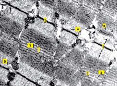
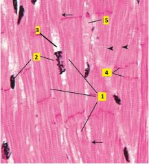
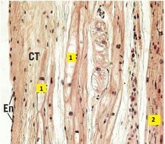
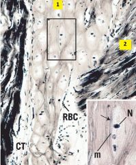
Identify where this image was taken from. Identify the labels. What type of tissue is 2?
|
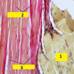
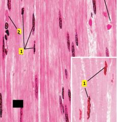
Muscle-tendon junction.
1 - skeletal muscle 2 - tendon (dense regular collagenous connective tissue) 3 - fibroblast nuclei |
|
Identify what this image is showing. Identify 1 and 2.
|
Muscle-tendon junction.
1 - skeletal muscle 2 - tendon (dense regular collagenous connective tissue) |
|
What type of tissue is this? Identify 1 and 2.
|
Smooth muscle cut longitudinally.
1 - smooth muscle nucleus (note the fusiform/corkscrew shape when they are contracted-inset). 2 - capillary |
|
What type of tissue is this? For 1-4, describe why the cells appear the way they do.
|
Smooth muscle in cross section (transverse section).
Smooth muscle fibers and their nuclei are spindle shaped; their nucleus is centrally located and at its greatest girth the nucleus is almost as wide as the cell. 1 - Cell cut at its greatest girth, appears as a round nucleus with a rim of cytoplasm. 2 - Cell cut at the tapered end of the nucleus. 3 - Cell cut in a region away from the nucleus. 4 - Cell cut at the tapered end of the cell. Transverse sections of smooth muscle appear to have a few cells containing nuclei of varying diameters. Most of the field is closely packed profiles of sarcoplasm containing no nuclei. |
|
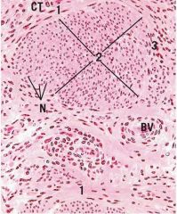
Image is of uterine myometrium. Identify the orientation that the smooth muscle cells were cut in area 1, 2 and 3.
|
1 - longitudinal
2 - transverse (cross section) 3 - oblique |
|
What type of tissue is this? Which side is the lumen, 1 or 2?
What are 3 and 4? |
Small intesting (duodenum).
2 is in the lumen. 3 - outermost layer of smooth muscle (parallel to length of intestine). 4 - innermost layer of smooth muscle (perpendicular to length of intestine). |
|
This image taken from the small intestine. What orientation was this tissue cut in?
|
Longitudinally. Outermost layer of smooth muscle runs longitudinally with the organ. If the innermost layer of the smooth muscle appeared longitudinally then it would have been cut in cross section.
|
|
This image is taken from a sample of the duodenum. In what orientation was it cut?
|
Cross sectionally. The outermost smooth muscle have been cut in cross section, therefore the organ has been cut that way. Outermost layer is parallel with organ.
|
|
Identify what 1 and 2 are.
What type of muscle is 3? What type of muscle is 4? |
1 - trachea
2 - esophagus 3 - smooth muscle 4 - striated upper third, smooth lower third, mixed in between. |
|

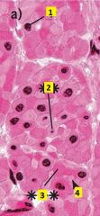
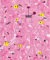
What type of tissue is this?
Identify region 1 and 2, and 3-5 within region 1. Identify 6-8. |
Adrenal (suprarenal gland).
1 - cortex, 2 - medulla 3 - zona glomerulosa, 4 - zona fasciculata, 5 - zona reticularis 6 - veins 7 - capsule 8 - adipose tissue |
|
What type of cells are found in region 2? Where are they derived from? What do they secrete?
|
Chromaffin cells, they are derived from neurons (they are modified sympathetic postganglionic neurons). They secrete norepinephrine or epinephrine.
|
|

What do cells in region 3 secrete?
What are cells in region 4 called? What do they secrete? What do cells in region 5 secrete? |
Mineralocorticoid hormones (major one is aldosterone) are secreted from cells in the zona glomerulosa.
Cells of the zona fasciculata are called spongiocytes because of their appearance due to the lipid droplets they possess. They secrete glucocorticoid hormones (cortisol and corticosterone. They are regulated by ACTH and CRF (CRF stimulates ACTH). Androgen hormones are secreted from cells in the zona reticularis. This area may be a zone of cellular degeneration. |
|
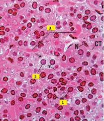
This is an image of the suprarenal gland. Identify 1-4. What type of cells are 5?
|
1 - capsule
2 - zona glomerulosa 3 - zona faciculata 4 - blood vessels 5 - spongiocytes |
|
This is an image of the suprarenal gland. Name the regions labeled 1-3.
What type of cells are in region 3? What type of cells are 4? What is 5? |
1 - zona faciculata
2 - zona reticularis 3 - medulla (chromaffin cells) 4 - spongiocytes 5 - veins |
|
Identify the regions labeled 1-3 in this image taken of a suprarenal gland sample.
|
1 - zona glomerulosa
2 - zona faciculata 3 - capsule |
|
Name the tissue labeled 1 and 2.
|
1 - parathyroid gland
2 - thyroid gland |
|
What type of tissue is this?
Identify 1-3? |
Thyroid gland.
1 - follicule 2 - follicular cells 3 - parafollicular cells |
|

Image of thyroid gland. Identify 1 and 2. What is 1 primarily composed of?
|
1 - colloid
2 - follicule The colloid is primarily composed of thryoglobumlin, the extracellular storage form of thyroid secretions. |
|
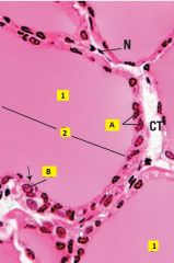
Image of thyroid gland tissue. Identify which cells are principal cells and which ones are C cells. What do they secrete?
|
A - principal cells (follicular cells); they secrete thyroid hormones (T3 and T4).
B - C cells (parafollicular cells); they secrete calcitonin. |
|
Identify the tissue type for 1 and 2. Identify 3 and 4. Identify the cell type for 5 and 6.
|
1 - thyroid gland, 2 - parathyroid gland.
3 - capsule, 4 - blood vessels 5 - chief (principal cells), 6 - oxyphil cells. |
|
High mag image of parathyroid tissue. Identify cell type of 1 and 2. Which one secretes hormones and which one?
Identify 3. |
1 - Chief (principal cells), secrete PTH. Variable staining intensity due to their level of activity.
2 - Oxyphil cells, function unknown (may be necrotic or inactive chief cells). Lighter and larger cytoplasm. 3 - Blood vessels. |
|
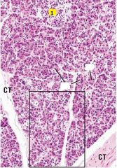
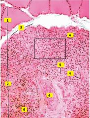
What type of tissue is this? Name the group of cells labeled 1.
|
Pancreas.
1 - islets of Langerhans CA - centroacinar cell AC - acinus |
|

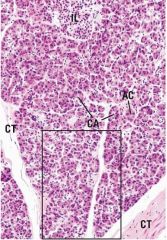
High mag image of a pancreas sample. Identify 1-3. What type of cells are 4.
|
1 - islet of Langerhans
2 - blood vessels 3 - acinar 4 - RBCs |
|
What do cells in the region labeled 1 secrete?
|
alpha cells - glucagon
beta cells - insulin delta cells - somatostatin (paracrine: inhibits horme release from alpha and beta cells; endocrine: reduces smooth muscle contraction in alimentary tract and gall bladder). G cells - gastrin PP cells (F cells) - pancreatic polypeptide (inhibitor of exocrine pancrease secretions). |
|
Which cell is this in erthropoiesis?
|
Erythrocyte.
When the reticulocyte loses its polyribosomes, it becomes a mature RBC. |
|
What are the layers in a test tube containing anticoagulent and blood after it has settled.
|
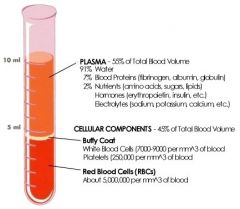
Red precipitate at the bottom is RBCs (45%), a thin light layer above that (~1%) which represents the leukocytes, and a yellowish fluid layer on top which is the plasma (~55%).
|
|
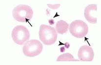
What are the arrowheads pointing at? What are the arrows pointing at?
|
Platlets (arrowheads). RBCs (arrows).
Platlets have a dense granular central region and clear peripheral region. RBCs have a central clear region. |
|
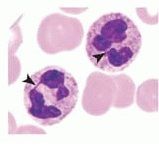
What are the cells with arrowheads on them?
|
Neutrophils.
The most numerous leukocyte, accounting for 60% to 70% of the total in circulation. Neutrophils are 12-15m in diameter and have a distinctive nucleus typically arranged into three lobes connected by small strands of chromatin (although it can have two to five lobes). The neutrophil cytoplasm has many small specific granules which are not obvious since their size is near the limit of the light microscope resolution. Azurophilic granules are also present in the cytoplasm. The cytoplasm stains lightly eosinophilic. |
|
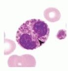
What type of cell is this?
|
Band neutrophil.
Neutrophil still in late stages of development (stab cell), in which the nucleus has not yet fully condensed and become lobed. Nucleus appears “horseshoe-shaped”. These comprise ~1% of neutrophils in a normal peripheral blood smear. |
|
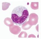
What type of cell is the arrow head on?
|
Eosinophil.
These cells derive their name from the numerous large, refractive granules that stain with eosin. Eosinophils only account for 2-4% of the leukocytes. They are about 12-15m in diameter and have a bilobed nucleus. The eosinophilic granules are membrane-bound, and their staining characteristic arises from a crystalline core composed of major basic protein. The crystalline core combined with the eosinophilia give the granules a somewhat ruby-like appearance in blood smears. |
|
What type of cell is this?
|
Basophil
Basophils comprise <1% of leukocytes. They are about 12-15m in diameter and have a lobed nucleus, but lobes are usually masked by the basophilic specific granules present in the cytoplasm. The intensely-stained, violet-colored specific granules are less numerous and more irregularly shaped than granules of other granulocytes. |
|
What type of cell is this?
|
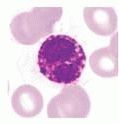
Monocyte
The largest circulating blood cell (~12 to 20m in diameter), monocytes account for 3% to 8% of the leukocytes. Monocytes typically have a large, eccentric kidney-shaped nucleus that stains light due to the less condensed chromatin. Cytoplasm stains lightly basophilic; azurophilic granules are present. |
|
What type of cell is this?
|
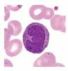
Lymphocyte
Round cells with a dense, round nucleus, constituting 20% to 25% of the leukocyte population. Small lymphocytes are by far the most common in circulating blood and appear as ~6-8m diameter cells with a thin rim of basophilic cytoplasm surrounding the predominant nucleus. Medium and large lymphocytes are up to 18m in diameter and are thought to represent lymphocytes that have been activated by specific antigens. |
|
What type of cells are the lines touching?
|
Megakaryocytes
Megakaryocytes are giant cells present in the bone marrow. They are not present in peripheral blood. The mature megakaryocyte is a giant cell (up to 150m in diameter) with a plethora of membrane invaginations throughout the cytoplasm. These cells appear to be multinucleate when observed in routine histological preparations, but each megakaryocyte actually has a single, large lobulated nucleus. |
|
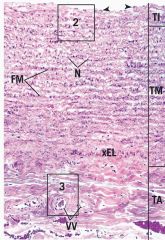
Name the tissue, the layers, and other features of it.
|
Elastic artery (aorta)
Layers: TI - tunica intima TM - tunica media TA - tunica adventitia Other features: FM- fenestrated membrane VV - vasa vasorum |
|
Identify A, B, C, and D of this section taken of tunica intima.
|
A - elastic fiber
B - smooth muscle cell C - nuclei of simple squamous epithelium D - nuclei of smooth muscle cell (corkscrew shape) |
|

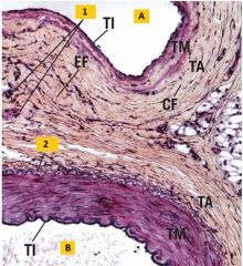
What type of tissue is this? What layers are A,B, and C?
Identify the features 1-5. What type of fibers are 6 and 7? |
Tissue: muscular artery (no fenestrated membranes)
A: tunica intima B: tunica media C: tunica adventitia 1 - subendothelial connective tissue; 2 - endothelial layer; 3 - internal elastic lamina; 4 - external elastic lamina; 5 - nucleus of smooth muscle; 6 - elastic fibers; 7 - collagen fibers. |
|
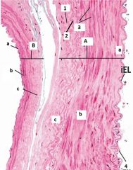
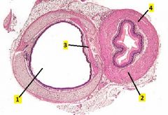
Identify tissue A and B.
What are the layers of a,b, and c of each? What type of fibers are 1 and 2? What type of cells are 3 and 4? |
A - muscular artery; B - medium vein.
a, b, c - tunica intima, media, adventitia, respectively. 1 - collagen; 2 - elastic 3 - smooth muscle; 4 - simple squamous endothelium |
|
Identify the lumen type of A and B.
Identify what 1 and 2 are. |
A - lumen of medium vein; B - lumen of muscular artery
1 - vaso vasorum; 2 - external elastic layer |
|
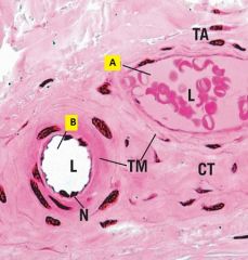
Identify the vessel labelled A and B.
Identify 1-5. |
A - venule; B - arteriole (observe lumen diameters and wall thickness).
1- nuclei of simple squamous endothelial cell; 2 - smooth muscle cells; 3 - duct; 4 - internal elastic layer; 5 - tunica adventitia |
|
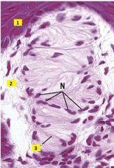
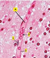
Identify the vessels labelled A and B.
Why is N bulging into the lumen of B? |
A - venule; B - arteriole (observe lumen diameter and wall thickness).
The nuclei of endothelial cells bulge into the lumen of arterioles due to the greater muscularity of its walls. |
|
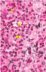
Identify A.
Identify 1-3. |
A - Capillary
1 - endothelial cell nuclei; 2- cytoplasm of endothelial cell (attenuates light, appears as thin dark lines bordering lumina of capillary); 3 - RBC. |
|
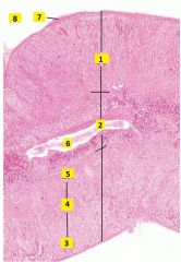
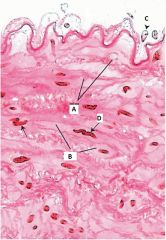
What type of tissue is this? Describe the different layers.
Identify 1. |
Large vein (inferior vena cava). Tunica intima – comparable in structure to that of medium veins, but subendothelial connective tissue is considerably thicker in the largest veins.
Tunica media – poorly defined or absent in most large veins. Tunica adventitia – thick and well-developed in most large veins. Innermost layer (1) has thick collagen bundles arrayed in spiral configuration. Middle layer (2) has smooth muscle longitudinally oriented. Outer layer (3) has thick bundles of collagen fibers interspersed with elastic fibers. 1 - vaso vasorum |
|
What type of tissue is this?
Identify 1-5. |
Cardiac muscle (longitudinal section).
1 - myofibrils 2 - nuclei (centrally located) 3 - perinuclear space 4 - intercalated discs 5 - capillary |
|
What type of tissue is this?
Identify 1-5. |
Cardiac muscle (cross section)
1 - blood vessel 2 - perinuclear space 3 - cardiac myocyte nucleus 4 - endothelial nuclei 5 - capillaries |
|
What type of tissue is this?
Identify 1 and 2. |
Endocardium. This lining of the heart consists of endothelium, a thin layer of subendothelial connective tissue with some collagen and elastic fibers, and a denser layer with elastic fibers and occasional smooth muscle cells
1 - Perkinje fibers 2 - Myocardium |
|
What type of tissue is this?
Identify 1 and 2. |
Ventricular myocardium.
1 - Perkinje fibers 2 - Cardiac myocyte |
|
What type of tissue is this?
Which cells are in cross-section and which are in longitudinal-section? |
Ventricular myocardium, example of myocardial layering.
Note the difference in the orientation of the cardiac myocytes. Left - cross section Right - longitudinal section |
|
What type of tissue is this?
Identify 1-3. |
Ventricular cardiac muscle.
1 - ventricular myocytes 2 - Perkinje fibers 3 - Fibrous skeleton of the heart (irregular collangenous tissue) |
|
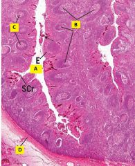
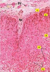
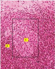
Identify this tissue.
Identify A, B, C, and D. |
Tonsil.
A - crypt B - lymphatic nodules (follicles) C - germinal center D - capsule |
|
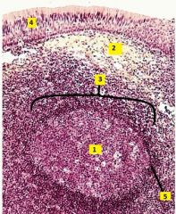
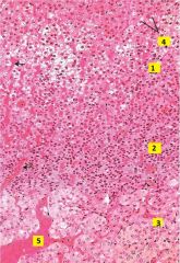
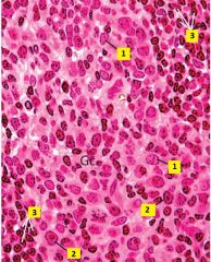
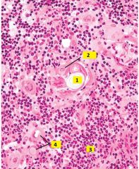
Tissue sample from a palatine tonsil. Identfiy 1-5. What type of cells are in the area of 1? The area where the 3 is?
|
1 - germinal center
2 - connective tissue 3 - secondary nodule (follicle) 4 - epithelium 5 - mantle (corona) Area 1 - lymphoblast, these mitotically-active lymphoblasts are aggregated in the center of the follicle and account for the light staining of the germinal center. They have undergone blast transformation to a larger cell with more cytoplasm and a larger, light nucleus. Area 3 - lymphocytes. |
|
Identify 1 and 2. Tissue taken from a lymph node.
|
1 - germinal center
2 - mantle (corona) |
|
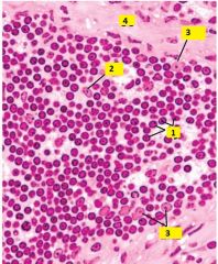
This image is of a germinal center in a lymph node. Identify the cell types of 1-3.
|
1 - lymphoblasts
2 - plasmablasts 3 - small lymphocytes |
|

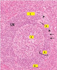
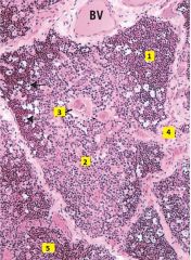
What type of tissue is this? Identify the regions 1-3. Identify what 4-9 are.
|
Lymph node.
1 - cortex, 2 - medulla, 3 - paracortex. 4 - trabeculae 5 - sinusoid 6 - medullary cord 7 - capsule 8 - lymphatic nodule 9 - germinal center |
|
This is the outer layer of a lymph node. What are 1 and 2?
|
1 - afferent lymphatic vessel
2 - subcapsular sinus |
|

Where are high endothelial venules found: 1, 2 or 3?
What type of cells are located in region 3? |
High endothelial venules are found in the paracortex (3). Cells present in the paracortex include llymphocytes, macrophages and interdigitating reticular dendritic cells.
|
|
What type of cells are located where the 6 is pointing?
What type of cells line the spaces 5 is pointing at? |
Medullary cords are dense aggregations of cells in the medulla; cells present in the cords are small lymphocytes, plasma cells and lymphoblasts.
Macrophages line the medullary sinuses which are extensions of the trabecular sinuses. |
|

Where is the point of entry and exit for blood vessels and exit for the efferent lymph vessels? What area would it extend from: 1, 2, or 3?
How would you distinguish efferent lymph vessels from veins? |
The hilum, extends from the medulla.
Veins have RBCs in them, efferent lymph vessels do not. |
|
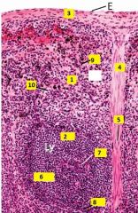
What type of tissue is this? Identify 1-4.
|
Spleen.
1 - white pulp 2 - red pulp 3 - capsule 4 - trabeculae |
|
This image is taken from spleen tissue. Identify 1-10.
|
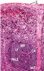
1 - red pulp. 2 - white pulp.
3 - capsule. 4 - trabeculae (septa) 5 - trabecular artery & veins (arteries arise from branches of the splenic artery and branch into the white pulp as central arteries; veins receive blood from pulp veins that collected filtered blood from the splenic sinuses. 6 - periarterial lymphatic sheath (PALS surround each central artery and are dense aggregates of lymphocytes, macrophages, and reticular cells. 7 - central arteries. 8 - marginal zone. 9 - splenic cords (of Bilroth) (areas between sinuses, packed with all of the formed elements of blood). 10 - Splenic sinuses |
|
This image is of an area of white pulp in spleen tissue. Identify 1-4.
|
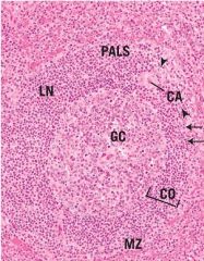
1 - PALS
2 - germinal center 3 - corona 4 - marginal zone (white pulp - red pulp border; this where branches of the central arteries end, dumping whole blood into splenic cords; this is therefore a major area for blood-borne antigen deposition). |
|
This image is of red pulp in spleen tissue. Identify 1-3.
|
1 - Splenic sinuses
2 - Splenic cords (of Bilroth) 3 - Endothelial lining of the sinuses |
|
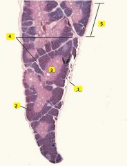
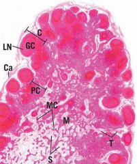
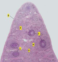
What type of tissue is this?
Identify 1-5. |
Thymus (multi-lobular; will undergo age involution and acquire more adipose tissue between lobules).
1 - capsule 2 - cortex 3 - medulla 4 - septum 5 - lobule (each lobule has an outer cortex and a inner medulla region). |
|
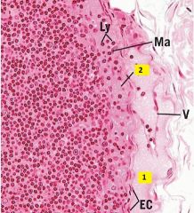
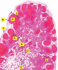
This image is taken from what type of tissue? Identify 1-5.
|
Thymus (only tissue with thymic corpuscles).
1 - cortex 2 - medulla 3 - thymic (Hassall's) corpuscle 4 - Connective tissue septa (trabeculae) 5 - lobule |
|
What type of tissue is this image from? Identify 1-4.
|
Thymic medulla (only tissue with thymic corpuscles).
1 - thymic corpuscle (Hassall's) 2 - epithelial reticular cell 3 - medulla (lymphocytes) 4 - blood vessels |
|
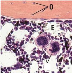
What area of thymic tissue is this image taken from? Identify type cells located at 1-3. What is 4?
|
Thymic cortex (adjacent to septum).
1 - lymphocyte 2 - macrophage 3 - epithelial reticular cell 4 - septum |