Reading...
![]()
Play button
![]()
Play button
![]()
Use LEFT and RIGHT arrow keys to navigate between flashcards;
Use UP and DOWN arrow keys to flip the card;
H to show hint;
A reads text to speech;
47 Cards in this Set
- Front
- Back
|
do microtubules grow and shrink in length during the lifetime?
|
yes....most of them
|
|
|
microtubules that form the spindle fibers during chromosome divsion are _________?
|
changing
|
|
|
microtubules in cilia are highly ______ in length
|
stable
|
|
|
where is there a lot of alpha-beta tubulin dimers ?
what does it do? |
in the cytoplasm
forms new microtubules at MTOC's by nucleation also, the pool of dimers also changes which dimer is on the positive end of the microtubule |
|
|
what is the growth or shrinkage of microtbule dependent on?
|
whether the rate of addition of new dimers is greater or less than the loss of old ones
|
|
|
dynamic instability
|
when no addition occurs to microtubules
they will shrink in length |
|
|
how long does it take for microtubules to shrink in length when no dimers are being added?
|
a couple of minutes
|
|
|
describe the position of microtubules in a plant cell during mitosis
|
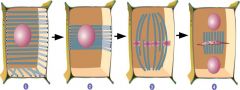
interphase: edge
approaching mitosis: no microtubules on cortex except pre-prophase band mitosis: spindle after mitosis: replaced by phragmoplast which separates daughter nuclei |
|
|
cilia
found in? movement? structure? |
hairlike motile structures
extensions of the plasma membrane with complex, microtubule-based cytoskeleton oar like movement epithelial cell lining : lungs and oviducts surface of ciliated protozoa |
|
|
oviduct
|
move particles, eggs along
|
|
|
epithelium
|
layers of cells that line the cavities and surfaces of cells throughout our body
|
|
|
flagella
structure in what? what does it do? |
hairlike motile structure
extensions of the plasma membrane with microtubule based cytoskeleton waves of motion run along the length of the flagellum: provide motile force to algea and protozoa |
|
|
how is the locomotion of an object determined by the movement of flaggella
|
it will always go the opposite direction of the power stroke
|
|
|
what happens after the power stroke in flagella and cilia?
|
there is a recovery stroke
|
|
|
where are the positive ends of microtubules in cilium?
|
at the tip of the cilium
|
|
|
motion of cilium requires what?
|
atp
|
|
|
dynein arms have what?
|
atp-ase activity
|
|
|
draw the structure of a cilia and flagella
|

corey joseph likes men
|
|
|
describe dyneins purpose in cilia and flagella structure
|
connect periphial microtubules
|
|
|
what family is the dynein in cillia?
|
axonemal dynein
|
|
|
where do cillia and flagella originate from?
|
basal body
|
|
|
why do cilia and flagella move?
|
1)dynein arm on alpha tubule attach to binding sites on beta tubule
2) a conformational change occurs in dynein which causes a power stroke 3) due to radial spokes this stroke can only go so far...(dyneins will release) 4) once |
|
|
size of intermediate filaments
|
intermediate in size between microtubules and actin microfilaminets
|
|
|
where have intermediate filaments been identified?
|
in animals only so far
|
|
|
plectins
|
wispy cross-bridging proteins
|
|
|
what are the filaments in cells linked by
|
plectins
sometimes microtubules |
|
|
what are filaments resistant to?
|
pulling
|
|
|
what is the general structure of intermediate filaments
|
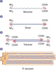
aa
|
|
|
do intermediate filaments have polarity?
|
no
|
|
|
where is keratinfound?
|
epidermal and epithelial cells
|
|
|
epidermal cells
|
outermost layer of skin
|
|
|
desmosins
|
join cells together
|
|
|
what kind of network do keratins form
|
elaborate cage like network
|
|
|
what is the outer layer of skin made from?
|
a waterproof airtight mat of keratin filaments
|
|
|
what happens to mice if they lack keratin
|
sensitve to mechanical pressure...skin blistering...human genetic condition ebs
|
|
|
name 3 types of intermediate filaments
|
keratin
neurofilaments desmin |
|
|
desmin
|
maintains aligment of myofibrils in smooth muscle cells
|
|
|
neurofilaments
|
run along nerve cell axons
|
|
|
what do neurofilament tangles cause
|
ALS....lou gehrigs disease
|
|
|
als
|
caused by neurofilament tangles
these impede normal vesicle transport down axons and lead to death of neurons |
|
|
what is the basis of the contractile cytoskeleton in muscle cells and non-muscle cells?
|
actin microfilaments
|
|
|
what is the structure of actin
|

composed of globular actin monomers G-actin
2 strands of monomeres are wound together into a stiff filament |
|
|
what supports the non-motile cellular structure like microvilli in forming the membrane skeleton beneath the plasma membrane?
|
actin microfilaments (F-actin)
|
|
|
is actin a major or minor protein in cells and how is it conserved
|
major
highly conserved |
|
|
how is F-actin polarized
|
has a + and - end
|
|
|
how can polarization of actin be visualized?
|
by adding myosin S1 head fragments which bind to and decorate the actin filaments
arrowhead appearence is created and the pointed end will be the minus end |
|
|
F-actin assembly
|
A pool of G-actin monomers exists in the cytoplasm.
G-actin binds ATP G-actin w/ATP bound can add onto both ends of filament, but much faster addition occurs at the (+) end. Since G-actin can also dissociate from the filament at both ends, net growth tends to occur at the (+) end and net loss is more likely, sometimes, at the (-) end. A dynamic equilibrium at both ends therefore determines filament length After addition, ATP is hydrolysed to ADP after some time. G-actins with ADP bound are more prone to dissociate from the filament. In fast-growing microfilaments a stabilizing “ATP-cap” is therefore created at the (+) end. |