Reading...
![]()
Play button
![]()
Play button
![]()
Use LEFT and RIGHT arrow keys to navigate between flashcards;
Use UP and DOWN arrow keys to flip the card;
H to show hint;
A reads text to speech;
21 Cards in this Set
- Front
- Back
- 3rd side (hint)
|
4 functions of cytoskeleton
|
1) maintain and direct cell structure
2)intracellular transport -organelle movement -vesicle movement 3)spatial organization of cell 4)contractility and mobility -actin/myosin contraction -movement of cilia and flagella -chromosome movement -cell migration...amoeboid movement |
|
|
|
3 major things that from cytoskeleton?
|
-microtubules
-intermediate filaments -microfilaments |
|
|
|
techniques to study cytoskeleton
|
1)Fluorescence microscopy
Immunofluorescence: Use of Antibodies against cytoskeletal proteins. Use of fluorescently-tagged drugs that bind to cytoskeletal proteins (e.g. phalloidin, binds to actin filaments) 2)Electron Microscopy (especially of freeze-etched surfaces) 3)Video-enhanced light microscopy – can detect presence of tubules and filament bundles, and follow movement of vesicles in living cells (but cannot resolve details of their structure) 4) genetic engineering -create knockout mutants in gene coding for cytoskeleton -ie...mouse embryos 5) GENETICALLY engineer cell line or organism to overexpress a dominant negative form of cytoskeletal protein -dominant negative forms are non functional and compete with native protein 6)siRNA -has complementary strand to mRNA so it will bind to mRNA and block translation |
|
|
|
what to microtubules form?
|
-mitotic spindles of dividing cells
-core of cilia and flagella -network of rigid tubules that radiate through the cytoplasm of all eukaryote cells |
3
|
|
|
what are microtubules formed from
|
alpha and beta tubulin
-tubulins are a diverse family with a 50KDA Mw |
|
|
|
microtubule structure
|
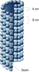
Microtubule structure:
-A rigid tube, whose wall is made from of approx. 13 protofilaments, composed of alternating alpha and beta tubulins. -Microtubules are approx 25 nm in diameter and up to a micron or more in length. -The filament is polarized, with a (+) and a (-) end. -NOTE this naming of the ends has NOTHING to do with electrical charge. The (+) end is composed of a row of beta tubulins, while the (-) end is composed of a row of alpha tubulins During microtubule assembly, the (+) end grows more rapidly than the (-) end |
|
|
|
proteins that bind to microtubules?
|
1) motor proteins- kinesins and dyneins
2)MAPS (microtubule associated proteins)- cross link MT's or regulate their assembly |
|
|
|
2 motor proteins
|
kinesins and dyneins
|
|
|
|
MAPs
|
MAPS (microtubule associated proteins)- cross link MT's or regulate their assembly
|
|
|
|
what is binding of MAPs to microtubules regulated by?
|
their own phosphorylation
|
|
|
|
Tau protein
|
a protein that regulates microtubule assembly in neurons: MAPS
over phosphorylation of tau may be involved in creating the tangled neuronal structure observed in alzheimers disease patients |
|
|
|
drugs that affect microtubules
|
-nocodazole: prevents microtubule assembly
-taxol-prevents microtubule disassembly ...both drugs prevent cell division sinse they disrupt the assembly of microtubules needed to allow chromosomes to seperate |
|
|
|
accepted chemotherapy agent for cancer treatment
|
taxol since it will prevent the rapid division of tumor cells...it also prevents mitosis in all other cells: hairfollicles>hair loss
|
|
|
|
what helps maintain the overall shape of many cell types?
|
rigid microtubule
|
|
|
|
microtubule bundles are importnat in maintaining the shape of
|
very elongated cell extensions or processes
ex. axons of nerve cells-treatment with microtubule-disorganizing drugs causes collapse of growing axons in the developing of nervous cells |
|
|
|
in plant cells, microtubule bundles at the edge of cytoplasm dirrect what?
|
direct cellulose deposition in the neighbouring cell wall by influencing the positioning of cellulose-synthesizing enzymes
|
|
|
|
if a drug is used on microtubules, what could happen to the golgi complex?
|
may become dispersed throughout the cell instead of being just outside of the nucleus.
|
|
|
|
draw the structure of a sensory nerve cell
|
..draw it
|
|
|
|
where are proteins synthesized in nerve cells
|
cel bodies...close to nucleus
|
|
|
|
do all areas of nerve cells require proteins?
|
yes even though the proteins are made in the cell body
|
|
|
|
describe fast axonal transport
|
Proteins, peptides are synthesized in the cell bodies of nerve cells, close to the nucleus but are required throughout the nerve cell – even in the distant nerve terminals at the end of long axons
Most of these materials are compartmentalized into vesicles in the ER and Golgi complex of the cell body which are then transported down the axon along microtubule “tracks”. Other vesicles are moving in the opposite direction, carrying waste materials and regulatory factors back from the nerve terminals to the cell body. This “fast axonal transport” can proceed at rates up to 5 microns per second (400 mm per day) This transport is mediated by motor proteins that move along the microtubules. It requires ATP |
|