Reading...
![]()
Play button
![]()
Play button
![]()
Use LEFT and RIGHT arrow keys to navigate between flashcards;
Use UP and DOWN arrow keys to flip the card;
H to show hint;
A reads text to speech;
41 Cards in this Set
- Front
- Back
|
What do alpha neurons innervate? Beta neurons? Gamma neurons?
|
Alpha - skeletal muscle
Beta - smooth/cardiac muscle and glands Gamma - muscle spindles |
|
|
Which membranes cover the brain?
|
Dura mater
Arachnoid Pia mater |
|
|
T or F:
Since white matter is the least susceptible to anoxia, brain stem is less susceptible to anoxia than the cerebral cortex. |
True!
|
|
|
From which embryonic layer is the nervous system derived?
|
Ectoderm
|
|
|
What does the neural tube become? The neural crest cells?
|
Neural tube - neurons and neuroglia
Neural crest cells - Spinal and autonomic ganglia and neurolemmocytes |
|
|
What are the three parts of the 3-vesicle stage of neurulation?
|
Prosencephalon
Mesencephalon Rhombencephalon |
|
|
What are the three parts of the 5-vesicle stage of neurulation? What is each derived from (from 3-vesicle stage)?
|
Telencephalon and Diencephalon (Prosencephalon)
Mesencephalon (Mesencephalon) Metencephalon and Myelencephalon (Rhombencephalon) |
|
|
What are the ultimate fates of regions of the 5-vesicle stage of neurulation?
|
Telencephalon > Cerebral hemispheres
Diencephalon > Thalamus, Hypothalamus, and Epithalamus Mesencephalon > Midbrain Metencephalon > Pons Myelencephalon > Medulla |
|
|
Failure of neural groove to close correctly leads to which condition(s)?
|
Anencephaly and spina bifida
|
|
|
What are the four functional divisions of the CNS?
|
1. Spinal cord
2. Brain stem 3. Cerebellum 4. Cerebral hemispheres |
|
|
What comprises the brain stem?
|
Medulla
Pons Midbrain Diencephalon |
|

Identify I thru V
|
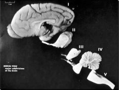
I - Telencephalon (Cerebral hemispheres)
II - Diencephalon (Thalamus/Hypothalamus/Epithalamus) III - Mesencephalon (Midbrain) IV - Metencephalon (Cerebellum and Pons) V - Mylencephalon (Spinal cord) |
|
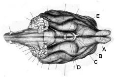
Identify these brainiac structures
|

A - Olfactory bulb (CN I)
B - Optic Nerve (CN II) C - Optic Chiasm D - Piriform lobe E - Optic tract |
|
|
T or F:
The Cisterna Magna and lumbosacral space are acceptable for a CSF tap in a dog. |
False.
The lumbosacral space is too far caudal. Should be done at L5 or L6. |
|
|
T or F:
The Cisterna Magna and lumbosacral space are appropriate regions for CSF taps in horses. |
True!
|
|
|
Where are two places to draw CSF from a horse and how should they be oriented in each situation?
|
1. lumbosacral space with the horse standing
2. Cisterna Magna with the horse sedated. Point needle toward lower jaw and NOT the eye! |
|
|
Where does the spinal cord end in the following species: man, dog, horse
|
Man - L2
Dog - L6 Horse - S2 |
|
|
What spinal cord segments lie under L3?
|
L3 and cranial L4
|
|
|
T or F:
the caudal portion of L4, L5, L6, and cranial L7 all lie under the L4 vertebra. |
True! Lotsa stuff under this one...
|
|
|
Which spinal cord segments are under L5?
|
Caudal L7, S1 thru S3
|
|
|
Which spinal cord segments are under L6?
|
Coccygeal 1 - 5
|
|
|
Name the 6 major FUNCTIONAL regions of the spinal cord and their approximate location (in the dog).
|
1. Upper cervical C1 - C5
2. Cervical enlargement C6 - T1 3. Thoracic & upper lumbar T2 - L3 4. Lumbar enlargement L4 - S2 5. Sacral S1 - S3 6. Caudal (coccygeal) Ca1 - |
|
|
Which spinal cord segments would be likely be found under L4 in a Boston Terrier?
|
L5 - L7 (all of L7). Small dogs are ~1 segment more caudal!
|
|
|
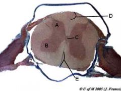
ID these parts of a spinal cord cross section
|
A - Dorsal horn
B - Ventral horn C - Central canal D - Dorsal median sulcus E - Ventral median fissure |
|

ID these parts of a spinal cord cross section
|
A - Dorsal horn
B - Ventral horn C - Central canal D - Dorsal median sulcus E - Ventral median fissure |
|
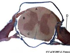
ID these spinal cord cross sectional regions
|

A - Dorsal funiculus
B - Lateral funiculus C - Ventral funiculus D - Dorsal root ganglion |
|
|
What do a ganglion and nucleus have in common? How do they differ?
|
Both are collections of nerve cell bodies. Ganglia are outside the CNS and nuclei are within the CNS
|
|
|
What structure produces CSF? What cells are responsible for this?
|
Choroid Plexus
Modified Ependymal Cells |
|
|
Through which structure does CSF flow from the lateral ventricles into the third ventricle?
|
The Interventricular foramen (Foramen of MOnro)
|
|
|
What structure connects the third and fourth ventricles?
|
Mesencephalic aqueduct (cerebral aqueduct of Sylvius)
|
|
|
Where does most CSF exit the ventricular system?
|
Via the lateral recesses
|
|
|
What is the region where the pia mater contacts ependymal tissue called? Add in the vessels contacting the pia mater and what do you have now?
|
Tela choroidea
Choroid plexus |
|
|
What is the only way to decrease CSF production?
|
Increase the osmolality of the blood
|
|
|
How long does it take to move drugs from the lateral ventricle to the lumbosacral space?
|
~40min (in humans)
|
|
|
What condition arises from an obstruction of internal CSF flow pattern?
|
Hydrocephalus
|
|
|
What are the four regions of the neocortex?
|
Frontal lobe
Parietal lobe Temporal lobe Occipital lobe |
|
|
In which meningeal space is CSF found?
|
Subarachnoid
|
|
|
Which glial cell myelinates axons in the CNS?
|
Oligodendrocyte
|
|
|
What is the main structure at the cellular level that makes the blood-brain barrier effective?
|
Nonfenestrated capillaries with tight junctions between endothelial cells
|
|
|
Which space inside the brain does not contain choroid plexus?
|
Cerebral aqueduct
|
|
|
What is the optimum location for a CSF tap in a golden retriever?
|
L5-L6
|