Reading...
![]()
Play button
![]()
Play button
![]()
Use LEFT and RIGHT arrow keys to navigate between flashcards;
Use UP and DOWN arrow keys to flip the card;
H to show hint;
A reads text to speech;
25 Cards in this Set
- Front
- Back
|
From which embryonic layer does the heart derive?
|
Splanchnic mesoderm
|
|
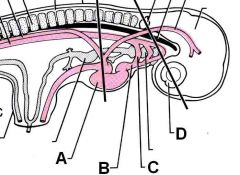
ID these embryonic structures:
|

A - Tubular heart
B - Ventral aorta C - Aortic arch D - Optic cup |
|
|
What gives rise to endocardial tubes in embryonic cardiac development?
|
cardiogenic plate or field (forms anterolateral to neural plate)
|
|
|
What forms the single endocardial tube?
|
L and R endocardial tubes fuse
|
|
|
T or F:
The mammalian heart forms in the cervical region then moves to the thoracic region. |
True!
|
|
|
What is the name for the defect where the heart is not in its correct location?
|
Ectopia cordis
|
|
|
What are the regions of the tubular heart (from cranial to caudal)?
|
Truncus arteriosus
Bulbis cordis Ventricle Atrium Sinus Venosus |
|
|
What is the fate of the embryonic regions of the tubular heart?
|
Truncus arteriosus - aorta and pulmonary trunk
Bulbus cordis - part of R ventricle Ventricle - L. Ventricle Atrium - R and L Atria Sinus Venosus - R side > coronary sinus; L side > R. atrium |
|
|
Describe the initial folding that occurs in the tubular heart.
|
Bulbis cordis and ventricle bend caudo-ventrally and to the right (fall to the right).
Atrium moves dorsocranially. |
|
|
What condition occurs when the bulbus cordis falls to the left?
|
Dextrocardia (R heart displacement)
|
|
|
What condition occurs when the body symmetry is flipped?
|
Situs inversus
|
|
|
What structure(s) develop(s) to divide the ventricle into two sides?
|
Interventricular septum
Also the endocardial cushions part of the spiral septum |
|
|
The failure of which structure to develop correctly causes a ventricular septal defect?
|
Failure of the endocardial cushion and/or the spiral septum to develop the membranous portion of the ventricular septum
|
|
|
T or F:
Perforations in septum I and septum II comprise the foramen ovale. |
False!
Only the perforation in septum II makes up the foramen. Septum I comprises the valve of the foramen ovale. |
|
|
What causes the foramen ovale to close?
|
Increased pulmonic venous pressure post pariturition closes the foramen.
|
|
|
What structure divides the truncus arteriosus? What does the truncus arteriosus divide into?
|
Spiral septum.
Aorta and pulmonary trunk |
|
|
T or F:
The spiral septum contributes to the interventricular septum. |
True!
If there is an issue with the spiral septum; VSD always occurs. |
|
|
What process creates cardiac valves?
|
Erosion
|
|
|
What are the features of the Tetralogy of Fallot?
|
Pulmonary stenosis
Overriding aorta (aorta too wide) VSD Hypertrophy (Also PDA) |
|
|
How many aortic arches are present (usually)? What is the fate of each arch?
|
5 to 6
Arches 1, 2, and 5 degenerate Arch 3 becomes internal carotid R arch 4 becomes R subclavian L arch 4 becomes aortic arch Arch 6 becomes pulmonary aa. and ductus arteriosus |
|
|
Which arch becomes the ductus arteriosus?
|
Left Arch 6
|
|
|
Why aren't two aortas better than one?
|
Double aortic arch squishes the trachea and esophagus
|
|
|
What closes the ligamentum arteriosum?
|
Nothing! The ligamentum arteriosum is ALREADY closed! However, it was closed by post-pariturition factors (oxygen concentration methinks).
|
|
|
What forms the L and R hepatic veins? What is the embryologic origin of these structures?
|
L and R vitelline veins from the yolk sac.
(vitelline is greek for yolk sac apparently) |
|
|
What is the fetal bypass structure for the liver?
|
Ductus venosus
|