Reading...
![]()
Play button
![]()
Play button
![]()
Use LEFT and RIGHT arrow keys to navigate between flashcards;
Use UP and DOWN arrow keys to flip the card;
H to show hint;
A reads text to speech;
140 Cards in this Set
- Front
- Back
|
Which CN's have their nuclei in the brainstem?
|
CN 3-12
|
|
|
What are the only two cranial nerves that come from the midbrain?
|
CN 3 and 4
|
|
|
What structures are in the brainstem?
|
midbrain, pons, medulla
|
|
|
What structures connects the brainstem with the rest of the brain?
|
the peduncles
|
|
|
What lobe do the cerebellar peduncles connect the brainstem to?
|
the frontal lobe
|
|
|
Now we talk only about cranial nerve exit points!
|
ok!
|
|
|
Why is it useful to know where the cranial nerves are exactly?
|
We can use this to make clinical inferences on which part of the brain is damaged.
|
|
|
What is CN 3 called?
|
occulomotor nerve
|
|
|
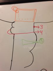
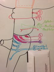
Draw where CN3 exits from the side.
|
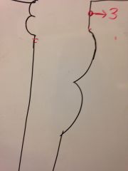
|
|
|
Draw where CN3 exits from the front and where it's nucleus is.
|
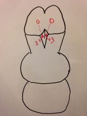
|
|
|
What is this little groove area called?
|
the interpeduncular fossa or cistern.
|
|
|
What is the only CN to exit out the back of the brainstem?
|
CN 4, trochlear nerve
|
|
|
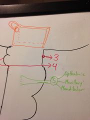
Draw where the CN4, trochlear nerve exits laterally.
|
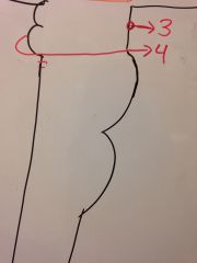
|
|
|
Narrate where the CN4 exits and goes.
|
it comes out of the inferior colliculus and then comes around to the front just under CN3, occulomotor.
|
|
|
Imagine where the 3rd ventricle and the pineal gland is in relation to the brainstem.
|
|
|
|
Why does CN4 come out the back?
|
it used to innervate the pineal gland, but lost it's function there.
|
|
|
What did the pineal gland used to do?
|
It was our very primitive version of the eye! (OMG the 3rd eye thing is true!)
|
|
|
What reminants of innervation goes to the pineal gland?
|
the modern visual system still has some connections there.
|
|
|
What info do our eyes give the pineal gland?
|
How much light/dark exposure there is.
|
|
|
What does the pineal gland do in response to this information?
|
It secretes melatonin
|
|
|
What cranial nerves come out of the pons?
|
only 3 and 4
|
|
|
What is the CN5 called and why?
|
trigeminal nerve because it is a massive nerve and gives off 3 branches
|
|
|
Draw what the 5th CN looks like from the side.
|
|
|
|
What level does CN5 come out of?
|
mid pons
|
|
|
Draw the CN 5 branches from the side and label them.
|
|
|
|
Is CN 5 largely sensory or motor? Mnemonic?
|
sensory because we can use it to detect dura lesions via referred pain.
|
|
|
What travels below the large sensory CN5? Draw it.
|
A trigeminal motor nerve.
|
|
|
Draw the frontal view of CN5.
|
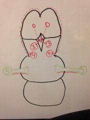
|
|
|
So what area of the brainstem is CN5 in?
|
The anterolateral part of the pons.
|
|
|
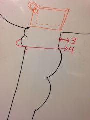
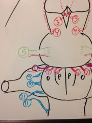
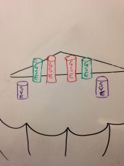
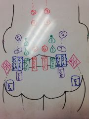
Draw the frontal view of the exits of CN 6, 7, and 8. Show how 7 and 8 attach.
|
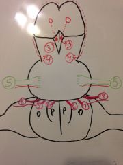
|
|
|
What can you deduce if CN 6 and 7 are both disturbed?
|
There is a lesion at the medial pontine-medullary junction
|
|
|
What can you deduce if CN 7 and 8 are both disturbed? Why?
|
that there is a lesion at the lateral pontine-medullary junction because they meet later on when CN7 travels laterally.
|
|
|
Draw the lateral view of CN 6, 7, and 8.
|
|
|
|
What are the names of CN 6, 7, and 8?
|
CN6- abducens
CN7- facial CN8- Vestibular/cochlear |
|
|
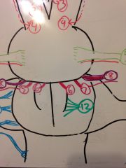
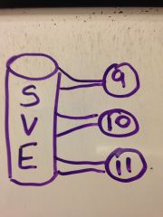
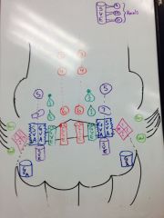
Draw the lateral view of CN 9, 10, and 11.
|
|
|
|
What defines the borders of CN 9,10, and cranial part of 11?
|
the inferior cerebellar peduncle and the olives
|
|
|
What is the name for CN 9?
|
glossopharyngeal
|
|
|
What is the name for CN 10?
|
Vagus nerve
|
|
|
What is the name "vagus" derived from?
|
vagabond because it is the nerve that loves to wander
|
|
|
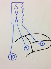
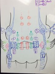
Draw the frontal view of CN 9,10, and 11.
|

|
|
|
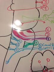
Draw the frontal view of CN 12
|
|
|
|
Draw the lateral view of CN 12
|
|
|
|
How big is CN 12?
|
very big, it comes out of multiple rootlets
|
|
|
What is the name for CN 12?
|
hypoglossal nerve
|
|
|
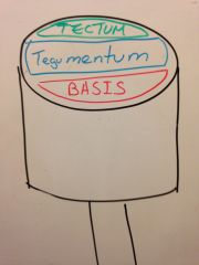
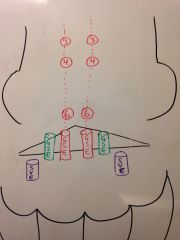
Draw the 3 major divisions (anterior to posterior) of the brainstem.
|
|
|
|
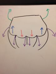
What are the three main types of systems going through the brainstem? (4 directions)
|
1. descending- motor
2. ascending- sensory 3. posterolateral- cerebellar 3. anterolateral- cranial nerves |
|
|
Draw the basic locations in the medulla that each system is going through. (use arrows instead of words)
|
|
|
|
What section do the descending fibers go through? Mnemonic?
|
the anterior part, the basis. (base for descending!)
|
|
|
Draw what happens to the transection of the spinal cord when it hits the medulla? How are the different parts being dragged?
|
|
|
|
Which are the lateral columns and which are the anterior?
|
lateral columns on the sides, anterior are medial
|
|
|
What does the central canal become when it is stretched into this triangle?
|
the 4th ventricle
|
|
|
What happens to the white matter in all of this? What do they create.
|
they end up crossing at the medulla and breaking up the grey matter into small pieces of nuclei.
|
|
|
What are these nuclei going to supply?
|
they become the motor and sensory nuclei of the cranial nerves.
|
|
|
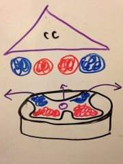
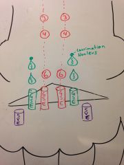
Draw a transection of the medulla when the tracts and nuclei first enter (before they are broken up). Divide into tegumentum and basis.
|
|
|
|
Narrarate what just happened.
|
only the descending pyrimidal tracts are in the basis.
Tegumentum- all the nuclei in the posterior side and the ascending tracts in front |
|
|
Why are the ascending tracts in this drawing purely ascending when they are mixed in the spinal cord?
|
Because the descending motor tracts have yet to mix with them. (they are either in the pyrimids or in their supplementary nuclei)
|
|
|
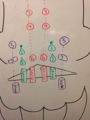
What does this mean for the location of all the cranial nerve nuclei?
|
they should all be located posteriorly in the brainstem.
|
|
|
What extra nuclei are present in the brainstem that didn't come from the spinal cord?
|
supplementary motor nuclei
|
|
|
What are some examples of supplementary motor nuclei? Mnemonic?
|
All the ones that help with motor tone like vetibular, reticular, and rubello.
Vip and Rubber Mat! |
|
|
Are the supplementary motor nuclei confined to the posterior brainstem?
|
No, they have their own unique nuclei not derived from the anterior horns.
|
|
|
The motor nuclei of the cranial nerves will always be ____ to the sensory nuclei.
|
medial
|
|
|
What is in all the space that wasn't filled in on the previous diagram?
|
the nuclei that got so broken up by tract crossing that they mixed aka the RETICULAR FORMATION
|
|
|
Show where the reticular formation is.
|
|
|
|
Draw in where the supplementary motor nuclei are.
|
|
|
|
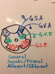
remind me what general means? (vs specific)
|
something the applies to the whole body (touch, somatic function)
|
|
|
What are the two types of tissue that can be innervated?
|
somatic and visceral
|
|
|
In the spinal cord, do we have general fibers or specific fibers?
|
general
|
|
|
Can you draw out all the general somatic/visceral fibers connected with the transection of the spinal cord?
|
|
|
|
What does CN 12 (hypoglossal) control?
|
the toungue movement
|
|
|
What type of nerve is CN 12? Which horn cell should it be from?
|
It is a general somatic efferent so it should be from the anterior horn cell.
|
|
|
Draw in the nuclei of CN12/the general somatic efferents
|
|
|
|
What is an alternate name for this large column/nucleus?
|
dorsal somatic column
|
|
|
What does the vagus control and what type of nerve is it?
|
a lot of smooth muscle and glands and cardiac tissue so it is general visceral efferent.
|
|
|
Draw in the nuclei of the vagus nerve aka the nuclei of general visceral efferents in green
|

|
|
|
What is an alternate name for this large column/nucleus?
|
dorsal visceral column
|
|
|
Are all things in the head either automatic (autonomic nuclei) or voluntary (motor nuclei)? Give examples.
|
No, some things like smiling, mastication, and vocalization are semiautomatic.
|
|
|
How are these actions semiautomatic?
|
you don't have to think about them and sometime you can't help doing them.
|
|
|
What cranial nerves do these functions correspond with?
|
smiling- facial nerve 7
mastication- trigeminal nerve 5 (mandibular) vocalization- |
|
|
What embryological thing are all the nerves for these semiautomat!c muscles called?
|
branchial arches
|
|
|
What is the categorization of these nerve fibers?
|
special visceral efferent
|
|
|
Draw in where the special visceral efferent nuclei would be.
|
|
|
|
What is this special visceral efferent nucleus in the medulla also called? Why?
|
nucleus ambiguous because scientists didn't know what to make of it.
|
|
|
Which CN's are general somatic efferent fibers? (control skeletal muscle voluntarily)
|
3, 4, and 6
|
|
|
Draw in the column with CN nuclei 3, 4, and 6.
|
|
|
|
What happens to the column of grey matter outside of these 3 nuclei?
|
they become broken up by crossing tracts
|
|
|
What kinds of functions will be general visceral efferent in the head?
|
crying and salivating
|
|
|
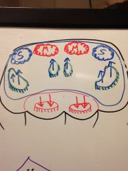
Draw in where the nuclei for lacrimation and salivation are.
|
|
|
|
Describe what was just drawn.
|
The inferior and superior salivary nuclei sit in the bottom/middle of the pons.
The lacrimation nucleus sits right at the tip of the SS nucleus. |
|
|
What does nucleus ambiguous give branches to below the medulla?
|
the layrnx and pharynx
|
|
|
What does the nucleus ambiguous have nuclei similar to above the medulla?
|
facial nerve- CN7
trigeminal- CN5 |
|
|
Draw in the nuclei for the special visceral efferent column.
|
|
|
|
What does nucleus ambiguous give branches to below the medulla?
|
CN 9- glossopharyngeal
CN 10- vagus CN 11- accessory |
|
|
Draw a side view of the nucleus ambiguous with these inferior nerves.
|
|
|
|
What is different about this connection from the other CN connections?
|
they are direct branches off the column!
|
|
|
Are the general visceral efferents considered parasympathetic or sympathetic?
|
parasympathetic (cranial-sacral)
|
|
|
How can I use numbers to remember the cranial nerves that are general somatic efferent?
|
3, 4, 6, 12
3 x 4 = 12 6 also goes into 12. |
|
|
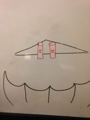
What nuclei are in the superior/middle pons?
|
CN5- trigeminal
superior salivary/lacrimal nucleus |
|
|
What is the very big nucleus in the medulla that supplies a lot of things?
|
dorsal nucleus of vagus
|
|
|
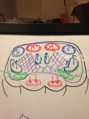
Draw in the sensory nuclei in the medulla. (don't label them yet)
|
|
|
|
What did I screw up in the last couple of pics?
|
I labeled the dorsal vagus nuclei as general somatic efferent instead of visceral.
|
|
|
The nucleus next to the vagus is very squiggly and happy. Why?
|
because it gets to taste many things!
|
|
|
What is in the medial part of the 2 part sensory nuclei? Why does this make sense?
|
general visceral afferent because you want all the things controlled by the vagus to come back and be sensed in the same area.
|
|
|
What is the main sensation going back to the general visceral afferent nucleus?
|
distension
|
|
|
What is the lateral part of the 2 part nucleus?
|
special visceral afferents
|
|
|
Why are these two considered visceral?
|
Because they are not getting sensation from skin, muscles, or joints.
|
|
|
Label the special visceral afferent nuclei.
|
|
|
|
What kinds of sensations go to this nuclei?
|
taste
|
|
|
what is this 2 part nucleus called? Why?
|
nucleus tractus solitarius because scientists found a lone nucleus, followed it up, and found this nucleus.
|
|
|
Do all the sensory taste fibers go here?
|
yes!
|
|
|
What are all the cranial nerves involved in taste?
|
CN 7, 9, and 10
|
|
|
What is the general rule for which CN innervates which part of the tongue?
|
the higher the number, the further back down in the tongue they go
|
|
|
Draw the connection of the nucleus tractus solitarius with all the sensory taste fibers on the tongue.
|
|
|
|
What are the specific regions controlled by 7, 9, and 10?
|
Anterior 2/3rds- CN 7
Posterior 1/3rd- CN 9 Back near esophagus- CN 10 |
|
|
a
|
a
|
|
|
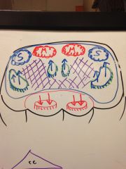
Label the remaining sensory nuclei in the medulla that are in the anterolateral side.
|
General somatic afferent
|
|
|
Are there any nuclei in the upward column of the viscersal afferents that are smushed together?
|
No
|
|
|
What type of sensation are the vestibulocochlear?
|
special somatic afferent
|
|
|
Draw in the vestibular nuclei and it's parts.
|
|
|
|
What are the two cochlear (hearing) nuclei called?
|
ventral and dorsal cochlear nuclei
|
|
|
Draw in the cochlear nuclei near the vestibular nuclei.
|
|
|
|
What is the landmark defining the ventral and dorsal cochlear nuclei?
|
The inferior cerebellar peduncle (ventral in front and dorsal in back.
|
|
|
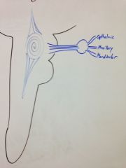
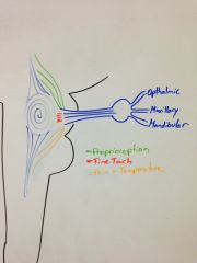
What nerve do touch, pain, and temperature of the head mostly come through?
|
The trigeminal nerve
|
|
|
WHat is that big swirley nucleus called?
|
Principle nucleus of trigeminal system
|
|
|
Narrarate what the principle nucleus gives off.
|
it gives off a big sensory branch that goes into a ganglion and then out comes the three branches of the trigeminal nerve.
|
|
|
Draw in where the motor part of the trigeminal nerve comes from
|
|
|
|
Draw in the superior and inferior extensions of the trigeminal system nuclei.
|

|
|
|
What were those upper and lower extensions called? Where do they each like to go?
|
Upper- mesocephalic nucleus of trigeminal system in the midbrain
Lower- spinal nucleus of trigeminal system to the cervical spine |
|
|
What does the spinal nucleus of the trigeminal system go through?
|
the general somatic afferent nuclei
|
|
|
Essentially, what does the trigeminal system do?
|
Gathers sensory info from it's 3 divisions, sorts them, and sends them along it's nuclei
|
|
|
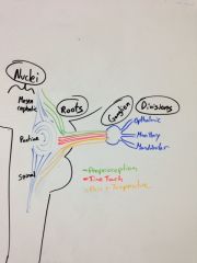
Draw out the trigeminal system from a lateral view.
|
|
|
|
Label the roots of the fibers going to different nuclei after they have been sorted.
|
|
|
|
Label the different parts of the system (roots, nuclei, etc)
|
|
|
|
What would happen if you had damage to the pontine trigeminal nuclei?
|
you would lose fine touch in your face
|
|
|
Where do fibers from the trigeminal go? What are they called?
|

|
|
|
How many trigeminal systems do we have?
|
2, one for each side of the face
|
|
|
What is the mnemonic to remember which cranial nerves are motor vs sensory?
|
Most Say Marry Money, But My Brother Says Big Brains Matter Most
|
|
|
What cranial nerve can I blame my tense shoulders on? Why?
|
I can blame it on the spinal part of CN 11 because it's only innervation is the sternocleidomastoid and the trapezius.
|
|
|
What's the deal with the cranial part of CN 11? Why is it such a wannabe?
|
It merges with the vagus nerve really early on so now scientists think it may actually just be part of the vagus nerve and are calling CN 11 the spinal accessory nerve.
|
|
|
How can I remember accessory nerve and the things it innervates? Mnemonic?
|
If you carry a lot of accessories on you, your shoulders are bound to hurt.
|
|
|
What functions do the vagus and glossopharyngeal have in common?
|
baroreceptor
tasting (7,9,10) vocals (9,10,11) |