Reading...
![]()
Play button
![]()
Play button
![]()
Use LEFT and RIGHT arrow keys to navigate between flashcards;
Use UP and DOWN arrow keys to flip the card;
H to show hint;
A reads text to speech;
129 Cards in this Set
- Front
- Back
- 3rd side (hint)
|
What does the Brainstem consist of?
|
Connects spinal cord to the cerebrum
Consists of : --medulla oblongata --pons --midbrain --reticular matter throughout location of cranial nerve nuclei |
|
|
|
What is the function of the medulla oblongata?
|
pathway for ascending and descending nerve tracts
center for several important reflexes heartrate breathing swallowing vomiting etc |
|
|
|
What is the function of the pons?
|
contains ascending and descending nerve tracts
relay between cerebrum and cerebellum reflex centers |
|
|
|
What is the function of the midbrain?
|
Contains ascending and descending nerve tracts
visual reflex centerj part of auditory pathway |
|
|
|
What is the function of the reticular formation?
|
brainstem pathway which receives sensory input of many types including vision, auditory and somatic senses.
directs these stimuli to the thalamus as part of Reticular Activating System controls cyclic activities, such as sleep wake cycle |
|
|
|
What is the function of the cerebellum?
|
Control of muscle movement and tone
balance regulates extent of intentional movement involved in learning motor skills |
|
|
|
What is the diencephalon?
|
Consists of:
Thalamus Hypothalamus Subthalamus Epithalamus |
|
|
|
What is the function of the thalamus?
|
major sensory relay center
influences mood and movement |
|
|
|
What is the function of the Subthalamus?
|
Contains several nerve tracts and nuclei
|
|
|
|
What is the function of the Epithalamus?
|
Contains nuclei responding to olfactory stimulation and contains pineal body
|
|
|
|
What should you think of when you see pineal body?
|
melatonin
recieves stimulus from hypothalamus. secretes melatonin during dark periods and establishes biological clock (circadian rhythm) which affects sleeping, eating, sexual desire, etc. |
|
|
|
What is the function of the Hypothalamus?
|
Major control center for maintaining homeostasis and regulating endocrine function
|
|
|
|
What does the Cerebrum consist of ?
|
Conscious perception
Basal nuclei Limbic System |
|
|
|
What is the function of the cerebrum?
|
Conscious perception, thought, and conscious motor activity
can override most other systems Basal nuclei function: control of muscle activity and posture Limbic System: Autonomic response to smell, emotion, mood, memory and other such functions |
|
|
|
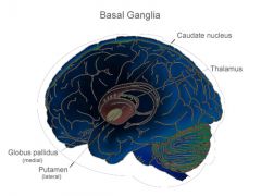
Discuss Basal nuclei
|
group of structures of gray and white matter which modify motor functions from cerebral cortex.
Includes: caudate nucleus putamen globus pallidus and connects to substantia nigra and amygdaloid nucleus Several circuits connect the basal nuclei with the motor association areas, sensory association areas and motor cortex in loops which provide both positive and negative feedback |
|
|
|
What happens when there is damage to basal nuclei?
|
Seeing how they function in association with corticospinal system to control complex patterns of motor activity, damage results in the motor cortex no longer providing patterns for skilled and repetitve actions
i.e. writing ABC's using scissors hammering nails shooting a basketball controlled eye movements etc. |
|
|
|
What neurotransmitters are associated with basal nuclei?
|
INHIBITORY:
--GABA (gamma amino butyric acid) --Dopamine BOTH: --ACh --Norepinephrine --Serotonin --Enkephalin EXCITATORY: --Glutamate |
|
|
|
What is the substantia nigra?
|
located in the midbrain
work with the caudate and lentiform nuclei to control movement |
|
|
|
What is the lateral ventricle?
|
Each cerebral hemisphere contains a relatively large cavity
separated by the septa pellucida (transluscent walls) which lie in the midline, just inferior to corpus callosum are usually fused with eachother |
|
|
|
Where are the 4 ventricles?
|
exist in:
--each cerebral hemisphere (lateral) --between the hemispheres (3rd) --beside the cerebellum (4th) |
|
|
|
What are the canals that connect the ventricles?
|
Interventricular foramen connects lateral with the 3rd ventricle
cerebral aqueduct connects the 3rd and 4th ventricles the 4th ventricle connects to the central canal of spinal cord and other canals called apertures connect to subarachnoid space |
|
|
|
COUP
|
brain trauma occuring at site of impact
|
|
|
|
CONTRECOUP
|
brain trauma occuring on the opposite side of the brain from the impact and resulting from movement of the brain within the skull
|
|
|
|
What is the most common type of traumatic brain injury?
|
Concussion
immediate, but transient, impairment of neural function, such as a loss of consciousness or blurred vision |
|
|
|
What is the difference between diffuse and focal traumatic brain injury?
|
Diffuse= shaking, not localized but damage to many small vessels and nerves, especially around the brainstem
Focal=direct impact injury, usually with cortical contusions, hemmorages. (usually only to gyri) |
|
|
|
What is hemorrahgic brain injury?
what are the different types? |
hemotoma is a collection of blood
Extradural (outside dura) Epidural (outside dura) Subdural (between dura and brain) Chronic subdural (slow bleeding over long period of time) Intracerebral (within the brain) |
|
|
|
INTERVENTRICLAR FORAMEN
|
connects the lateral with the 3rd ventricle
|
|
|
|
CEREBRAL AQUEDUCT
|
connects the third and fourth ventricles
|
|
|
|
GRAY MATTER ARRANGEMENT
(spinal cord and cerebrum) |
In spinal cord, makes the H
In brain it diverges passes through center of cerebrum and terminates in the cortex |
|
|
|
GRAY MATTER ARRANGEMENT
(cerebellum, basal nuclei) |
arranged similar to cortex
|
|
|
|
GRAY MATTER IN BRAIN
|
Similar to spinal cord
It is the site of connections between neurons and contains the cell bodies of motor and interneurons. Composed of unmyelinated neurons. |
|
|
|
WHITE MATTER IN THE BRAIN
|
like spinal cord
composed of myelinated fibers in tracts which carry information from one place to another. |
|
|
|
How is CSF produced?
|
by filtration from blood capillaries located in tissue of choroid plexuses
filtered substance processed by ependymal cells and released into CSF Waste is also removed and put back in blood stream |
|
|
|
What does filtration of CSF remove?
|
water, electrolytes and nutrients are removed from the blood
|
|
|
|
What is the purpose of CSF in the brain?
|
bathes the brain and provides buoyancy
|
|
|
|
Where is the CSF reabsorbed into the bloodstream?
|
arachnoid granulations or arachnoid villi
pockets of arachnoid tissue invagniated in the superior sagittal sinus |
|
|
|
Hydrocephaly
|
To maintain balance, CSF must be absorbed at the same rate of production.
This condition occurs if there is insufficient absorption shows up in childhood and can damage brain and lead to abnormal development. Shunt used to drain fluid to nearby vein |
|
|
|
What does the brain need to function?
|
15-20% of the blood is sent to the brain.
needs a constant supply of glucose |
|
|
|
How does blood reach the brain?
|
internal carotid arteries
vertebral arteries |
|
|
|
What is the cerebral arterial circle (circle of willis)?
|
basilar artery (vertebral arteries)
internal carotid arteries |
|
|
|
LONGITUDINAL FISSURE
|
divides the cerebrum into 2 hemispheres
|
|
|
|
GRYUS
|
raised area between grooves or sulci
|
|
|
|
Cerebral cortex
|
composed of grey matter, outer layer
area of conscious thought and perception "thinking cap" lobes correspond with bone above it |
|
|
|
central sulcus
|
separates the parietal from the frontal lobe
|
|
|
|
lateral fissure
|
divides parietal lobe from occipital lobe
|
|
|
|
parieto-occiptal fissure
|
marks upper delineation of the parietal from occipital lobe.
|
|
|
|
longitudinal fissure
|
divides the cerebrum into two hemispheres
|
|
|
|
CORPUS CALLOSUM
FORNIX |
main fiber tracts permiting the 2 hemispheres to communicate with eachother.
|
|
|
|
principle of lateralization and dominance
|
hemispheres normally divide up the tasks and one hemisphere is dominant; usually the left.
|
|
|
|
LEFT BRAIN
|
logic, mathematics and language
|
|
|
|
RIGHT BRAIN
|
emotions, artistic endeavors.
|
|
|
|
Pre-Central Gyrus
|
Primary Motor Cortex
--voluntary control of skeletal muscles --picture humoncolus feet at the top and progress downward to head |
|
|
|
In the pre-central gyrus, what parts of the body take up the majority of area of the cortex here?
|
facial expression,
speech hand movement |
|
|
|
Where do the corticospinal tracts originate?
|
pre-central gyrus
|
|
|
|
Post-Central Gyrus
|
Primary Somatosensory area
--conscious sensations from the musculocutaneous regions of the body pain temp touch pressure |
|
|
|
What supplies the sensation to the Post-Central Gyrus?
|
Spinothalamic tract
Fasciculus gracilis Fasciculus cuneatus |
|
|
|
What are the main areas in the Post-central gyrus occupied with?
|
Hands
face mouth |
|
|
|
Where is pre-motor area and what is it associated with?
|
in front of pre-central gyrus
motor association area for learned reflexes |
|
|
|
Frontal eye field
|
synchronizes eye movements
|
|
|
|
Broca's area
|
motor speech area for muscle control of speech
|
|
|
|
Prefrontal cortex
|
1. planning complex movement
2. planning and elaboration of thoughts 3. emotions and stress response self control, reasoning 4.personality and some memory |
|
|
|
Wernicke's Area
|
language comprehension
Interpretive area |
|
|
|
Primary auditory area
|
receives and perceives hearing
|
|
|
|
Auditory Association Areas
|
association of hearing with other functions such as speech and memory, necessary to speak and to understand speech
|
|
|
|
Primary visual area
|
perceives visual stimuli and constructs a 3-D image using stimuli from both eyes
|
|
|
|
Visual association area
|
interprets the image and relates it to images in memory for recognition
|
|
|
|
Corona radiata
|
fiber tracts and gray matter areas deep to the cortex
fibers which bring impulses to and from the cerebral cortex from the thalamus |
|
|
|
Internal capsule
|
fibers deep to cortex which connect the cortex with the basal nuclei and corticospinal tracts
|
|
|
|
Parkinson's Disease
|
caused by destruction of dopamine secreting cells in the substantia nigra which send impulses to the caudate nucleus and putamen.
Without inhibitory impulses, tremor and rigidity occur. Akinesia (inability to perform willful movements( |
|
|
|
What are the treatments for Parkinson's Disease?
|
1. use of L-Dopa which is converted into dopamine in the brain
2. transplanted fetal dopamine cells and genetically engineered dopamine cells 3. MAO inhibitor Deprenyl 4. growth factors which stimulate recovery or block deterioration of the dopmaine cells. |
|
|
|
What is MAO?
|
mono amine oxidase
breaks down dopamine |
|
|
|
Huntington's Disease
|
Begins with flicking movements at joints which progress to severe distortional movements of the entire body leading eventually to severe dementia.
caused by genetic mutation of enzyme producing gene Results in loss of GABA secreting neurons in teh basal nuclei and in a loss of ACH neurons in many parts of the brain |
|
|
|
Alzheimer's Disease
|
caused by the development of neurofibrillar tangles and beta-amyloid plaques in teh brain. The tangles occur when a support protein called 'tau' breaks down and therefore the protiens intertwine and loose their functional transport structure.
Without these transport structures neurons cannot function or survive plaques accumulate and cytokines attack the plaque, killing the neurons they are supposed to protect |
|
|
|
What are treatments for Alzheimer's?
|
attempting to stop the breakdown of 'tau', preventing the tangles
antibodies to beta amyloid plaque have been developed which show promise |
|
|
|
septum pellucidum
|
membrane which covers the opening into the lateral ventricle
|
|
|
|
THALAMUS
|
RELAY CENTER
synapse spot for spinothalamic, fasciculs gracilis and fasciculus cuneatus before going to cortex lies at top of reticular formation and acts as alert of reticular activating system filter unwanted stimuli |
|
|
|
reticular formation
|
controls cyclic activities
(sleep wake cycle) |
|
|
|
intermediate mass
|
connects the two halves of the thalamus
|
|
|
|
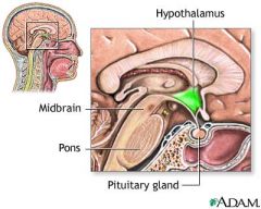
HYPOTHALAMUS
|
control mechanism for endocrine glands
directs pituitary secretions through infundibulum autonomic visceral functions such as blood glucose, heart rate, respiration in stress, thermoregulation, hunger, thirst, electrolyte balance, water balance and sleep wake cycle |
|
|
|
pineal gland
|
melatonin
part of the epithalamus, but stimulated by hypothalamus circadian rhythms Seasonal affective disorder |
|
|
|
Midbrain is composed of...
|
...corporal quadrigemina superior and inferior colliculi)
cerebral peduncles (descending tracts carrying motor info to brainstem and spinal cord) Tegmentum (ascending tracts carrying sensory info) Red nuclei (unconscious regulation and coordination of motor activites) Substantia nigra (muscle tone and coordination of movements) |
|
|
|
Corporal quadrigemina
|
(four bodies, twins) are the superior colliculi
center for visual reflexes (blinking, accommodation of the lens) inferior colliculi auditory reflexes are centered (contraction of the stapedius muscle) |
|
|
|
Pons
|
= bridge
bridge to higher brain main functions are: --regulation of rate and depth of respiration --CNS connection for cranial nerves V, VI, VII, VIII |
pons is like ponds
what do you need to walk over it? if you fall in what may be hard to regulate? and one more thing |
|
|
Medulla
|
center for vital functions of heart rate
respiration blood pressure CNS connections for VIII, IX, X, XI, XII. contains pyramids stimulates muscles of respirations and controls breathing rhythm regulates heart rate and volume controls blood pressure and overall blood distribution |
|
|
|
Cerebellum
|
coordinates with skeletal muscles
recieves unconscious proprioception as well as input from all higher motor centers. Monitors muscle contraction and planned muscle contractions and maintains a constantly adapting system to coordinate them |
|
|
|
cingulate gyrus
|
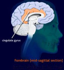
lies above the corpus callosum
connects limbic system to cerebral cortex |
|
|
|
hypothalamus of limbic system
|
contains pleasure, reward, or satiety center.
Also mediates hormonal responses to stress |
|
|
|
mamillary body of limbic system
|
contains memory pathways, especially for olfaction
|
|
|
|
hippocampus of limbic system
|
short term and long term memory
learning |
think of "campus"
What do you need to use to get the most out of a college campus? |
|
|
amygdaloid nucleus of limbic system
|
center for many emotional response pathways
|
|
|
|
thalamus of limbic system
|
sensory center for input which stimulates emotions
|
|
|
|
fornix of limbic system
|
provides connecting pathways for the limbic system from one hemisphere to the other
|
|
|
|
Reticular formation
|
brainstem pathway which receives sensory input of many types including vision, auditory and somatic senses.
directs these stimuli to the thalamus as part of Reticular Activating System |
|
|
|
Reticular Activating System
|
alert system for the cortex.
Filters unwanted and unimportant stimuli to be filtered out, while making us aware of important and critical stimuli |
think filter
|
|
|
basal nuclei
|
control of muscle activity and posture
|
|
|
|
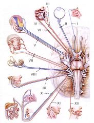
What are the 12 cranial nerves?
|
Oh, Oh, Oh, To Touch A Female Vagina Gives Virgins Amazing Happiness
Olfactory I Optic II Oculomotor III Trochlear IV Trigeminal V Abducent VI Facial VII Vestibulocochlear VIII Glossopharyngeal IX Vagus X Accessory XI Hypoglossal XII |
|
|
|
What are the sensory cranial nerves
|
I Olfactory
II Optic VIII Vestibulochoclear |
|
|
|
What are the somatic motor cranial nerves?
|
IV Trochlear
VI Abducent XI Accessory XII Hypoglossal |
|
|
|
What are the somatic motor and sensory cranial nerves?
|
V Trigeminal
|
|
|
|
What are the somatic motor and parasympathetic cranial nerves?
|
III Oculomotor
|
|
|
|
What are the somatic motor, sensory and parasympathetic cranial nerves?
|
VII Facial
IX Glossopharyngeal X Vagus |
|
|
|
What is hydrocephalus?
|
internal = blockage which cause CSF to accumulate and cause pressure
treatment is a shunt to relieve pressure, infection is a complication external =subarachnoid hemorrhage blocks return of CSF to circulation causing pressure on brain externally both types cause brain damage |
|
|
|
What are the 3 meninges that surround the CNS?
|
dura mater
arachnoid mater pia mater |
|
|
|
describe dura mater
|
in spinal cord has epidural space, but in brain only has potential space.
has 2 layers--one continuous with the periosteum of cranial bones (periosteal dura) and the other is continuous with the dura of the spinal cord (meningeal dura) contains dural folds and sinuses |
|
|
|
what is the largest dural venous sinus?
|
superior sagittal sinus
forms between the falx cerebri and periosteal dura on median plane transport blood and CSF away from brain |
|
|
|
Where does the blood draining from the brain go?
|
dural venous sinuses.
eventually sinuses drain into internal jugular veins which take it back to heart |
|
|
|
where does the brain get its blood from?
|
internal carotid and vertebral arteries
|
|
|
|
where do internal carotid arteries enter the brain?
|
ascend head on anterior-lateral part of neck
enter thru carotid canals meets up with the verterbral arteries to create the cerebral arterial circle (circle of willis) |
|
|
|
Where do vertebral arteries enter the brain
|
ascend neck along posterior
enter through foramen magnum meet up with internal carotid artery to form cerebral arterial circle (circle of willis) |
|
|
|
Where are the arteries located in the brain?
|
subarachnoid space
|
|
|
|
Describe the flow of CSF
|
1. CSF produced in choroid plexuses of the 4 ventricles
2. CSF flows from lateral ventricle through interventricular foramina to 3rd ventricle 3. CSF flows from the 3rd ventricle thru cerebral aqueduct to 4th ventricle 4. CSF exits 4th ventricle through lateral and median apertures and enters subarachnoid space. Some goes down central canal of spinal cord 5. CSF flows thru subarachnoid space to arachnoid granulations in superior sagittal sinus where it goes back into venous circulation |
|
|
|
cranial nerves and region
|
|
|
|
|
cranial nerves and functions
|
I Olfactory directly to temporal lobe
II Optic Retina through thalamus to occipital lobe III Oculomotor eyeball muscle Eyelid and eyeball muscles, lens (focusing), pupil (constriction) IV Troclear pulley eyeball muscle proprioception V Trigeminal Chewing muscles proprioreception, cutaneous touch and pain receptors for face, jaw and teeth chewing muscles VI Abducens eyeball muscle proprioreception VII Facial Facial muscle proprioreception, tongue taste receptors facial expression muscles, saliva and tear glands VIII Vestibulocochlear Auditory Hearing and equilibrium organs IX Glossopharyngeal arterial chemoreceptors and baroreceptors, tongue taste receptors, tongue muscle proprioreception muscles (swallowing), (aids in regulation of breathing and BP) X Vagus arterial chemoreceptors and baroreceptors, pharangeal taste receptors, pressure/pain receptors in thoracic and abdominal organs Swallowing and speaking muscles, smooth muscle and secretory glands in GI tract, Heart muscle, (aids in regulation of breathing, BP and slows HR) XI Accessory Neck and shoulder muscle proprioreceptors swallowing muscles, head and neck muscles XII Hypoglossal Swallowing and speaking muscle proprioreceptors speaking muscles |
|
|
|
Olfactory nerve
|
I
smell |
|
|
|
Optic nerve
|
II
vision |
|
|
|
Oculomotor Nerve
|
III
motor to eye muscles proprioceptive to eye muscles parasympathetic constriction and accomodation of pupil and llens |
|
|
|
Trochlear
|
IV
motor to superior oblique proprioception from that muscle |
|
|
|
Trigeminal
|
V
sensory: opthalmic--scalp, forehead, nose upper eyelid and cornea maxillary--palate, upper jaw, upper teeth and gums, nasopharynx, nasal cavity, skin/ mucous membrane of cheek, lower eyelid and upper lip, mandibular--lower jaw, lower teeth and gums, anterior 2/3 of tongue, lower lip, auricle, temporal region MOTOR: mandibular--mastication |
|
|
|
Abducent nerve
|
VI
motor to lateral rectus eye muscle proprioceptive to that muscle |
|
|
|
Facial nerve
|
VII
Sensory: taste from anterior 2/3 tongue external ear palate Motor: facial expression throat middle ear Parasympathetic: salivary glands lacrimal glands nasal and palantine glands |
|
|
|
Vestibulocochlear
|
VIII
Hearing and balance |
|
|
|
Glossopharengeal
|
IX
Sensory: taste from posterior 1/3 of tongue pharynx tonsils middle ear carotid sinus and body blood pressure gas in blood motor: pharyngeal muscle parasympathetic: salivary gland and glands of posterior tongue |
|
|
|
Vagus nerve
|
X
Sensory: pharynx, larynx, thoracic and abdominal organs taste from posterior tongue blood pressure gas levels in blood motor: soft palate, pharynx, laryngeal muscles tongue muscles parasympathetic: thoracic and abdominal viscera |
|
|
|
What is the function of the frontal lobes of the cerebrum?
|
smell
voluntary motor function motivation aggression mood |
|
|
|
What is the function of the parietal lobes of the cerebrum?
|
major sensory areas recieving general sensory input, taste, and balance
|
|
|
|
What is the function of the occipital lobes of the cerebrum?
|
visual centers
|
|
|
|
What is the function of the temporal lobes of the cerebrum?
|
olfactory and auditory
memory abstract thought judgement |
|
|
|
What is the blood brain barrier?
|
formed from the endothelial cells of the capillaries in the brain, the astrocytes in the brain tissue, and the basement membrane in between.
|
|
|
|
What is the function of the limbic system?
|
controls visceral functions through the autonomic nervous system and the endocrine system and is involved in emotions and memory
|
|