![]()
![]()
![]()
Use LEFT and RIGHT arrow keys to navigate between flashcards;
Use UP and DOWN arrow keys to flip the card;
H to show hint;
A reads text to speech;
281 Cards in this Set
- Front
- Back
|
Chapter 1: Define Anatomy? |
Anatomy is the scientific discipline that investigates the structure of the body. Anatomy means to dissect parts of the body for study. |
|
|
Explain the relationship between structure and function? |
Structure assists in the functions of the human body. Ex. Bones are good for support cause they are strong, due to the bone cells secreting hard, mineralized substances. |
|
|
Two basic approaches to studying anatomy 1. Systemic anatomy 2. Regional anatomy |
Systemic: study of the body by systems, such as, skeletal and nervous. Approach is used for teaching purposes. Regional: study of the organization of the body by areas. Regions such as the head, abdomen, or mouth. Approach is used by medical schools. |
|
|
Two ways to examine internal structures 1. Surface anatomy 2. Anatomical imaging |
Surface Anatomy: study of external features, such as bony projections, which serve as landmarks for locating deeper structures. Anatomical Imaging: Involves x-rays, ultrasound, MRI, etc. to create a picture of internal structures. |
|
|
Define physiology? Define Human physiology? |
Physiology: scientific discipline that deals with the process or functions of living things. Human Physiology: study of specific organisms, the human, whereas cellular physiology and systemic physiology are subdivisions that focus on specific organizational levels |
|
|
State two major goals of physiology |
1. To understand and predict the body's responses to stimuli 2. To understand how body maintains conditions within a narrow range of values in the presence of continually changing internal external environments |
|
|
Six Levels of Organization 1. Chemical Level |
Chemical level determines how atoms, such as hydrogen and carbon interact and combine into molecules. The function of a molecule is intimately related to its structure. Ex. Collagen fibres give skin its structural strength |
|
|
Six Levels of Organization 2. Cell Level |
Cells are basic structural and functional units of organisms, such as plants and animals. Molecules can combine to form organelles, which make up cells. Ex. Mitochondria produces ATP. |
|
|
Six Levels of Organization 3. Tissue Level |
Tissue is a group of similar cells and the materials surrounding them. Characteristics of the cells and surrounding materials determine the function of the tissue. 4 Types: Epithelial, Connective, Muscle, and Nervous |
|
|
Six Levels of Organization 4. Organ Level |
Organs are composed of two or more tissue types that together perform one or more functions. Ex. Heart, Bladder, Skin |
|
|
Six Levels of Organization 5. Organ System Level |
Organ system is a group of organs classified as a unit because of a common function or set of functions. The coordinated activity of the organ systems is necessary for normal functions. Ex. Digestive system processes food into nutrients, which is carried by blood in the cardio vascular system to the cells of other systems. |
|
|
Six Levels of Organization 6. Organism Level |
Organism is any living thing considered as a whole, can be unicellular or multicellular. |
|
|
Eleven Major Organ Systems |
1. Cardiovascular 7. Endocrine 2. Muscular 8. Lymphatic 3. Skeletal 9. Reproductive 4. Urinary 10. Integumentary 5. Nervous 11. Respiratory 6. Digestive |
|
|
Six Characteristics of Life 1. Organization |
Refers to the specific interrelationships among the parts of an organism and how those parts interact to perform specific fxns. |
|
|
Six Characteristics of Life 2. Metabolism |
Ability to use energy to perform vital fxns, such as growth, movement, and reproduction. Ex. Plants capture energy from sunlight and humans obtain it from food. |
|
|
Six Characteristics of Life 3. Responsiveness |
Ability of an organism to sense changes in the environment and make the adjustments that help maintain its life. Responses include movement toward food or water and away from danger or poor environmental conditions. |
|
|
Six Characteristics of Life 4. Growth |
Refers to an increase in size of all or part of the organism. It can result from an increase in cell number, cell size, or the amount of substance surrounding cells. |
|
|
Six Characteristics of Life 5. Development |
Includes the changes an organism undergoes through time; it begins with fertilization and ends at death. The greatest developmental changes occur before birth, but continue after birth, and some continue throughout life. |
|
|
Six Characteristics of Life 6. Reproduction |
is the formation of new cells or new organisms. Without reproduction of cells, growth and tissue repair are impossible. Without reproduction the species would become extinct. |
|
|
What is Homeostasis? |
Homeostasis is the existence and maintenance of relatively constant environment or body despite fluctuations in either the external environment or the internal environment. |
|
|
Homeostatic Mechanisms |
Examples include sweating or shivering, normally maintain body temperature near an ideal normal value, or set point. |
|
|
Negative Feedback |
Negative means that any deviation from the set point is made smaller or is resisted. Negative feedback does not prevent variation but maintains variation within a normal range. Ex. The maintenance of normal body temp. is an example of a negative feedback mechanism. |
|
|
Three components to Negative Feedback 1. Receptor 2. Control Center 3. Effector |
Receptor: Monitors the value of a variable, such as body temp. Control Center: such as part of the brain, establishes the set point around which the variable is maintained Effector: such as the sweat glands, can change the value of the variable. |
|
|
Positive Feedback |
Occurs when the initial stimulus further stimulates the response. The deviation from the set pint becomes even greater. Ex. During blood loss, chemical responsible for clot formation stimulates production of itself. |
|
|
Chapter 2: Atom |
the smallest particle if an element that gas the chemical characteristics of that element. |
|
|
Atom nucleus (Composed of?) |
composed of protons and neutrons. Surrounding the nucleus are electrons, move around it. |
|
|
Matter, Mass, and Weight |
1. Matter is anything that occupies space and has mass 2. Mass is the amount of matter in an object 3. Weight results from the gravitational attraction between the earth and the object |
|
|
Ions |
When an atom loses or gains electrons, the numbers of protons and electrons are no longer equal, and a charged particle called an ion is formed Ex. Na to Na+ (loses an electron) |
|
|
Ionic Bonding |
occurs when electrons transfer between atoms, between oppositely charged ions. Ex. Na+ and Cl- |
|
|
Covalent Bonding |
Forms when atoms share one or more pairs of electrons. The resulting combination of atoms is called a molecule. Ex. Sharing of electrons between to Hydrogens |
|
|
Polar Covalent Bond and Non Polar Covalent Bonds |
Polar: Unequal sharing of electrons results in one end (pole) of the molecule having a partial charge Non-polar: equal sharing of electrons between atoms |
|
|
Hydrogen Bond |
A positive polar end of one molecule is attracted to a negatively charged end of another polar molecule. This attraction is called a hydrogen bond. Ex. (H+) of one H20 molecule is attracted to (O-) of another H20 molecule |
|
|
Molecule |
A molecule is formed when two or more atoms chemically combine to form a structure that behaves as an independent unit. Ex. H20 |
|
|
Dissociation |
Is the separation of ions in an ionic compound by polar water molecules |
|
|
Chemical Reactions |
Ina chemical reaction, atoms, ions, molecules, or compounds interact either to form or to break chemical bonds. Reactants are substances that enter the reaction, then products are the result of it. |
|
|
Synthesis Reactions |
When two or more reactants combine to form a larger, more complex product, the process is called synthesis reaction, represented as A+B = AB |
|
|
Synthesis reaction in the body |
Called anabolism. Includes, Growth maintenance, and repair of the body. |
|
|
Decomposition Reactions |
In a decomposition reaction, reactants are broken down into smaller, less complex products. A decomposition reaction is the revers of a synthesis AB = A+B |
|
|
Decomposition reactions in the body |
Called catabolism, include the digestion of food molecules. |
|
|
Metabolic Reactions |
Combination of all anabolic and catabolic reaction the body |
|
|
pH Scale |
Indicates the [H+] in solution. Scale ranging from 0-14. |
|
|
Neutral Solution |
A neutral solution has an equal number of H+ and OH- making pH 7.0. |
|
|
Acidic Solution |
Has a greater [H+] than OH- making pH less than 7 |
|
|
Basic Solution |
Has less a [H+] than OH- making pH greater than 7 |
|
|
Why do Humans require Oxygen? |
Humans require O2 in the final step of a series of chemical reaction in which energy is extracted from food molecules. |
|
|
Why do Humans require Water? |
H2O is an inorganic molecule, that provides roles such as: 1. stabilizing body temperature 2. Providing Protection 3. Facilitating chemical reactions 4. Transportation |
|
|
4 major groups of organic molecules |
1. Carbohydrates 2. Lipids 3. Proteins 4. Nucleic Acid |
|
|
Carbohydrates (Structure and Function) |
carbs are composed of carbon, hydrogen, and oxygen. For 1 carbon there are 2 hydrogen and 1 oxygen. Function: Energy |
|
|
Carbohydrates 1. Monosaccharides |
Smallest carb, (1 sugar) simple sugars. Ex. Glucose or Fructose |
|
|
Carbohydrates 2. Disaccharides |
Two monosaccharides joined together through covalent bonds are called disaccharides (2 sugar) Ex. Maltose (2 glucoses) |
|
|
Carbohydrates 3. Polysaccharides |
long chains of monosaccharides. In animal or plants when energy is needed polysaccharides are broken down into individual glucose. Ex. Glycogen (animals) or, Starch (plants) |
|
|
Lipid (Structure, Example, and Functions) |
Lipids are substances that dissolve in non-polar solvents such as, alcohol or acetone, but don't in polar solvents Ex. Fats, phospholipids, eicosanoids, and steroids Function: Energy, Structure, Regulation |
|
|
Phospholipids |
Phospholipids are similar to trigycerides, except that one of the fatty acids bound to the glycerol is replaced by a molecule containing phosphorus. Contains a hydrophilic polar head that is water-loving and a hydrophobic non-polar head that is water-fearing |
|
|
Protein (Structure, Example, and Functions) |
consists of many amino acids joined together to form a chain. Shape determines their ability to perform functions. |
|
|
Denaturation |
Occurs when the protein hydrogen bonds in the protein are broken, protein becomes nonfunctional. Ex. pH changes or High temp. |
|
|
Enzyme |
Is a protein catalyst that increase the rate of a chemical reaction. This is done through lowering activation energy. |
|
|
Nucleic Acids (DNA) |
Deoxyribonucleic acid is the genetic materal of cells, copies of DNA are transferred from one generation of cells to the next. DNA contains info that determine structure of proteins. Look like a double helix |
|
|
Nucleic Acids (RNA) |
Ribonucleic Acid is structurally related to DNA, except it is only one helix. RNA is used for transcription. |
|
|
Nucleic Acids (What are they made of?) |
The nucleic acids are large molecules composed of carbon, hydrogen, nitrogen and phosphorus. |
|
|
Nucleotides (Building blocks of DNA) |
composed of one sugar to which a nitrogen organic base and a phosphate are attached. Sugar in DNA= deoxyribose, Sugar in RNA= Ribose, Bases: Thymine - Guanine Adenine - Cytosine (Urasil in RNA) |
|
|
Adenine Triphosphate (ATP) |
important organic molecule found in all living organisms. Consists of adenosine and three phosphates. Adenosine diphosphate is the breakdown of ATP to provide energy for chemical reactions |
|
|
Potential Energy Kinetic Energy Chemical Energy Mechanical Energy |
Potential Energy: is stored energy Kinetic Energy: is energy of motion Chemical Energy: potential energy stored in chemical bonds Mechanical Energy: is energy resulting from the position or movement of objects |
|
|
Chapter 3: Cell |
Cell is the basic living unit of all organisms |
|
|
Organelles |
located inside the cell, perform specific functions. |
|
|
Nucleus |
contains genetic material |
|
|
Cytoplasm |
surrounds organelles and inside the cell, suspending them there |
|
|
Cell membrane |
surrounds the whole cell. Providing protection and a gateway for molecules and elements to move in and out of the cell. |
|
|
4 functions of a cell |
1. Cell metabolism and energy use 2. Synthesis of molecules Ex. Protein and Lipids 3. Communication Ex. Receptor Proteins 4. Reproduction and inheritance |
|
|
Describe the structure of the cell membrane |
Is the outermost component of the cell. Encloses the cell cytoplasm. Substances outside are extracellular, while inside are intracellular. Is made up of phospholipids, 2 layers, non-polar ends face towards each other. Proteins, Carbs, and cholesterol are suspended in it. |
|
|
Fluid-Mosaic Model |
Phospholipids form a double layer. They have a liquid quality. Cholesterol within the phospholipid gives it strength and flexibility. Protein molecules float among the lipid layer. Carbs may be bound to some proteins, these proteins act as channels, carriers, receptors, enzymes, and support. |
|
|
Define: Solution Solute Solvent |
Solution: A mixture of two or more dissolved substances (solute + solvent = solution) Solute: the component of the solution in lesser amount than the solvent Solvent: the component of the solution in greater amount; the substance in which the solute dissolves |
|
|
Define Diffusion |
The movement of molecules from an area of [high] to an area of [low] |
|
|
Define Concentration Gradient |
Is the difference in the concentration of a solute in a solvent between two points divided by the distance between the two points. |
|
|
Define Osmosis |
Osmosis is the diffusion of water across a selectively permeable membrane, from a region of [higher] to an area of [low] |
|
|
Osmotic Pressure |
The force required to prevent movement of water across a selectively permeable membrane. Water tends to move towards more concentrated solutions. |
|
|
Why is Osmotic pressure important in cell membranes? |
It is needed to prevent the movement of water to move out of the cell into more concentrated solutions. Or, it is needed to help prevent water from entering the cell. |
|
|
Solutions: Hypotonic, Isotonic, and hypertonic |
Hypotonic: When a cell is immersed in a solution, the solution has a lower [ ] of solutes and a high [ ] of water. Water will move into the cell causing lysis. Isotonic: When a cell is immersed in a solution, the [ ] of all the solutes in the cell and the solution are equal. Cell doesn't change. Hypertonic: When a cell is immersed in a solution, the solution has a high [ ] of solutes and a low [ ] of water. Water will move out of the cell causing crenation. |
|
|
Carrier-Mediated Transport |
Carrier proteins move large, water-soluble molecules across the membrane. |
|
|
Facilitated Diffusion |
Carrier-mediated transport process that move substances across the cell membrane from an area of [high] to [low]. Moves with concentration gradient, no ATP required. |
|
|
Active Transport |
Carrier-mediated process that moves substances across cell membrane from region of [low] to [high]. Movement is against concentration gradient, requires ATP. |
|
|
Secondary Active Transport |
Involves active transport of one substance, such as the Na/K pump. |
|
|
Endocytosis |
Endocytosis: is the uptake of material through the cell membrane through vesicles. -requires ATP -Phagocytosis: cell eating, solid particles are ingested by the cell membrane. -Pinocytosis: cell drinking, liquid is ingested by the cell membrane. |
|
|
Exocytosis |
Exocytosis: The movement of secretory vesicles out of the cell. Vesicles are absorbed by the cell wall then released into the environment. -Requires ATP |
|
|
Nucleus (Location, Structure, Function) |
Location: Center of the cell Structure: Bound by nuclear envelope, which has nuclear pores in it for material to go in and out. Function: Contain 23 pairs of chromosomes, which consist of DNA and protein. Directs the whole Cell (Brain). |
|
|
Nucleolus (Location, Structure, Function) |
Location: Inside the nucleus Structure: Subunits of ribosomes with no surrounding membrane Function: Brings together rRNA and protein to form Ribosomes |
|
|
Rough ER and Smooth ER (Location, Structure, Function) |
ER: Location: Around the Nucleus Structure: series of membranes forming sacs Rough ER (Function): Has ribosomes attached to it. Ribosome undergo protein synthesis to produce protein. Smooth ER(Function): The site of lipid synthesis and participates in detoxification of chemicals. |
|
|
Golgi Apparatus (Location, Structure, Function) |
Location: Various Structure: Curves membrane-bound sacs Function: Collects, modifies, packages, and distributes proteins and lipids made by the ER. Forms secretory vesicles for transport of lipid and protein out of the cell, through exocytosis. |
|
|
Secretory Vesicles (Location, Structure, Function) |
Location: Various Structure: small, membrane-bound sac Function: Transport or stores material within cells. |
|
|
Lysosomes and Peroxisomes (Structure, Function) |
Lysosomes Structure: membrane-bound vesicles from Golgi Body. Contain enzymes. Function: Intracellular digestion, fuse with vesicles taken in by endocytosis. Peroxisomes: Structure: small, membrane-bound vesicles. Contain enzymes. Function: Enzymes breakdown hydrogen peroxide. |
|
|
Mitochondria (Structure, Function) |
Structure: small,has inner and outer membranes Function: Aerobic Respiration, O2 + Food = CO2 + H20 + ATP |
|
|
Cytoskeleton (Structure, Function) |
Structure: Made of proteins; intermediate filaments, microtubules, and microfilaments Function: Support, hold organelles in place, enable cell to change shape. |
|
|
Centriole (Structure, Function) |
Structure: small cylindrical organelle made of nine triplets; each triplet contains 3 parallel microtubules Function: Cell division (Mitosis) |
|
|
Cilia, Flagella, and Microvilli (Function) |
Cilia: In respiratory system these hair like structures move mucus upward and away from the lungs Flagella: Tails that propel sperm cells forward Microvilli: shaggy hairs, they increase the surface area of cell membranes for increased absorption. |
|
|
Gene Expression Transcription |
Takes place in the nucleus. Double strand of DNA separate, DNA pair with mRNA nucleotide. Pair with A-T and A-U and G-C. Once mRNA segment has been transcribed, portions of mRNA are removed. Info in mRNA is carried in groups of 3 called codons. |
|
|
Gene Expression Translation |
The synthesis of proteins based on info in mRNA, occurs at ribosomes. mRNA travel through nuclear pores to ribosomes. Ribosomes and mRNA combine. mRNA and tRNA molecules align to pair anticodon with codon. Ribosome moves down the mRNA 1 codon at a time, allowing tRNA to move into position each time. As the process continues a polypeptide chain forms. Translation ends when mRNA stop codon is reached. Peptide chain is released forming a 3D protein molecule. |
|
|
Chapter 7: List the function of the muscular system1-4 |
1. Movement of the body: contraction of skeletal muscle to produce a movement 2. Maintenance of posture: skeletal muscles constantly maintain tone 3. Respiration: respiratory muscles carry out movement needed for respiration 4. Production of body heat: When skeletal muscles contract, heat is given off |
|
|
List the function of the muscular system 5-7 |
5. Communication: Speaking, writing, typing, etc. 6. Constriction of organs and blood vessels: smooth muscle constrict to assist organs 7. Contraction of heart: cardiac muscles contract to push blood throughout body |
|
|
Skeletal Muscle (4 major functions) |
1. Contractibility: produces force through contraction to overcome an opposing force. 2. Excitability: response to a stimulus. 3. Extensibility: after contraction muscles can be stretched to their resting length and beyond. 4. Elasticity : recoil to their original resting length after they have been stretched |
|
|
Structure of Skeletal muscles 3 layers |
Epimysium: a connective tissue that surrounds the who muscle. Perimysium: each muscle is separated by this layer of loose connective tissue Endomysium: loose connective tissue that houses the muscle fiber (Cells) Fascicle: Make up Perimysium and Endomysium |
|
|
Structure of Muscle Fiber
|
-Cell membrane is called sarcolemma. -many nuclei -cell is divided into sections called T-tubules, made of smooth ER called sarcoplasmic reticulum -Sacroplasm(cytoplasm) contains myofibrils -myofibrils contain two major proteins actin and myosin -actin and myosin are highly ordered, repeating units called sarcomeres |
|
|
Actin Myofilaments 3 components, 3 functions |
1. Actin (thin filament) made up of three components: 1)actin, have attachments for myosin 2)troponin, have binding sites for Calcium 3)tropomyosin, block actin binding sites so myosin can't bind in unstimulated muscles |
|
|
Myosin Myofilaments 1 component, 3 function |
2. Myosin (thick filament) made up of one components: The head, 1) can bind to actin 2) can bend and straighten during contraction 3) can break down ATP, releasing energy |
|
|
Sacromeres |
-The sarcomere is the basic structural and functional unit of skeletal muscle because it is the smallest portion of skeletal muscle capable of contracting. -It is part of the myofibril extending from Z-disk to Z-disk |
|
|
Describe the Excitability of Muscle Fibers? (Polarization and Resting Potential reasons) |
-Polarization occurs when the inside of the membrane is negative and the outside is positive. -Resting Potential, occurs because there is an uneven distribution of ions across the cell membrane |
|
|
3 Reasons for Resting Potential |
1) [K+] inside cell is higher than outside the cell 2) [Na+] outside cell is higher than inside the cell 3) Cell membrane is more permeable to K+ than Na+ |
|
|
Action Potential (Depolarization) |
-Nerve impulse is felt causing Na+ channels to open. Na+ moves into the cell making the inside of the cell more positively charged. -Muscle contraction is achieved |
|
|
Action Potential (Repolarization) |
Na+ channels close, and Na+ movement into cell stops. K+ channels open. K+ moves out of the cell, making the inside of the cell negatively charged. |
|
|
Action Potential (Refractory Period or Hyperpolarization) |
Na/K pump pumps Na+ out and K+ back into the cell. This brings it back to resting potential. |
|
|
Motor Neuron |
Specialized nerve cells that stimulate muscles to contract. Generate action potentials in skeletal muscles. |
|
|
Neuromuscular Junction |
A synapse between the cell of nerve and a muscle cell. They summate motor units. |
|
|
Motor Unit |
-Can be a small precisely controlled muscle, such as in the hand, may have very few fibers per-unit
-Can be large muscles, such as the bicep that have many fibers per motor unit |
|
|
How does the synapse work? (4 steps) |
1) An action potential reaches the pre-synapse, it causes Ca2+ channels to open. 2) Calcium ions enter the pre-synapse causing synaptic vesicles to release ACh into the synaptic cleft by exocytosis. 3) Diffusion of ACh across the synaptic cleft and binding of ACh to receptors on the post-synaptic muscle, membrane opens Na+ channels 4) Na+ diffuse down their [gradient], which results in depolarization of muscle; once threshold has been reached, an action potential results |
|
|
Sliding Filament Model |
The sliding of actin myofilaments past myosin myofilaments. During contraction neither actin nor myosin shorten. The H zones and I bands shorten during contraction, A bands stay the same. |
|
|
Muscle Contraction |
Skeletal muscle contracts by actin and myosin filaments slide past one another, causing the Sacromeres to shorten. Sacromeres shortening causes the the myofibrils to shorten. This causes the whole muscle to shorten. |
|
|
Chemical Level of Contraction (refer to page 158 for pictures) |
Ca2+ diffuses into sacroplasm. The Ca2+ binds to troponin attached to actin filaments. The binding causes tropomyosin to expose myosin attachment sites. The exposed sites attach to myosin heads to form cross-bridges. ATP (energy) needed for the contraction, is released. ATP breaks down to ADP, forming energy. This energy is then used to move the heads of myosin toward the center of the sarcomere, causing the contraction. When Ca2+ leaves the sacroplasm the muscle relaxes. |
|
|
Power Stroke |
Upon the release of ADP, myosin head pulls the thin filament towards the centre of the Sacromere. |
|
|
Shivering (Raising Body Temp?) |
The act of shivering raises body temperature. This is due to the contraction of muscles which produce heat. |
|
|
Muscle Twitch |
Contraction of a muscle fiber in response to a stimulus. Because most muscle fibers are grouped into motor units, summates the whole unit. |
|
|
How to Increase the force of muscle contraction? (2 ways) |
*Strength of a contraction varies from weak to strong 1) Summation, involves increasing the force of contraction of the muscle fibers with a muscle 2) Recruitment, involves increasing the # of fibers contracting |
|
|
Tetanus |
a sustained contraction that occurs when the frequency of stimulation is so rapid that no relaxation occurs |
|
|
Summation |
-contain type I myosin as the predominant type -slow contractions -can sustain contractions much longer -mainly respirate aerobically -Dark red in colour Ex. Distance Running |
|
|
Recruitment |
-contain type IIa or type IIb (fastest) myosin -quick contractions -can't sustain contractions as long -mainly respirate anaerobically -white in colour Ex. Sprints |
|
|
List 4 ways ATP is created in skeletal muscles |
1) Aerobic production of ATP during exercise 2) Anaerobic production of ATP during intensive short-term work 3) Conversion of creatine phosphate to ATP 4) Conversion of two ADP to one ATP and one AMP during heavy exercise |
|
|
Aerobic Respiration *Also know as cellular respiration |
occurs in mitochondria, requires O2 and breaks down glucose to produce ATP, CO2, and H20 O2 + glucose = ATP (energy) + CO2 + H2O |
|
|
Anaerobic Respiration |
does not require O2, breaks down glucose to produce ATP and lactate. |
|
|
Fatigue |
Fatigue is a temporary state of reduced work capacity. Protective response to prevent muscles from getting damaged from overworking. |
|
|
Lactate converts to ________. |
Converts to glucose |
|
|
Fatigue: 3 mechanisms 1. Acidosis and ATP Depletion |
Anaerobic respiration results in breakdown of glucose to lactate and protons, accounting for lowered pH has several cellular effects including decreased effectiveness of Ca2+ on actin. |
|
|
Fatigue: 3 mechanisms 2. Oxidative Stress |
During intense exercise, increases in ROS (reactive oxygen species) production cause the breakdown of proteins, lipids, or nucleic acids. |
|
|
Fatigue: 3 mechanisms 3. Inflammation |
ROS signals immune system to releases chemical IL-6 which causes inflammation and muscle soreness. |
|
|
Isometric and Isotonic contractions |
Isometric: the length of the muscle does not change, but the amount of tension increases during the contraction process. Isotonic: the amount of tension produced by the muscle is constant during contraction, but the length of the muscle decreases. |
|
|
Concentric and Eccentric Contractions |
Concentric: are isotonic contractions in which muscle tension increases as the muscle shortens Eccentric: are isotonic contractions in which tension is maintained in a muscle, but the opposing resistance causes the muscle to lengthen |
|
|
Muscle Tone |
is the constant tension produced by body muscles over long periods of time. Muscle tone is responsible for keeping back and legs straight. |
|
|
Skeletal Muscle (Location, Control, and Function) |
-Are attached to bones -voluntary control to move the whole body. -Move bones in the body |
|
|
Cardiac Muscle (Location, Control, and Function) |
-Make up the heart -under involuntary control. -They function to contract the heart to propel blood in the body. -Special feature called intercalated disks -Have Autorhythmicity |
|
|
Smooth Muscle (Location, Control, and Function) |
-Located in walls or hollow organs, blood vessels, or glands -involuntary control. -They function to compress organs, ducts, tubes, etc. -Special feature called Cell-to-cell attachments -Have Autorhythmicity |
|
|
Chapter 8: Functions of the Nervous System (1-3) |
1. Receiving sensory input: Sensory receptors monitor numerous external and internal stimuli 2. Integrating Info: The brain and spinal cord are major organs for processing sensor input and initiating responses. 3. Control Muscles and Gland: Skeletal muscles contract with stimulated by N system. As well, contracts cardiac and smooth muscles. |
|
|
Functions of the Nervous System (4-5) |
4. Maintaining Homeostasis: N. system responds to stimuli in internal and external conditions. Will inhibit or stimulate activities accordingly. 5. Establishing and Maintaining Mental Activity: Brain is center of mental activity: consciousness and memory |
|
|
Central Nervous System |
Consists of the brain and spinal cord |
|
|
Peripheral Nervous System |
consists of all the nerve tissues outside the CNS. PNS functions to link the CNS with various parts of the body. Carries info about the different tissues of the body to CNS (afferent neurons) and CNS carries out commands through it (efferent neurons) |
|
|
Sensory Division (afferent neurons) |
conducts action potentials from sensory receptors to the CNS. The neurons in this system are called sensory neurons |
|
|
Motor Division (efferent neurons) |
conducts action potentials from the CNS to effector organs, such as muscle and gland. The neurons in this system are called motor neurons. |
|
|
Somatic Nervous System |
Transmits action potentials from CNS to skeletal muscles (Under voluntary control) |
|
|
Autonomic Nervous System (Name the two subdivisions) |
Transmits action potentials from the CNS to cardiac muscle, smooth muscle, and glands (Under involuntary control). It is divided into sympathetic and parasympathetic divisions |
|
|
Enteric Nervous System |
a unique subdivision of the peripheral nervous system. The ENS has both sensory and motor neurons contained wholly within the digestive tract. |
|
|
Neurons (Neuron fxns, Cell body, Axon, Dendrite) |
Receive stimuli, conduct action potentials, and transmit signals to other neurons or effect organs. Cell body: contains nucleus Axon: vary in length, conduct info away from cell body Axon Hillock: area where axon leaves neuron cell body Dendrites: short, highly branching cytoplasmic extensions. Transmit info to cell body |
|
|
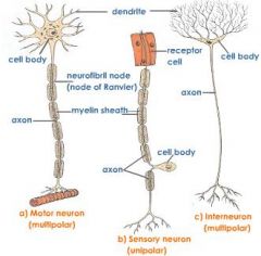
Types of Neurons 1. Multipolar Neurons 2. Bipolar Neurons 3. Pseudounipolar Neurons |
Multipolar Neurons: Have many dendrites and a single axon. Ex. CNS neurons and motor neurons Bipolar Neurons: have two processes: one dendrite and one axon. Ex. Sensory organs, such as retina of the eye and nasal cavity Pseudounipolar Neurons: have a single process extending from the cell body. Process divides into two processes a short distance from cell body. Two processes fxn as an axon with small dendrite like receptors at periphery. Axon receives sensory info at periphery and transmit it to CNS |
|
|
Types of Neurons (Pictures) |
|
|
|
Glia Cells |
Supportive cells of CNS and PNS, these cells do not conduct action potentials. Make up most of the nervous system. |
|
|
5 types of Neurolgia Cells 1. Astrocytes 2. Ependymal 3. Microglia (refer to page 198 for pics) |
Astrocytes: major supporting cells in CNS. Stimulate or inhibit signalling activity of nearby neurons. Also, form blood-brain barrier, between blood and CNS which help limit damage to neural tissue. Ependymal: cells that line the fluid-filled cavities within CNS. Produce cerebrospinal fluid, and others, with cilia on surface move cerebrospinal fluid through CNS Microglia: act as immune cells of the CNS. They help protect the brain by removing bacteria and cell debris |
|
|
4. Oligodendrocytes 5. Schwann cells |
Oligodendrocytes: Insulating material that surrounds axons. Make myelin CNS. Schwan cells: Insulating material that surrounds axons. Make myelin PNS. |
|
|
Myelin Sheaths (refer to page 198 for pics) |
Myelinated axons are covered by Insulation that surrounds an axon in segments. They function to speed the rate of conduction. The gaps between segments are called nodes of Ranvier. |
|
|
White and Gray matter |
Both CNS and PNS contain area of gray matter and white matter. Gray matter is unmyelinated. White matter is myelinated. |
|
|
Ganglion |
A cluster of nerve cells |
|
|
Continuous Conduction |
Stimulation of adjacent parts of the cell membrane on unmyelinated axons. Local currents in adjacent membrane produce and action potential. |
|
|
Saltory Conduction |
Jumping form node to node of Ranvier on a myleinated sheath causes local current to flow through surrounding extracellular fluid through cytoplasm of the axon to the next node, stimulates an action potential. |
|
|
The Synapse |
A junction where the axon of one neuron interacts with another neuron or with an effector organ (Muscle). |
|
|
Acetylcholine (Ach) |
Site of release: CNS synapse, ANS synapse, neuromuscular junctions Effect: Excitatory or inhibitory Ex: decreases in ACh is called Alzheimer disease |
|
|
Norepinephrine (NE) |
Site of release: CNS synapse and ANS synapse Effect: Excitatory Ex: Cocaine increases the release, blocks reuptake of NE causing overstimulation |
|
|
Serotonin |
Site of release: CNS synapse Effect: Inhibitory Ex: Involved with mood, anxiety and sleep induction. Elevated levels in schizophrenia |
|
|
Dopamine |
Site of release: CNS synapses and ANS synapses Effect: Excitatory and Inhibitory Ex: Parkinson disease results in destruction of dopamine |
|
|
Gamma-aminobutyric acid (GABA) |
Site of release: CNS synapse Effect: inhibitory Ex: Drugs that increase GABA fxn have been uses to treat epilepsy |
|
|
Glycine |
Site of release: CNS synapse Effect: Inhibitory Ex: Glycine receptors are inhibited by the poison strychnine, it increases the excitability of certain neurons by blocking inhibition. |
|
|
Endorphins |
Site of release: Descending pain pathways Effect: Inhibitory Ex: The opiates morphine bind to endorphins receptors on presynaptic neurons and reduce pain by blocking the release of it |
|
|
Reflex and Reflex arc |
reflex is an involuntary reaction in response to a stimulus applied to periphery and transmitted to the CNS. They allow people to act to stimuli more quickly. A reflex arc is a neuronal pathway by which a reflex occurs. |
|
|
5 components of a reflex |
1) sensory receptor 2) Sensory neuron 3) Interneurons 4) Motor Neurons 5) effector organ |
|
|
Example of a reflex (refer to page 205 for pic) |
person puts finger on hot stove. Heat stimulates sensory receptors, and action potentials are produced. Sensory neurons conduct impulse toward spinal cord where interneurons transmit info to motor neurons. Motor neurons stimulate effector muscles and fingers contract to move away from stove. |
|
|
Neuronal Pathway (refer to page 205 for pic) |
Neurons are organized in CNS to form pathways form simple to complex. Two simplest pathways are converging and diverging pathways |
|
|
Converging Pathway (Spacial Summation) |
-Two or more neurons synapse with the same neuron. Allows info transmitted in more than one neuronal path to converge into a single path. -Spacial Summation occurs when originate from different locations. |
|
|
Diverging pathway (Temporal Summaton) |
-Axon from one neuron divides and synapses with more than one neuron. Allows info transmitted in one neuronal oath to diver into two paths. - Temporal summation occurs when local potentials overlap in time. |
|
|
Where does the Spinal Cord start and finish? What is the cauda equina? (page 206 for pic) |
Extends form the foramen magnum at base of skull to second lumbar vertebra. The inferior end of spinal cord and spinal nerves exiting there resemble a horses tail and are called cauda equina. Spinal cord consist of Gray and white matter. |
|
|
Name the 3 columns of the spinal cord |
Dorsal Ventral Lateral |
|
|
What are Ascending and descending tracts? |
Ascending: axons that conduct action potentials toward the brain Descending: axons that conduct action potentials away from the brain. |
|
|
What are the Dorsal Root and Ventral Root? What do they form when they combine? (refer to page 207 for pic) |
Ventral (anterior): contain effector (motor) neurons Dorsal (posterior): contain afferent (sensory) neurons, dorsal root is characterized by its dorsal ganglion (knot) -They form a spinal nerve when they combine |
|
|
Look at page 207 for info on horns and neuron types |
page 207 |
|
|
What is a stretch reflex and knee-jerk reflex? |
Stretch: muscles contract in response to a stretching force applied to them Knee: is a classic example of a stretch reflex. When patellar ligament is tapped, quads contract. |
|
|
What is a Withdrawal Reflex? |
Is to remove a limb or another body part from a painful stimulus. Following painful stimuli, sensory neurons conduct action potentials through dorsal roots to spinal cord, where the sensory neurons synapse with interneurons, which in turn synapse with motor neurons. Muscles stimulated my motor neurons and remove limb from painful source. |
|
|
Brain stem (fxn and parts) (pics on page 213) |
connects the spinal cord to the remainder of the brain. Consists of the medulla oblongata, pons, and midbrain |
|
|
Brain stem: Medulla Oblongata |
Location: The most inferior portion of the brainstem. Function: regulate heart rate and blood vessel diameter, breathing, swallowing, vomiting, coughing, sneezing, balance, and coordination |
|
|
Brain stem: Pons |
Location: superior to medulla Function: Bridge btwn cerebrum and cerebellum. In lower pons, controls breathing, swallowing, and balance. |
|
|
Brain stem: Midbrain |
Location: superior to pons Function: Auditory nerve pathways, visual reflexes, receive touch and auditory input. |
|
|
Brain Stem: Reticular formation |
Location: Scattered throughout brainstem, basically group of nuclei Function: Regulation |
|
|
Cerebellum |
Location: Connected to brain stem Function: Motor Functions, such as, maintaining balance and muscle tone and in coordinating fine motor movement. |
|
|
Diecephalon |
Part of the brain between the brainstem and cerebrum. Components are thalamus, epithalamus, and hypothalamus |
|
|
Diecephalon: Thalmus (pic on page 214) |
Location: Superior to midbrain Function: Collects info from brainstem and sends it to correct area of the brain. Also, influences mood, and pain perception |
|
|
Diecephalon: Epithalamus |
Location: Superior and posterior to Thalamus Function: Emotional and visceral response to odors, and pineal gland |
|
|
Diecephalon: Hypothalamus |
Location: inferior to Thalamus Function: Maintaining homeostasis in body: body temp, hunger, and thirst |
|
|
Cerebrum (function of 4 lobes) |
Location: Largest part of the brain Frontal: voluntary control of motor fxns, motivation, aggression, mood Parietal: Receiving sensory info, such as touch, pain temp., and balance Occipital: Receiving Visual sensory info Temporal: Receiving auditory and smell sensory info |
|
|
Sensory Functions: Ascending Tract |
-Transmit info to the brain from sensory nerves. -Spinothalamic tract transmits info dealing with pain and temp to thalamus then to cerebral. -Spinocerebellar tract transmits info about body position to cerebellum. |
|
|
Sensory Functions: Sensory Areas of Cerebral Cortex (Look at page 217 for pic) |
Look at fig 8.27 |
|
|
Motor Functions: Motor system of brain and spinal cord |
responsoible for maintaining the body's posture and balance, as well as moving the trunk, head, limbs, tongue, and eyes and communicating though facial expressions and speech |
|
|
Involuntary movements and voluntary movements |
Involuntary: reflexes through spinal cord and brainstem that occur without conscious thought Voluntary: consciously activated to achieve a specific goal, such as running. |
|
|
Upper and lower motor neurons |
Upper: have cell bodies in cerebral cortex. Lower: have cell bodies in anterior horn of spinal cord gray matter. |
|
|
Motor Areas of Cerebral Cortex: Primary motor cortex and Premotor area |
Primary: Located in posterior porton of frontal lobe. Controls voluntary movements of skeletal muscles. Premotor Area: where motor functions are organized before they actually initiate. |
|
|
Descending Tract |
-Transmits info from brain to motor neurons -Lateral corticospinal control speed and precision of skilled movements of the hands |
|
|
Autonomic Nervous System |
comprises of motor neurons that carry action potentials from the CNS to the periphery.This is an involuntary system that innervates: smooth and cardiac muscle, and glands. ANS is split up into sympathetic (Fight or flight) and parasympathetic (Rest and digest) divisions. |
|
|
How does the ANS extend from the CNS to effector organs? |
Two neurons extend from the CNS to effector organs by pre-ganglionic neuron and post-ganglionic neuron, they are named this because they synapse together (Page 228). |
|
|
Sympathetic Division |
Axons of preganglionic neurons extend through ventral roots and project to either sympathetic chain ganglia or collateral ganglia. |
|
|
Sympathetic Division: Preganglionic and Postgaglionic (Location and innervates) (page 229) |
-Preganglionic cell bodies lie in the thoracic or upper lumbar regions of the spinal cord -Postganglionic cell bodies are located in the sympathetic chain ganglia or collateral ganglia |
|
|
Parasympathetic: Preganglionic and Postgaglionic (Location and innervates) (page 229) |
-Preganglionic cell bodies of the PS division are associated with cranial and sacral nerves -Postganglionic cell bodies are located in terminal ganglia, either near or within target organ |
|
|
Autonomic Neurotransmitters (Name the division and NTs it secretes) |
-All autonomic preganglionic and parasympathetic postganglionic neurons secrete ACh -Most sympathetic postganglionic neruons secrete norepinephrine |
|
|
Functions of the Autonomic Nervous System (Name the division and it's functions) |
1. Sympathetic division prepares a person for action by increasing heart rate, blood pressure, respiration, and release of glucose for energy 2. The parasympathetic division is involved in involuntary activities at test, such as digestion, defecation, and urnination. |
|
|
Enteric Nervous System (What does it form and how does it act independently of CNS?) |
- ENS forms plexuses in digestive tract wall - Enteric neurons are sensory, motor, or interneurons; they receive CNS input but can also function independently through local reflexes. Ex. Digestive tract stretching is detected by enteric sensory neurons, they stimulate enteric interneurons. They then stimulate motor neurons which stimulate glands to secrete. |
|
|
What are some Effects of aging on Nervous system |
1. In general, sensory and motor function decline with age 2. Mental functions, including memory, may decline with age, but varies form person to person |
|
|
Chapter 9: Define: Sense Sensation Perception |
Sense: the ability to perceive stimuli from the environment Sensation: the process initiated by stimulating sensory receptors Perception: the conscious awareness of stimuli |
|
|
Historically what are the 5 senses? |
1. Smell 2. Taste 3. Sight 4. Hearing 5. Touch |
|
|
Two Basic Sense Groups: 1. General Senses (Somatic and Visceral senses) |
-have receptors distributed over a large part of the body. Divided into two groups: somatic senses and visceral senses. Somatic: provide sensory info about the body and environment, such as Touch pressure, Temp, pain. Visceral: provide info about various internal organs, mainly pain and pressure |
|
|
Two Basic Sense Groups 2. Special Senses (Name examples) |
more specialized in structure and localized to specific parts of the body. The special senses are smell, sight, hearing, and balance |
|
|
Sensory Receptors |
are sensory nerve endings or specialized cells capable of responding to stimuli by developing action potentials. 5 types of receptors. |
|
|
5 type of receptors 1. Mechanoreceptors 2. Chemoreceptors 3. Photoreceptors 4. Thermoreceptors 5. Nociceptors |
1. Mechanoreceptors: respond to mechanical stimuli, bending/stretching 2. Chemoreceptors: respond to chemicals, such as odor molecules 3. Photoreceptors: respond to light 4. Thermoreceptors: respond to temp changes 5. Nociceptors: respond to stimuli that result in sensation of pain |
|
|
General Senses: Types of receptors 1. Free Nerve Endings (Structure and Response) |
relatively unspecialized neuronal branches similar to dendrites. They are distributed throughout almost all parts of the body. Respond to temp. itch and movement |
|
|
General Senses: Types of receptors 2. Temp. Receptors: cold & warm receptors (Response) *Why is it hard to tell if something is very hot or cold? |
Cold: respond to decreasing temp., stop responding at temp. below 12 C Warm: respond to increasing temp., stop responding at temp. above 47C -Hard to distinguish between Very hot and cold because only pain receptors are active <12C and >47C |
|
|
General Senses: Types of receptors 3. Touch receptors (5 sub types) |
Merkel Disks: small superficial nerve endings involved in detecting light touch and superficial pressure Hair Follicles: Associated with hairs, are also involved in detecting light touch. Very sensitive but not discriminative. |
|
|
General Senses: Types of receptors 3. Touch receptors (5 sub types) (Page 241 for pic) |
Meissner Corpuscles: located just deep to epidermis. They are very specific in localizing tactile sensation.
Ruffinin Corpuscles: deeper tactile receptors that play an important role in detecting continuous pressure in the skin. Pacinian Corpuscles: Deepest receptor, relay info regarding deep pressure, vibration, and position. |
|
|
Describe Pain (2 types) |
Characterized by a group of unpleasant perceptual and emotional experiences. Two Types: 1) Localized: sharp, pricking, or cutting pain resulting from rapidly conducted action potentials 2) Diffuse: Burning, or aching pain resulting from action potentials that are propagated more slowly |
|
|
Superficial Pain vs. Visceral pain |
Superficial pain sensation is highly localized as a result of stimulation of pain receptors and tactile receptors. Deep or visceral pain sensation not highly localized due to absence of tactile receptors in deep structures. |
|
|
Controlling Pain: |
Local: a treatment where chemical anesthetics are injected near sensory receptors, resulting in reduced pain sensation. General: suppressing pain through loss of consciousness. A treatment where chemical anesthetic that affect reticular formation is administered |
|
|
Controlling Pain: Gate Control Theory |
The gate control theory of pain states that non-painful input closes the "gates" to painful input, which prevents pain sensation from traveling to the central nervous system. Therefore, stimulation from another sensation suppress pain. Ex. Rubbing an injury |
|
|
Describe Referred Pain |
is perceived to originate in a region of the body that is not the source of the pain stimulus. Referred occurs when internal organs are damaged or inflamed. This is because the superficial area (pain is felt) and deeper visceral area (pain originates) converge onto the same ascending neuron in the spinal cord. Brain cannot distinguish it so the most superficial structure is referred to as the pain source, such as skin. |
|
|
Special Senses: Describe Olfaction |
The sense of smell, called olfaction, occurs in response to airborne molecules, called odorants, that enter the nasal cavity |
|
|
Describe Olfactory Neurons |
are bipolar neurons within the olfactory epithelium, which lines the superior part of the nasal cavity. The dendrites of the olfactory neurons extend to the epithelial surface, and their ends are modified with long, specialized cilia that lie in a thin mucous on the epithelial surface. Mucus traps and dissolves airborne molecules. |
|
|
Explain how airborne molecules can stimulate action potentials in the olfactory nerves |
Airborne odorants dissolve in the mucus on the surface and bind to receptor molecules on the membranes of specialized cilia. This binding causes an action potential, which is then conducted to the olfactory cortex. There are 400 olfactory receptors that allow us to smell 10,000 different smells. |
|
|
Neuronal Pathway for Olfaction |
Cilia > Olfactory neuron > Olfactory nerve > Olfactory Bulb > Ofactory tract > Olfactory cortex (in frontal lobe) |
|
|
Special Senses: Taste |
taste buds detect taste stimuli, they are oval structures located on surface of papillae, which are enlargements on the surface of the tongue. Also on other areas of mouth and pharynx. |
|
|
Explain how taste buds are stimulated |
consist of two type of cells: specialized epithelial cells form the exterior supporting capsule and the interior consist of 40 taste cells. Each taste cell has taste hairs, they then have tiny opening called taste pores. Dissolved molecs bind to receptors on taste hairs and initiate action potentials which then travel to the insula of cerebral cortex. |
|
|
What are the 5 types of taste and describe how olfaction inffluences taste |
-Sour, salty, bitter, sweet, and umami (savoury) - Taste sensation are strongly influenced by olfactory sensations |
|
|
Special Senses: Structure of the eye |
Include the eye, accessory structures, and sensory neurons. The eye is housed within bony cavities called orbits. |
|
|
Accessory Structures: Eyebrows |
Function: protect the eyes by preventing perspiration from running down the forehead and into the eyes |
|
|
Accessory Structures: Eyelids + eyelashes |
Function: protect eye form foreign objects. Eye will close quickly then open quickly if a foreign object approaches. Also, keep eye lubricated. |
|
|
Accessory Structures: Conjuctiva |
Structure: thin, transparent mucous membrane covering inner surface of eyelids and anterior surface of eye. Function: Keep eye lubricated. |
|
|
Accessory Structures: Lacrimal Apparatus |
Structure: consists of lacrimal gland situated in the superior lateral corner of orbit & nasolacrimal duct in inferior medial corner of orbit. Function: Produces tears, tears lubricate and cleanse the eye. |
|
|
Accessory Structures: Extrinsic Eye muscles |
Function: move the eye ball |
|
|
Anatomy of the eye: Fibrous Tunic (Sclera and Cornea) |
Sclera: Structure: Sclera is the firm, white, outer connective tissue layer. Function: Maintain shape of the eye, protect internal structures and attachment sites for muscle Cornea: Structure: transparent part of eye Function: allows light to enter. Bends or refracts entering light to focus it |
|
|
Anatomy of the eye: Vascular Tunic (coracoid) |
- Called vascular tunic because contains most of the blood vessel Choracoid: Structure: Very thin structure consists of vascular network and many melanin pigment cells causing it to appear black. Function: black colour absorbs light, so that it is not reflected inside the eye. |
|
|
Anatomy of the eye: Vascular Tunic (Ciliary body and Iris) |
Ciliary body: Structure: continuous with choroid, contains smooth muscles called ciliary muscles, which attach to perimeter of lens. Iris: Structure: coloured part of the eye. It is attached to the ciliary body. Function: Controls diameter of pupil |
|
|
Pupil and Lens |
Lens: flexible, biconvex, transparent disc Pupil: Light passes through it controls amount of light entering eye |
|
|
Anatomy of the eye: Nervous Tunic |
- is the innermost tunic and consists of retina Retina: Structure: retina covers the back of the eye. Consists of outer pigmented retina and inner sensory retina Pigmented retina: along with choroid, keeps light from reflecting back into the eye Sensory retina: contains photoreceptor layer which has rods and cones and interneuron layer |
|
|
Rods and Cones |
Rods: very sensitive to light and can function in dim light, do not provide colour Cones: require more light, and provide colour vision. 3 types: Blue, Green, and Red cones |
|
|
What is Rhodopsin? |
in rod cells, a photosensitive pigment which consists of a protein opsin loosely bound to a yellow pigment called retinal. |
|
|
Effect of light of Rhodopsin 6 steps How does light change into an electrical signal? |
1) Rhodopsin is composed of opsin and retinal 2) Light causes retinal to change shape, which activates rhodopsin 3) Activated Rhodopsin stimulates cell changes that result in vision 4) Following activation, retinal detaches to its original form 5) Energy from ATP is required to bring retinal back to original form 6) Retinal recombines with opsin to form rhodopsin |
|
|
Fovea and Optic Disc |
Fovea: part of the retina where light is most focused when the eye is looking directly at an object. The fovea centralis contains only cone cells and are very tightly packed Optic Disc: white spot just medial to macula through which a number of blood vessels enter the eye and spread over the retina. This sis also where the optic nerve is. There are no photoreceptor cells so it is called the blind spot. |
|
|
Chambers of the eye 3 chambers and humours |
anterior chamber & posterior chamber are filled with aqueous humour. Vitreous chamber is filled with vitreous humour. The humours keep the eye inflated, refract light and provide nutrients to the inner surface of the eye |
|
|
Functions of the eye: Light Refraction & focusing Images on the retina |
1. Light passing through a concave lens diverges. Light passing through a convex surface converges 2. Converging light rays cross at the focal point and are said to be focused 3. The cornea, aqueous humour, lens, and vitreous humour all refract light. The cornea is responsible for most of the convergence, where as the lens can adjust the focus by changing shape depending how far the object is |
|
|
Neural pathway for vision |
1. Axons pass through optic nerve to optic chiasm, where some cross. Axons from the nasal retina cross, those form the temporal retina do not 2. Optic tracts from chiasm lead to thalamus 3. Optic radiations extend from thalamus to visual cortex in occipital lobe |
|
|
Anatomy and Function of the Ear: 1. External Ear |
Structure: Auricle is the fleshy part of the external ear on the outside of the head. It opens up to the external auditory canal. Auricle collects sound waves and directs them toward external auditory canal which transmits them to the tympanic membrane. THe auditory canal is lined with hair and ceruminous glands which produce cerumen (earwax). They help prevent foreign objects from reaching the delicate tympanic membrane (eardrum). |
|
|
Anatomy and Function of the Ear: 2. Middle Ear |
Medial to tympanic membrane is the air-filled cavity of middle ear. Two covered openings on the medial side of the middle ear, oval window and round window connect the middle ear with inner ear. Middle ear contains 3 auditory ossicles: Malleus, Incus, and Stapes. These bones transmit vibrations from tympanic membrane to oval window. As these vibrations pass along the ossicles the force of them are amplified 20-fold. This is to dampen the vibrations to protect middle ear. |
|
|
Anatomy and Function of the Ear: 3. Inner Ear |
Interconnecting tunnels and chambers within temporal bone, called the bony labyrinth. Inside there is a chamber called the membranous labyrinth, it is filled with a clear fluid called endolymph, and the space between membranous and bony labyrinth is filled with a fluid called perilymph. Bony labyrinth is divided into 3 regions: cochlea (hearing), vestibule (balance), and semicircular canals (balance). |
|
|
Anatomy and Function of the Ear: 3. Inner Ear (Cochlea) |
Structure: shaped like a snail shell & contains a bony core shaped like a screw. Divided into 3 channels: scala vestiboli, scala tympani, and cochlear duct. Cochlear duct is specialized structure called spiral organ and contains sensory cells called hair cells, which sense vibrations. |
|
|
Hearing 8 steps |
1) Sound waves are collected by auricle and conducted through external auditory canal 2) Sound waves strike tympanic membrane and cause it to vibrate 3) Vibrations on tympanic cause the malleus, incus, and the stapes to vibrate 4) Base of stapes vibrates in oval window 5) Vibrations of the base of the stapes causes perilymph in the scala vestibuli to vibrate |
|
|
Hearing 8 steps |
6) vibrations of perilymph causes the vestibular membrane to vibrate, which causes vibrations in the endolymph 7) Vibrations on endolymph causes displacement of basilar membrane. short waves (high pitch) causes displacement near oval window, and longer waves (low pitch) cause displacement of basilar membrane some distance from oval window. Movement of basilar is detected by hair cells 8) Vibrations in the perilymph of the scala tympani are transferred into round window where they are dampened. |
|
|
Neural pathway for hearing |
From vestibulocochlear nerve, action potentials travel to cochlear nucleus and on to the cerebral cortex |
|
|
Balance/Equilibrium (Static and Dynamic) |
Has two components: Static and dynamic equilibrium Static: associated with the vestibule and is involved in evaluating the position of the head relative to gravity Dynamic: associated with the semicircular canals and is involved in evaluating changes in direction and rate of head movements |
|
|
Describe how equilibrium is detected in both |
1. Static equilibrium evaluates the position of the head relative to gravity 2. Maculae, located in the vestibule, consists of hair cells with the microvilli embedded in a gelatinous mass that contains otoliths. The gelatinous mass moves in response to gravity. 3. Dynamic equilibrium evaluates movements of the head 4. The inner ear contains three semicircular canals, arranged perpendicular to each other. The ampulla of each semicircular canal contains a crista ampullaris, which has hair cells with microvilli embedded in a gelatinous mass, the cupula |
|
|
Neural Pathways for balance |
Axons in vestibular portion of vestibulocochlear nerve project to the vestibular nucleus and on the cerebral cortex |
|
|
Chapter 10: Chemical Messengers |
Allow cells to communicate with each other to regulate body activities. |
|
|
Classes of Chemical Messengers: 1. Autocrine Chemical Messengers |
-Stimulates cell that originally secreted it and similar cells of same type around it Ex. Chemical messengers secreted by white blood cells to stimulate their own replication |
|
|
Classes of Chemical Messengers: 2. Paracrine Chemical Messengers |
-Act locally on nearby cell. -Secreted by one cell type into extracellular fluid and affect surrounding cells of a different type Ex. Histamine, released by certain white blood cells during allergic reaction. They stimulate vasodilation in nearby blood vessels |
|
|
Classes of Chemical Messengers: 3. Neurotransmitters |
-Chemical messengers secreted by neurons that activate an adjacent cell, whether it be another neuron, a muscle cell, or gland. -They are secreted into synaptic cleft Ex. Acetycholine, Epinephrine |
|
|
Classes of Chemical Messengers: 4. Endocrine Chemical Messengers |
-Messengers secreted into bloodstream by certain glands and cells, which together constitute the endocrine system -Affect cells that are distant from their source |
|
|
10 Regulatory Functions of Endocrine System |
1. Metabolism 6. Heart Rate and Blood Pressure 2. Control of food intake 7. Control of blood glucose & nutrients 3. Tissue Development 8. Control of reproductive functions 4. Ion Regulation 9. Uterine Contractions 5. Water Balance 10. Immune system regulation
|
|
|
Describe the Endocrine system and its function |
Endocrine system is composed of endocrine glands and cells. These glands and cells secrete small amounts of chemical messengers called hormones into the blood. Hormones then travel to target tissue where they produce a response on that target tissue. |
|
|
Distinguish Between endocrine and exocrine glands |
Exocrine: have ducts that carry their secretions to outside the body, such as stomach or intestines (Secretions of digestive enzymes) Endocrine: secrete into blood and carry secretions inside the body, such as muscles |
|
|
Describe Lipid-Soluble Hormones |
-Non-polar molecules -Include: Steroid, thyroid, and fatty acid derivative hormones. -Low solubility and small size allows them to travel in blood -Must bind to binding proteins to prevent diffusion out of blood at capillaries |
|
|
Describe Water-Soluble Hormones |
-Polar molecules -Include: Protein, peptide, and amino acid derivative hormones -Solubility allows them to dissolve in blood, circulating as free hormones -Are large enough so they don't need binding proteins to travel |
|
|
Describe three ways of regulating hormones |
-All hormones are destroyed either in the circulation or at their target cells, lysosomal enzymes degrade them -Water-soluble have a short half-life because they are rapidly degraded by enzymes called protease -Some Hormones are filtered out in the capillaries |
|
|
3 ways of regulating hormone release: 1. Control by Humoral Stimuli (Describe & Example) |
-Blood-borne chemicals that directly release hormones Ex. If a runner finishes a long race he may no produce urine for 12 hrs, this is due to [high] of blood solutes stimulates the release of water conservation hormone Antidiuretic hormone (ADH) |
|
|
3 ways of regulating hormone release: 2. Control by Neural Stimuli (Describe & Example) |
-After an action potential, neurons release a NT into the synapse with the cells that produce the hormone, NT stimulate cells to increase hormone secretion Ex. Autonomic NS stimulates adrenal gland to secrete epinephrine and norepinephrine, which help body respond to a stimulus, during exercise. (raise heart rate) |
|
|
3 ways of regulating hormone release: 3. Control by Hormone Stimuli (Describe & Example) |
-Occurs when a hormone is secreted that, in turn, stimulates the secretion of other hormones Ex. Hormones from anterior pituitary gland, called tropic hormones. Hypothalamus sends hormones to secrete hormones from the pituitary gland. |
|
|
3 ways of Inhibiting hormone release: 1. Inhibition of Humoral Stimuli (Describe & Example) |
-A companion hormone's effects oppose those of secreted hormone and counteract the secreted hormone's action. Ex. To raise blood pressure, the adrenal cortex secretes the hormone aldosterone in response to low blood pressure. However, if blood pressure goes up, the atria of the heart secrete the hormone atrial natriuretic peptide, which lowers blood pressure. Therefore, the two hormones work together to maintain homeostasis of blood pressure. |
|
|
3 ways of Inhibiting hormone release: 2. Inhibition of Neural Stimuli (Describe & Example) |
-Neurons inhibit targets just as often as they stimulate targets. If neurotransmitter is inhibitory, the target endocrine gland does not secrete the hormone. |
|
|
3 ways of Inhibiting hormone release: 3. Inhibition of Hormonal Stimuli (Describe & Example) |
-some hormones prevent the secretion of other hormones Ex. Hormones from the hypothalamus that prevent the secretion of tropic hormones from pituitary gland. |
|
|
Negative Feedback of hormones |
Hormone secretion is inhibited by the hormone itself once blood levels have reached a certain point and there is adequate hormone to activate the target cell. Ex. Thyroid hormones inhibit the secretion of their releasing hormone from the hypothalamus and their tropic hormone from anterior pituitary |
|
|
Positive Feedback of hormoens |
Some hormones, when stimulated by tropic hormones, promote the synthesis and secretion of the tropic hormone in addition to stimulating their target cell. This promotes secretion of original hormone Ex. prolonged estrogen stimulation promotes a release of anterior pituitary hormone |
|
|
Classes of Receptors 1. Lipid-soluble hormones bind to... |
nuclear receptors located inside the nucleus of the target cell. |
|
|
Classes of Receptors 2. Water-soluble hormones bind to... |
membrane-bound receptors, which are integral membrane proteins |
|
|
Action of Nuclear Receptors (What happens when it binds?) |
1. Nuclear receptors have proteins that allow them to bind to DNA in the nucleus once the hormone is bound - Hormone activates genes, which in turn activate DNA to produce mRNA -mRNA increases the synthesis of certain proteins that produce the target cell's response 2. Nuclear receptors cannot respond immediately because it takes time to produce mRNA and protein |
|
|
Actions of Membrane-Bound Receptors (What happens when they bind?) |
1) receptors activate a cascade if events once hormone binds 2) receptors are associated with membrane proteins called G proteins. When hormone binds to receptor, G proteins are activated. G proteins bind to ion channels and cause them to open or change the rate of synthesis of intracellular fxns, such as cAMP (activates protein kinase) 3) Second-messenger systems act rapidly because they act on already existing enzymes and produce amplification effect that produces cells response |
|
|
Hormonal Control of Anterior Pituitary Gland (page 277 for pic) |
1) Neurons of hypothalamus produce neuropeptides that act on cells of anterior pituitary. Act as either releasing or inhibiting hormones. 2) Releasing hormones and inhibiting hormones pass through hypothalamic-pituitary portal system to anterior pituitary 3) Releasing hormones and inhibiting hormones leave capillaries and stimulate or inhibit the release of hormones from anterior pituitary 4) In response to releasing hormones anterior pituitary hormones travel in blood to their target tissues, which in some cases, are other glands. |
|
|
Hormonal Control of Posterior Pituitary Gland |
1) Stimuli in nervous system cause hypothalamic neurons to either increase or decrease their action potential frequency 2) Action potentials are conducted by axons of hypothalamic neurons through hypothalamohypophysical tract to posterior pituitary. 3) In the posterior pituitary gland, action potentials cause the release of neurohormones from axon terminals through circulatory system 4) The neurohormones pass through the circulatory system and influence the activity of their target tissues |
|
|
Type 1 Diabetes (Cause & Effect) |
Causes: Occurs when too little insulin is secreted from pancreas Effect: -Tissues cannot take up glucose effectively, blood glucose levels become very high, a condition called hyperglycaemia. -Excessive glucose in blood causes increased urine production, resulting in dehydration. -Ketoacidosis can occur |
|
|
Type 2 Diabetes (Cause & Effect) |
Cause: insulin insensitivity that develops overtime Effect: similar to type 1 diabetes |